
Abstract
E2 enzymes in ubiquitin-like conjugation pathways are important, highly challenging pharmacological targets, and despite significant efforts, few noncovalent modulators have been discovered. Small-molecule microarray (SMM)–based screening was employed to identify an inhibitor of the “undruggable” small ubiquitin-like modifier (SUMO) E2 enzyme Ubc9. The inhibitor, a degradation product from a commercial screening collection, was chemically synthesized and evaluated in biochemical, mechanistic, and structure-activity relationship studies. Binding to Ubc9 was confirmed through the use of ligand-detected nuclear magnetic resonance, and inhibition of sumoylation in a reconstituted enzymatic cascade was found to occur with an IC50 of 75 µM. This work establishes the utility of the SMM approach for identifying inhibitors of E2 enzymes, targets with few known small-molecule modulators.
Introduction
The posttranslational attachment of the small ubiquitin-like modifier (SUMO) protein plays a critical role in cellular homeostasis. Although ubiquitin conjugation traditionally targets proteins for degradation, modification of a protein substrate with SUMO modulates a variety of factors such as protein stability, localization, or function. 1 SUMO conjugation occurs through an enzymatic cascade similar to ubiquitination, involving E1-activating, E2-conjugating, and E3-ligating enzymes. But unlike ubiquitination, which employs more than 30 E2s, the SUMO pathway uses a single E2 enzyme, Ubc9. Abnormal sumoylation is associated with several diseases, including cancers such as ovarian carcinoma, lung adenocarcinoma, and melanoma, as well as ischemia. Ubc9 is therefore a desirable target for the development of anticancer therapeutics. 2
Ubiquitin-like E2s are difficult drug targets due to their overall rigid conformations, lack of “druggable” pockets, and numerous protein-protein interaction sites. Consequently, identifying inhibitors of Ubc9 has been challenging, with few small-molecule ligands reported to date. Existing Ubc9 inhibitors include the natural product spectomycin B1 (IC50 = 4.4 µM) 3 as well as a series of small molecules computationally predicted to bind to Ubc9, although limited structural evidence for their binding modes exists. 4 Our group previously reported the inhibitor 2-D08 (IC50 = 6.0 µM), which prevents the transfer of SUMO from the Ubc9~SUMO thioester complex to its substrate. 5 A more recent fragment-based approach revealed an allosteric small-molecule binding site on Ubc9 that inhibits thioester formation, with fragments having IC50 values in the low millimolar range. 6 Inhibitors that target the SUMO E1 enzyme, including ginkgolic acid and tannic acid, have also been reported. To date, these existing inhibitors of sumoylation suffer from weak potency in biochemical or cell-based assays, polyphenolic structures, redox activity, pleiotropic mechanisms of action, or a combination of these issues. The difficulty with identifying selective, reversible inhibitors of sumoylation is highlighted by a study from GlaxoSmithKline that did not find any valid noncovalent inhibitors from a screen of over two million compounds. 7 New approaches are thus needed to uncover novel Ubc9-binding ligands and develop chemical probes for the important sumoylation pathway. Small-molecule microarray (SMM) high-throughput screening provides a unique strategy for the discovery of small molecules that bind directly to Ubc9 and inhibit sumoylation.
SMMs allow for profiling small-molecule/protein interactions and have been used to identify compounds that inhibit various “undruggable” targets.8–12 To generate the microarrays, a robotic microarrayer prints glass microscope slides with an arrangement of duplicate spots, with each pair containing a unique small molecule covalently linked to the glass surface. Arrays can be incubated with a fluorescently tagged protein of interest or a fluorescent antibody that recognizes the protein. The slides are then analyzed for fluorescence at each of the printed spots, where an increase in fluorescence reports on protein binding. Although SMM technology has been employed for a variety of macromolecular targets, it has never been applied to the recalcitrant class of E2 enzymes. SMMs, with the ability to screen thousands of structures simultaneously and success in identifying ligands for challenging targets, present a unique, high-throughput platform well suited for the discovery of novel Ubc9-binding compounds. We present herein the first example of applying SMM technology to any E2 enzyme with screening and validation methods that could be broadly applicable to other members of this enzymatic class.
Materials and Methods
General Remarks
Adenosine 5′-triphosphate (ATP) magnesium salt, dithiothreitol (DTT), and tris(2-carboxyethyl)phosphine hydrochloride (TCEP) were purchased from Sigma-Aldrich (St. Louis, MO). AlexaFluor 647 C2-Maleimide (AF-647) was purchased from ThermoFisher Scientific (Waltham, MA). Compounds identified from the small-molecule microarray screen were purchased from ChemBridge (San Diego, CA) or ChemDiv San Diego, CA). The following recombinant proteins were purchased and used without further purification in biochemical assays: SUMO E1 (E-315; Boston Biochem, Cambridge, MA), Ubc9 (BML-UW9320; Enzo Life Sciences, Farmingdale, NY), RanGAP1 fragment (GST-tag, BML-UW9755; Enzo Life Sciences), SUMO-1 (His-tag, UL-715; Boston Biochem), SUMO-1 Fluorescein (UL-735; Boston Biochem), UBE1 (UB101; Life Sensors, Malvern, PA), UBE2B (His-tag, E2-613, Boston Biochem), UbcH5b (BML-UW0565; Enzo Life Sciences), and Ubiquitin N-terminal Fluorescein (U-580; Boston Biochem). See supplementary material for additional experimental procedures.
Protein Labeling
Ubc9 was buffer exchanged into 100 mM Tris (pH 8) via centrifugal filtration (3000 Da MWCO; EMD Millipore, Billerca, MA) and concentrated to ~1.5 mg/mL. To 50 µL of the protein solution was added TCEP (0.5 µL of 100 mM) and AF-647 (1.5 µL of 10-mM DMSO stock) followed by incubation in the dark at 4 °C overnight. Unreacted dye was quenched by the addition of DTT (5 µL of 100 mM), and the labeled protein was purified by gel filtration through a Sephadex PD-10 (GE Life Sciences, Pittsburgh, PA) column preequilibrated with phosphate-buffered saline (PBS, pH 7.4) and then concentrated by centrifugal filtration. Labeling and purity were assessed using sodium dodecyl sulfate polyacrylamide gel electrophoresis (SDS-PAGE) as well as absorbance measurements collected on a NanoDrop 2.0 Spectrophotometer (NanoDrop, Wilmington, DE).
SMMs
SMMs were printed, and screening and analysis were performed as previously described.8,10,11 Briefly, microarray slides were incubated with 5 mL blocking buffer (1% bovine serum albumin [BSA] in PBS with Tween 20 [PBST]) in Nunc 4-well plates (ThermoFisher Scientific) wrapped in aluminum foil with gentle shaking for 30 min. Slides were washed three times with 0.1% BSA in PBST and then incubated with labeled Ubc9 (500 nM in 0.1% BSA/PBST) for 2 h at room temperature using the parafilm technique. In this technique, a 4 × 4–in. square of parafilm was taped to a smooth, flat surface. Labeled protein (500 µL) was pooled on the parafilm, and the slide was slowly inverted into the buffer, printed side down, beginning with the end opposite the barcode and allowing the protein solution to spread over the slide as it was lowered. Aluminum foil or a small cardboard box was placed over the slide to protect it from light during the incubation. After 2 h, the slide was gently lifted, taking care not to scratch the surface, and washed in a Nunc plate three times with PBST and once briefly with deionized water followed by centrifugation at 1000 rpm for 2 min to dry. Fluorescence of the small-molecule screening slide was immediately measured using a GenePix (Molecular Devices, Sunnyvale, CA) 4000a Microarray Scanner and analyzed for hits by comparison to a slide incubated with buffer alone.
Sumoylation Assays
The in vitro sumoylation assay was performed in 20 µL reaction buffer (50 mM Tris [pH 9], 5 mM MgCl2, 1 mM DTT) containing SUMO E1 (0.1 µM), Ubc9 (0.15 µM), SUMO-1 (His-tag, 1.4 µM), and a fluorescent peptide (FL-AR) substrate 5 (1 µM) incubated with various concentrations of small molecule in DMSO or DMSO alone (4% final concentration). Experiments with mutant Ubc9, obtained as previously described, 6 were conducted according to the same procedure using K59A or E42A Ubc9 in place of the wild-type E2. The reaction was then initiated by the addition of ATP (2 mM final concentration) and incubated at room temperature for 90 min. Following incubation, samples were quenched by the addition of NuPAGE 4X LDS sample buffer (ThermoFisher Scientific), heated at 95 °C for 5 min, loaded onto NuPAGE Novex 4-12% Bis-Tris Protein Gels (ThermoFisher Scientific), resolved by electrophoresis, and visualized by an in-gel fluorescence method using an ImageQuant LAS 4000 (GE Life Sciences) imaging system at an appropriate wavelength (488 nm excitation with blue Epi-RGB light, Y515 Di emission filter, and F0.85 iris).
Alternatively, the sumoylation assay was performed using a recombinant RanGAP1 fragment as the substrate with SUMO E1 (0.05 µM), Ubc9 (0.08 µM), SUMO-1 Fluorescein (0.1 µM), and the RanGAP1 fragment (0.25 µM) incubated with small molecules or DMSO in a 20-µL reaction buffer. Following ATP addition and incubation at room temperature for 1 min, reactions were quenched by the addition of LDS sample buffer and analyzed as described above.
Thioester Bond Formation Assay
The E1~SUMO-1 thioester bond formation assay was performed in a 20-µL reaction buffer (50 mM Tris [pH 8], 5 mM MgCl2) containing SUMO E1 (0.35 µM) and SUMO-1 Fluorescein (0.5 µM) incubated with various concentrations of small molecule in DMSO or DMSO alone (4% final concentration). The reaction was initiated by the addition of ATP (2 mM final concentration). Following incubation at 37 °C for 20 min, samples were quenched by the addition of NuPAGE 4X LDS sample buffer, resolved by SDS-PAGE, and visualized by in-gel fluorescence imaging.
Sumoylation Electrophoretic Mobility Shift Assay
The in vitro sumoylation assay was executed as previously described.5,6 The assay was performed in a 384-well plate format in 20 µL Tris buffer (50 mM Tris [pH 9], 5 mM MgCl2, and 1 mM DTT) with SUMO E1 (0.1 µM), SUMO-1 (His-tag, 1.4 µM), Ubc9 (0.15 µM), fluorescent peptide FL-AR (1 µM), and small molecule (4% DMSO final). For experiments with detergent, either Triton X-100 or Tween 20 (0.01%, v/v) was incorporated into the assay buffer as well. The reactions were initiated by the addition of ATP (2 mM final concentration). After 90 min, EDTA (0.25 M, 8 µL) was added to each well to quench the reactions. Samples were analyzed using a LabChip EZ Reader II (Caliper Life Sciences, Waltham, MA) with the following run conditions: downstream voltage of −500 V, upstream voltage of −2500 V, and pressure of −1.0 psi. The sumoylation reactions were analyzed by quantifying percent conversion, defined as 100 × P/(P + S), where P is peak height of sumoylated product SUMO-1-FL-AR and S is peak height of peptide substrate FL-AR. Data were normalized to positive and negative controls (ginkgolic acid and DMSO, respectively).
Carr-Purcell-Meiboom-Gill Nuclear Magnetic Resonance
Ubc9 was buffer exchanged into 100 mM sodium phosphate buffer (pH 6) by centrifugal filtration (3000 Da MWCO; EMD Millipore) and concentrated. Samples of
Results and Discussion
SMMs were used to screen a collection of 19,200 commercially available compounds for binding to fluorescently tagged Ubc9 ( Fig. 1 ). This library of compounds, described previously, was assembled to represent a chemically diverse collection that broadly fits within modern definitions of “druglike” chemical space.10,11 SMMs were prepared using isocyanate surface chemistry to immobilize the small molecules to the glass surface via an amine or alcohol handle. 8 Ubc9 was labeled with a thiol-reactive, maleimide-containing AlexaFluor 647 dye to achieve a 1:1 dye/protein stoichiometry. Ubc9 contains two free cysteine residues, the catalytic cysteine (C93) and a remote cysteine (C138), of comparable reactivity. We anticipated that the entire surface of the protein would be available for potential ligand binding by screening Ubc9 with a distribution of labeled cysteines, enabling the discovery of molecules that bind to allosteric sites or the active site. We prepared a similarly labeled UbcH5b, a ubiquitin E2 enzyme that contains only one reactive cysteine in the active site, to screen in parallel. SMM slides were then incubated with each fluorescently labeled protein. Protein-incubated slides were compared to buffer-incubated slides, and composite z scores were generated for each printed compound on the array. This approach yielded 133 hits for Ubc9 with a z score greater than 4, for an overall hit rate of 0.69%. Among these, 34 of the most promising hits were selected based on high z scores, lack of binding to UbcH5b, and visual inspection of array data and chemical structures then purchased for evaluation of biochemical activity ( Suppl. Fig. S1 ).
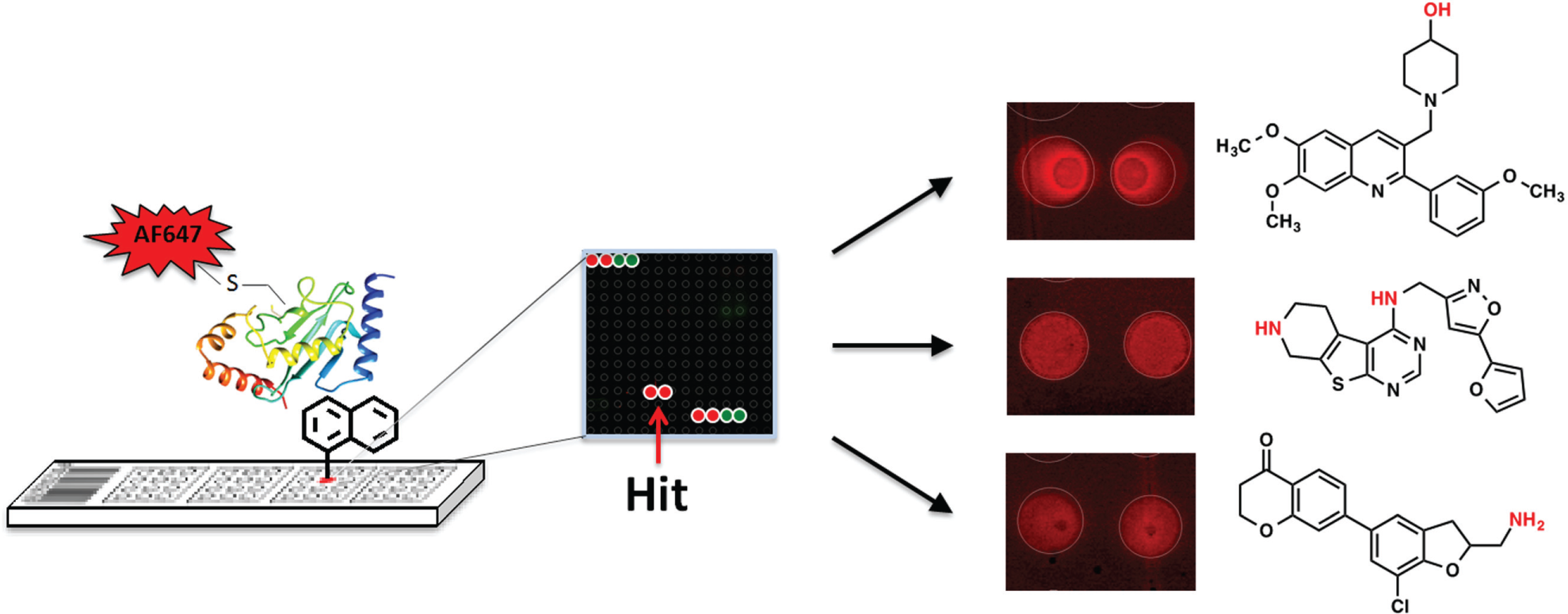
Small-molecule microarray screening approach for identifying compounds that bind to fluorescently tagged Ubc9. Structural points of attachment to the glass slide are indicated in red.
The ability of each compound to inhibit sumoylation in a reconstituted enzymatic cascade was measured at a single concentration through monitoring the conjugation of SUMO-1 to a fluorescently labeled peptide substrate by microfluidic electrophoretic mobility shift using an assay previously developed in our laboratory (
Fig. 2A
and
Suppl. Fig. S2
).
5
Compounds that caused at least a 25% decrease in sumoylation activity compared to controls at this single concentration were investigated in dose-response format to obtain full inhibitory curves (
Suppl. Fig. S3
). Several possible leads exhibited either poor curves or poor solubility and were not pursued further. However, one compound with the reported structure
(
The purity of the commercial sample of
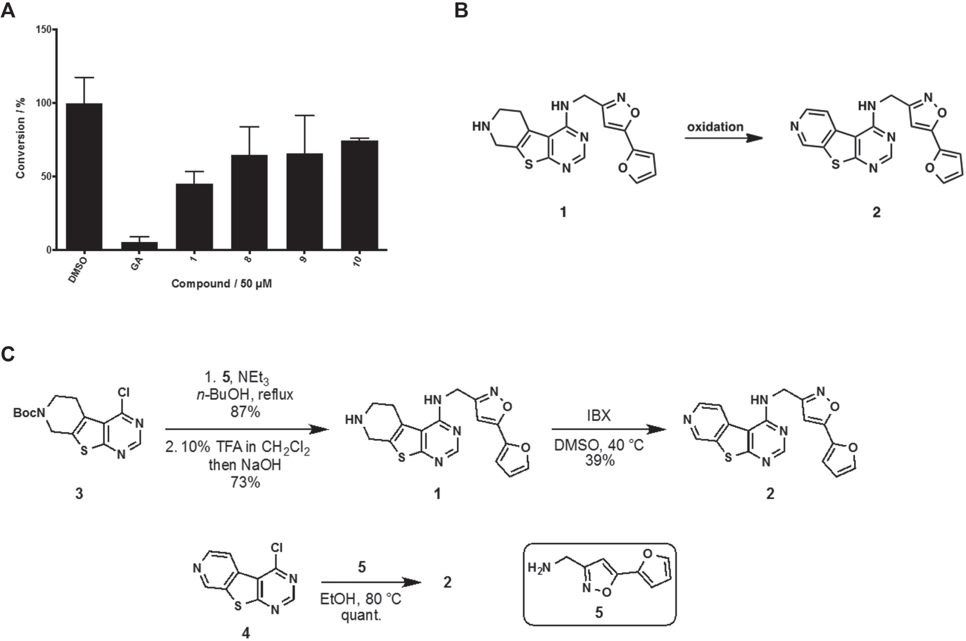
The syntheses of
Evaluation of synthetic samples of both
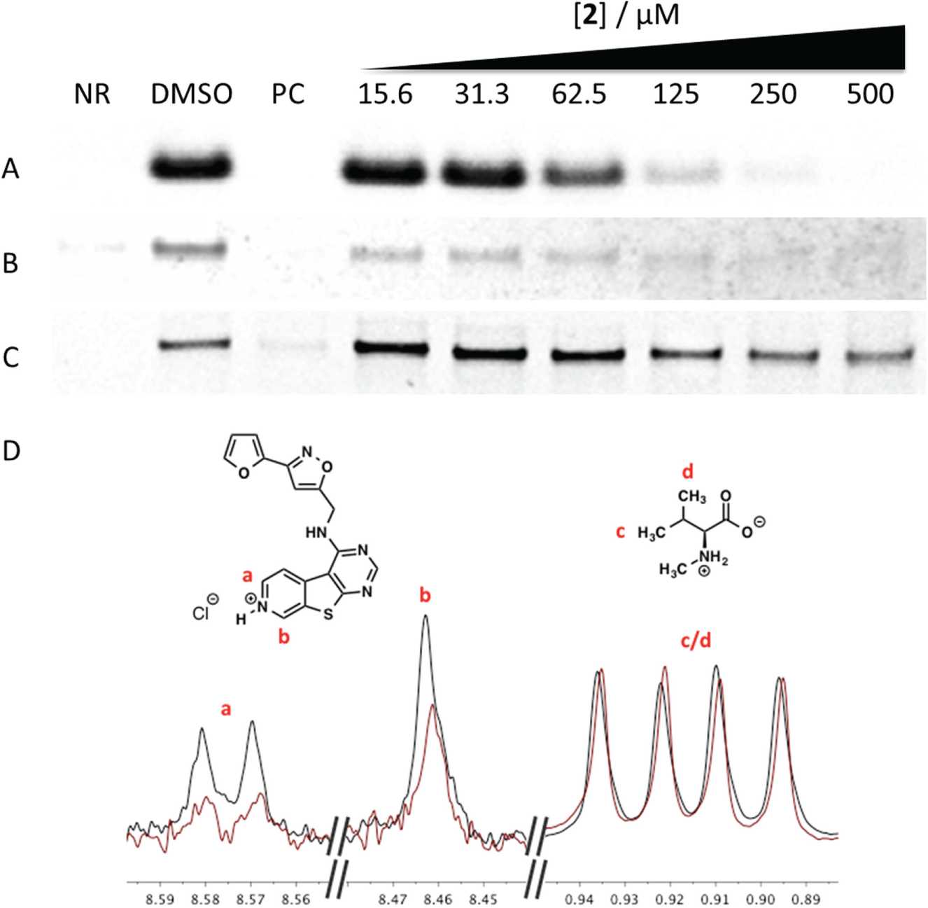
Inhibition of small ubiquitin-like modifier 1 (SUMO-1) conjugation to (
The structure-activity relationship (SAR) of the molecule was then studied. A series of truncated analogues were investigated in the microfluidic sumoylation assay (
Table 1
). Chloropyrimidine
Inhibition of Sumoylation by the Small-Molecule Microarray Hit
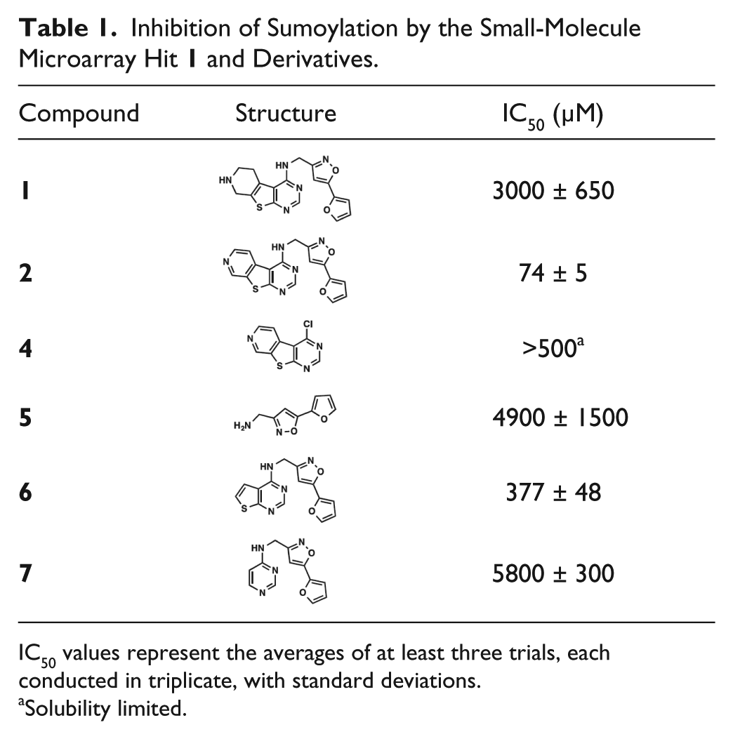
IC50 values represent the averages of at least three trials, each conducted in triplicate, with standard deviations.
Solubility limited.
A major concern with small molecules identified in high-throughput screening is the prevalence of false positives that nonspecifically inhibit enzymes due to aggregation in aqueous solution. One method of detecting artifactual inhibition is to incorporate nonionic detergent into the assay buffer. Compound
We performed ligand-observed CPMG (LO-CPMG) T2 relaxation NMR experiments to confirm that
To further study the biochemical behavior of
In summary, we have reported the first example of an SMM screening approach used to identify small molecules that bind to the SUMO E2 enzyme Ubc9. The applicability of this approach is evidenced through the discovery of
Footnotes
Acknowledgements
We thank Dr. S. Tarasov and M. Dyba (Biophysics Resource, SBL, NCI at Frederick) for assistance with high-resolution mass spectrometry (HRMS) and DLS measurements as well as Dr. M. Hoffmann (Department of Chemistry and Biochemistry, The College at Brockport, SUNY) for providing the 1D CPMG with soft excitation sculpting pulse sequence.
Supplementary material is available online with this article.
Declaration of Conflicting Interests
The authors declared no potential conflicts of interest with respect to the research, authorship, and/or publication of this article.
Funding
The authors disclosed receipt of the following financial support for the research, authorship, and/or publication of this article: This project has been funded in whole or in part with federal funds from the National Cancer Institute, National Institutes of Health, under contract HHSN26120080001E. The content of this publication does not necessarily reflect the views or policies of the Department of Health and Human Services, nor does mention of trade names, commercial products, or organizations imply endorsement by the U.S. Government. This Research was supported in part by the Intramural Research Program of the NIH, National Cancer Institute, Center for Cancer Research.
References
Supplementary Material
Please find the following supplemental material available below.
For Open Access articles published under a Creative Commons License, all supplemental material carries the same license as the article it is associated with.
For non-Open Access articles published, all supplemental material carries a non-exclusive license, and permission requests for re-use of supplemental material or any part of supplemental material shall be sent directly to the copyright owner as specified in the copyright notice associated with the article.