
Abstract
Pelizaeus-Merzbacher disease, a rare X-linked recessive disease occurring predominantly in males, is a disorder of proteolipid protein expression in myelin formation in the central nervous system. The disease is clinically manifested by neurodevelopmental delay, ataxia, hypotonia, and pendular eye movement. It is best confirmed by genetic study. A 4-year female child presented with ataxia, neuroregression, decreased scholastic performance, slurred speech, loss of bladder and bowel control, and hypotonia. MRI brain showed generalized hypomyelination and atrophy of the cerebrum and cerebellum. This case highlights that Pelizaeus-Merzbacher disease can be considered even in a female child who presented with neurodevelopmental delay and neuro regression, ataxia, and decreased scholastic performance and further confirmed by MRI showing diffuse demyelination along with cerebral and cerebellar atrophy.
Introduction
Pelizaeus–Merzbacher Disease (PMD) is a rare X-linked disease of proteolipid protein gene (PLP1) expression (locus at Xq22) in the central nervous system (CNS) myelin formation which was described clinically by Pelizaeus in 1885 and later in 1990, while clinical presentation and pathological features of cases belonging to the same family was documented by Merzbacher. 1 The product of the PLP1 gene affects the development of oligodendrocytes in CNS for myelin formation. The lack of normal white matter myelination is important pathology in PMD. 2 This disease clinically presents with neurodevelopmental delay and progressive hypotonicity in infancy. 3 Symptoms initially start before 3 months of age and gradually progress. 4
Brainstem auditory and somatosensory evoked potential tests are frequently abnormal in PMD. 5 Brain MRI finding is highly suggestive of a diagnosis of PMD which showed the low signal intensity of unmyelinated white matter structures on the T1 sequence and high signal intensity of these structures on the T2 sequence, along with diffuse cerebral and cerebellar atrophy. 2
Case Presentation
A 4-year female, born of non-consanguineous marriage presented to the emergency department with complaints of difficulty in walking followed by slurring of speech for 4 to 5 months. Initially the patient had difficulty in walking, followed by swaying of the body sidewards. With progression of the disease, she started to fall. Gradually she developed slurring of speech. After that, she also lost bladder and bowel control. Mother had also noticed a decrease in understanding and scholastic performance. The child had no history of any head trauma, convulsion, or exanthematous fever. The child had a normal vaginal delivery at term gestation, with an immediate cry. The child had normal development up to 3 years of age in all four domains.
On examination, the child was conscious with microcephaly (head circumference of 45 cm i.e. below 3rd percentile) without any ocular finding. The child had the power to lift both the upper and lower limbs against gravity but not against resistance. The child had hypotonia in all limbs with bilateral planter extensor with increased deep tendon reflexes and cerebellar Romberg sign positive (Figure 1).

Four-year female with microcephaly.
In the investigation, routine complete blood count (CBC), kidney function test (KFT), liver function test (LFT), and electrolytes were normal. In MRI Brain, mild T2/FLAIR hyperintensity with T1 isointensity in bilateral periventricular deep white matter, bilateral internal capsule were observed. Similar changes in the cerebral hemisphere, brain stem, and cerebellum were present (Figures 2 and 3).

T1 iso intensity in bilateral periventricular deep white matter in the hemisphere.
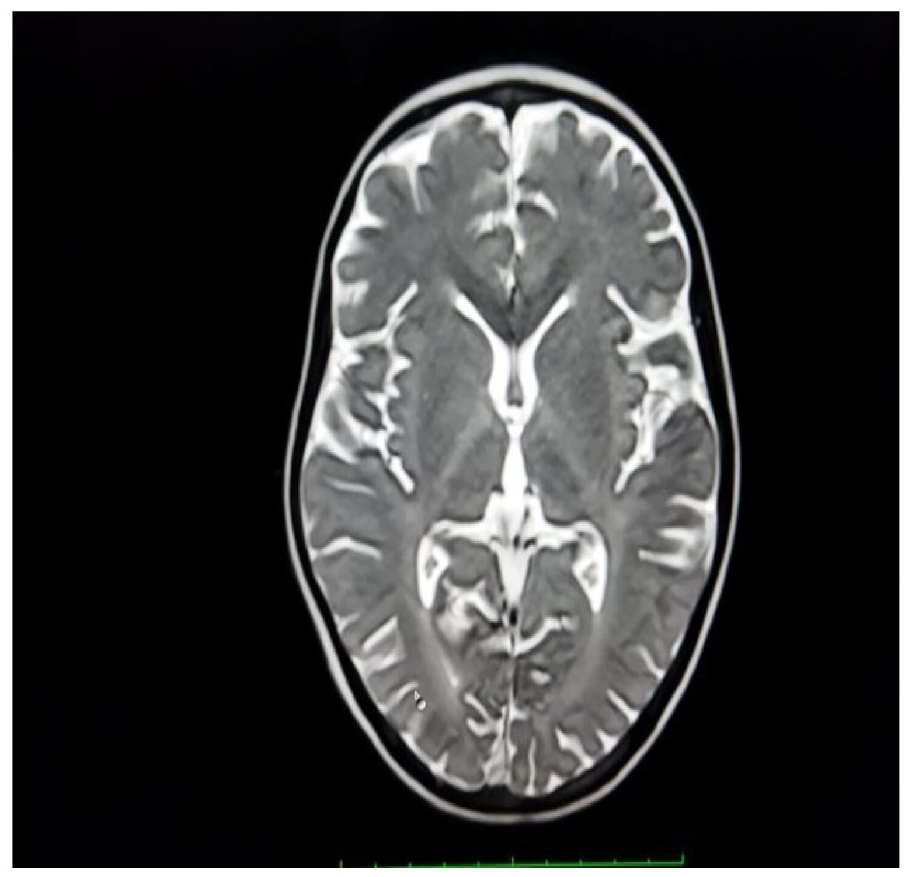
T2/FLAIR hyperintensity in bilateral periventricular deep white matter in the hemisphere.
Discussion
This case highlights a rare presentation of PMD in a female patient. PMD is an X-linked recessive disease that predominantly occurs in males and rarely occurs in females. In female patients, X-linked recessive inheritance pattern by gene mutation and skewed X inactivation needs to take place for his disease to present.6,7 Also, in a remote area like in this case, where genetic testing is not available and not affordable for families, PMD can be reliably diagnosed by MRI imaging as unique features of PMD are distinctively observed on imaging. 8
PMD can be categorized into four subgroups—classic and conatal (rare and severe), transitional, and adult (spastic paraplegia) forms. The most common classic form is presented with nystagmus, hypotonia, ataxia, dystonia, and cognitive delay. 9 This case is an example of the classic form. The MRI of this case revealed mild T2/FLAIR hyperintensity with T1 iso intensity in bilateral periventricular deep white matter in the cerebral hemisphere, B/L internal capsule, cerebrum hemisphere, brain stem, and cerebellum; which is similar to the MRI diagnostic criteria for PMD. 2
In conclusion, even a female child with microcephaly, neurodevelopmental delay, neuro regression, ataxia, and decreased scholastic performance can be considered as PMD and subsequently confirmed by MRI showing diffuse demyelination along with cerebral and cerebellar atrophy. It is best confirmed by the genetic study. Treatment of patients with PMD is symptomatic and not specific and the disease is almost always fatal.
Supplemental Material
sj-jpg-1-gph-10.1177_2333794X231157979 – Supplemental material for Pelizaeus-Merzbacher Disease in a 4-Year-Old Female Child: “A Rare Case Report”
Supplemental material, sj-jpg-1-gph-10.1177_2333794X231157979 for Pelizaeus-Merzbacher Disease in a 4-Year-Old Female Child: “A Rare Case Report” by Devesh Gagan, Sudesh Kumar, Piyali Bhattacharya and Simranjit Kaur in Global Pediatric Health
Supplemental Material
sj-jpg-2-gph-10.1177_2333794X231157979 – Supplemental material for Pelizaeus-Merzbacher Disease in a 4-Year-Old Female Child: “A Rare Case Report”
Supplemental material, sj-jpg-2-gph-10.1177_2333794X231157979 for Pelizaeus-Merzbacher Disease in a 4-Year-Old Female Child: “A Rare Case Report” by Devesh Gagan, Sudesh Kumar, Piyali Bhattacharya and Simranjit Kaur in Global Pediatric Health
Supplemental Material
sj-jpg-3-gph-10.1177_2333794X231157979 – Supplemental material for Pelizaeus-Merzbacher Disease in a 4-Year-Old Female Child: “A Rare Case Report”
Supplemental material, sj-jpg-3-gph-10.1177_2333794X231157979 for Pelizaeus-Merzbacher Disease in a 4-Year-Old Female Child: “A Rare Case Report” by Devesh Gagan, Sudesh Kumar, Piyali Bhattacharya and Simranjit Kaur in Global Pediatric Health
Supplemental Material
sj-jpg-4-gph-10.1177_2333794X231157979 – Supplemental material for Pelizaeus-Merzbacher Disease in a 4-Year-Old Female Child: “A Rare Case Report”
Supplemental material, sj-jpg-4-gph-10.1177_2333794X231157979 for Pelizaeus-Merzbacher Disease in a 4-Year-Old Female Child: “A Rare Case Report” by Devesh Gagan, Sudesh Kumar, Piyali Bhattacharya and Simranjit Kaur in Global Pediatric Health
Footnotes
Author Contributions
Devesh Gagan: Data acquisition, manuscript preparation
Sudesh Kumar: conceptualization, design, editing, manuscript review
Piyali Bhattacharya: conceptualization, design, editing
Simranjit Kaur: Data acquisition, manuscript preparation
Declaration of Conflicting Interests
The author(s) declared no potential conflicts of interest with respect to the research, authorship, and/or publication of this article.
Funding
The author(s) received no financial support for the research, authorship, and/or publication of this article.
Supplemental Material
Supplemental material for this article is available online.
References
Supplementary Material
Please find the following supplemental material available below.
For Open Access articles published under a Creative Commons License, all supplemental material carries the same license as the article it is associated with.
For non-Open Access articles published, all supplemental material carries a non-exclusive license, and permission requests for re-use of supplemental material or any part of supplemental material shall be sent directly to the copyright owner as specified in the copyright notice associated with the article.