
Abstract
Background:
Uniplanar coronal tibiofemoral subluxation (UCTFS) in the setting of multiple ligament knee injury (MLKI) or knee dislocation (KD) has rarely been discussed, and the potential for missed diagnosis may significantly impact long-term outcomes.
Purpose:
To describe the presentation, injury patterns, possible mechanical barriers for reduction, and management for isolated UCTFS after MLKI/KD.
Study Design:
Case series; Level of evidence, 4.
Methods:
A retrospective chart review was conducted at 4 institutions to identify patients with KD or MLKI who were evaluated with or developed subsequent UCTFS from January 2001 to January 2024. UCTFS was defined as medial or lateral translation of the tibial plateau in reference to the femoral condyle as seen on coronal imaging (radiograph, computed tomography scan, or magnetic resonance imaging scan), with normal alignment seen on the sagittal imaging. Medical records were reviewed for demographic data, clinical presentation, physical examination, diagnostic imagining, examination under anesthesia, surgical finding, and procedures performed.
Results:
A total of 15 cases were included. Of these, 12 patients were subluxed laterally and 3 medially. UCTFS was diagnosed at different time points with 10 patients within 1 week, 2 patients between 1 and 6 weeks, and 3 patients after 6 weeks from injury. The most common mechanism of injury was a fall (33%), and the most common pattern of injury was a KD-3L (26.6%). A mechanical blockage to reduction was found in 9 (60%) cases. These included medial soft tissue sleeve incarceration (n = 4), bucket-handle meniscal tears (n = 3), concomitant patellar dislocation (n = 2), and a displaced tibial spine fracture (n = 1). Some patients experienced subluxation due to several sources of mechanical block. Uniplanar external fixation was utilized in 7 patients for management of their initial ligamentous injuries, coronal instability, or a traumatic vascular injury. Hinged external fixation was utilized in 2 patients who presented in a chronic fashion to counter the propensity to subluxation while allowing early motion.
Conclusion:
UCTFS is a rare event that has several potential factors contributing to its cause, and ≥1 of these factors may need to be surgically addressed. Tibiofemoral subluxation can be found at various time points from injury, and awareness and monitoring for its development should be factored into the clinical decision-making. UCTFS is a challenging clinical dilemma that may require multiplanar or hinged external fixation to maintain reduction.
Multiple ligament knee injuries (MLKIs) are complex and challenging injuries that can occur in the context of knee dislocation (KD) or independently of KD.5,19 As a result of an MLKI or KD, radiographic evidence of maintained or irreducible multiplanar tibiofemoral joint subluxation, for example, posterolateral subluxation, has been described. 12 However, tibiofemoral subluxation isolated to the coronal plane, uniplanar coronal tibiofemoral subluxation (UCTFS) in the setting of MLKIs, with or without a history of KD, has rarely been discussed. 3 The origin of this phenomenon, treatment options, and the potential for missed diagnosis may significantly impact the long-term outcome of patients with this condition.2,3
Hence, we conducted a retrospective multicenter study involving patients with MLKIs to identify those with UCTFS after KD and MLKI. The objective of this study was to describe the presentation, injury patterns, possible mechanical barriers for reduction, and treatment management associated with UCTFS. Additionally, this study aimed to enhance recognition, decision-making, and overall management of this rare injury.
Methods
Institutional review board (IRB) approval was obtained for this multicenter study (Yale IRB 2000028912). A retrospective chart and imaging review was conducted at 4 institutions—Yale Department of Orthopedics and Rehabilitation, NYU Langone Department of Orthopedic Surgery, Stanford Department of Orthopedic Surgery, and the University of Thessaly Department of Orthopedic Surgery—to identify patients with KD and MLKI who were evaluated with or developed subsequent tibiofemoral subluxation purely in the coronal plane (UCTFS) between January 2001 and January 2024.
UCTFS was defined as medial or lateral translation of the tibial plateau in reference to the femoral condyle as seen on anteroposterior or coronal imaging (includes radiographic images, computed tomography [CT] scans, or magnetic resonance imaging [MRI] scans with normal alignment seen on the lateral or sagittal view). Presently, there does not appear to be a consensus in the literature regarding which radiographic view is most advantageous or landmarks to use. We used the medial femoral condyle to tibial plateau alignment as our reference, and any medial or lateral alignment discrepancy was considered subluxation. 5 Radiographic images were assessed for coronal subluxation on the initial injury radiograph, postreduction radiograph, or any subsequent imaging during the follow-up visits. Inclusion criteria were patients with KD or MLKI who were evaluated with or developed UCTFS, confirmed by radiograph, CT scan, or MRI scan. Exclusion criteria were history of previous surgery on the ipsilateral knee and subluxation in the sagittal plane or a combined rotatory biplanar subluxation. 7
Patient medical records were reviewed for demographic data, clinical presentation, injury history, physical examination, diagnostic imagining, concomitant polytrauma injuries, examination under anesthesia, surgical findings, and procedure(s) performed, as well as complications.
A board-certified musculoskeletal fellowship-trained radiologist (L.D.K.) reviewed the MRI scans to assess the presence and degree of ligamentous injuries. The MRI findings were cross-referenced with the operative notes. Injuries to the anterior cruciate ligament (ACL), posterior cruciate ligament (PCL), medial collateral ligament (MCL), lateral collateral ligament (LCL), posteromedial corner, posterolateral corner (PLC), quadriceps tendon, patellar tendon, biceps femoris, iliotibial band, popliteofibular ligament, mid-third capsule, and meniscus were classified based on the severity and grades indicating no tear, a partial tear, or a complete tear.
The management approach, whether operative or nonoperative for each patient, was documented. For those who underwent surgical intervention, the use of external fixation, ligamentous repair and/or reconstruction, meniscal repair or debridement, or fracture treatment was recorded.
KD and MLKI Classifications
Patients who were evaluated with KD were categorized based on the Schenck classification.4,15 Patients with MLKI who presented without dislocation were categorized according to the pathoanatomic MLKI classification proposed by Poploski et al, 13 a modification of the Schenck classification designed to encompass MLKIs separately from KDs. This classification expanded MLKI to include collateral ligamentous injuries occurring alongside isolated cruciate ligamentous injuries. Additionally, it designates clinically incompetent ligaments as third-degree injuries for MLKI classification purposes. However, it is important to note that the classification does not include a class 5 or KDV-equivalent category, as it relies on the involvement of ligamentous structures to define injury patterns and not a KD.9,10
Statistical Analysis
In this study, descriptive statistics were used to present frequency count and percentage for nominal variables, while mean and standard deviation were used for continuous variables.
Results
Among the collection of 680 cases of MLKI treated by the authors over a collective experience of 58 years, 15 (2.2%) cases met the inclusion criteria of UCTFS after MLKI or KD (Figure 1). The mean age of the patients was 45.26 ± 13.32 years (range, 21-67 years), with 67% male involvement. The mean body mass index (BMI) was 32.41 ± 11.91 kg/m2. The most common mechanism of injury was a fall (33%), followed by pedestrian being struck (20%), motor vehicle accident (20%), and sports (20%).

Coronal tibiofemoral subluxation seen on anteroposterior radiographic imaging, with normal alignment seen on the lateral radiograph for the 15 cases included in this case series. Lateral subluxation and dislocation seen in cases 1 to 12; medial subluxation seen in cases 13 to 15.
Injury Patterns and Associated Injuries
Eight of the 15 patients were initially evaluated with a KD, subsequently demonstrating UCTFS after reduction, whereas 7 of the 15 patients sustained a nondislocated MLKI and were evaluated with acute or delayed UCTFS. The most common direction of subluxation was lateral (12/15; 80%), with 3 of 15 (20%) patients demonstrating medial subluxation. UCTFS was diagnosed in 10 (67%) patients within a week of injury, in 2 patients between 1 and 6 weeks from injury, and in 3 patients 6 weeks after injury.
The most common patterns of injury were KD-3L (26.6%) and MLK-3M (20%). The injury characteristics of all patients are shown in Table 1.
Patient Characteristics (n = 15) a

Data are presented as mean ± SD or n (%) unless otherwise indicated. BMI, body mass index; MVA, motor vehicle accident.
Meniscal injury was observed in 73.3% of the patients, with 40% presenting both medial and lateral meniscus injuries. Meniscus root injuries and bucket-handle injuries were identified in 26.7% and 26.7% of the patients, respectively (Table 2).
Injury Characteristics a

ACL, anterior cruciate ligament; AH, anterior horn; BFT, biceps femoris tendon; BHLMT, bucket-handle lateral meniscus tear; BHMMT, bucket-handle medial meniscus tear; CPN, common peroneal nerve injury; Fx, fracture; ITB, iliotibial band; KD, knee dislocation; LCL, lateral collateral ligament; LFC, lateral femoral condyle; LM, lateral meniscus; LTP, lateral tibial plateau; MCL, medial collateral ligament; MLKI, multiple ligament knee injury; MM, medial meniscus; MPFL, medial patellofemoral ligament; MTP, medial tibial plateau; NV, neurovascular; PCL, posterior cruciate ligament; PH, posterior horn; PFL, popliteal fibular ligament; PLC, posterolateral corner; PTP, posterior tibial plateau.
An intra-articular mechanical blockage to reduction was found in 9 of 15 (60%) cases. These included medial soft tissue sleeve incarceration (n = 4), bucket-handle meniscal tear (n = 3), concomitant patellar dislocation (n = 2), and displaced tibial spine fracture (n = 1). In 6 cases, the mechanical block was identified at the initial presentation after obtaining both radiographs and MRI scans to evaluate knee subluxation/dislocation. This issue was addressed during the initial surgery. In 3 cases, the patients developed UCTFS after the initial closed reduction with or without external fixation. The deformity was identified through biplanar radiographs during follow-up visits, and MRI scans were subsequently performed to reevaluate the injured structures and identify potential intra-articular blockages. Some patients experienced subluxation due to several sources of mechanical blockage.
No source of any intra-articular mechanical blockage was seen in 6 of 15 (40%) patients. In 4 patients, the occurrence of UCTFS was presumed to be attributable to collateral ligament laxity resulting from partial injury additive to underlying cruciate ligament injury. One patient was evaluated with UCTFS while in an external fixator, suggesting that malpositioning of the external fixator could have been a contributing factor. After external fixator removal and examination under anesthesia, the subluxation was improved; however, complete reduction required manual relocation.
Complete peroneal nerve injury was evident in 5 (33%) patients, with no sciatic or saphenous nerve injuries. All patients with peroneal nerve injury were evaluated with a KD. Surgery was performed for 3 of the 5 patients with peroneal nerve palsy, all being neurolysis/decompression. One patient experienced partial nerve recovery at the 2-year follow-up.
Popliteal artery injury occurred in 2 (13%) patients, both of whom were initially evaluated with KD (Table 2). Popliteal artery thrombectomy was performed for both patients with popliteal artery injury. One of the 2 patients with vascular injury also had interposition bypass graft with greater saphenous vein and 4-compartment fasciotomies.
Treatment
Staged surgery was performed in 14 of 15 patients, and a high incidence of external fixator use was seen among these injuries (Table 3). Uniplanar external fixators were used in 9 cases for the initial injuries or coronal instability. Four patients demonstrated a stable knee after closed reduction or primary ligamentous repair/reconstruction and, therefore, did not require external fixation during the course of treatment. Multiplanar hinged external fixation was used in 2 of the chronic cases, primarily due to the concern of being able to control the tibiofemoral joint in the coronal plane, given the chronically subluxed state (Figure 2). The limited ability of a ligament reconstruction to be able to maintain a chronically subluxed joint led to the decision to use this device in 1 case (case 5). The second case was one in which the lateral ligaments had already been repaired and the joint appeared to be subluxed over a period of 7 weeks because of a bucket-handle lateral meniscus tear (case 13).
Treatment Summaries a
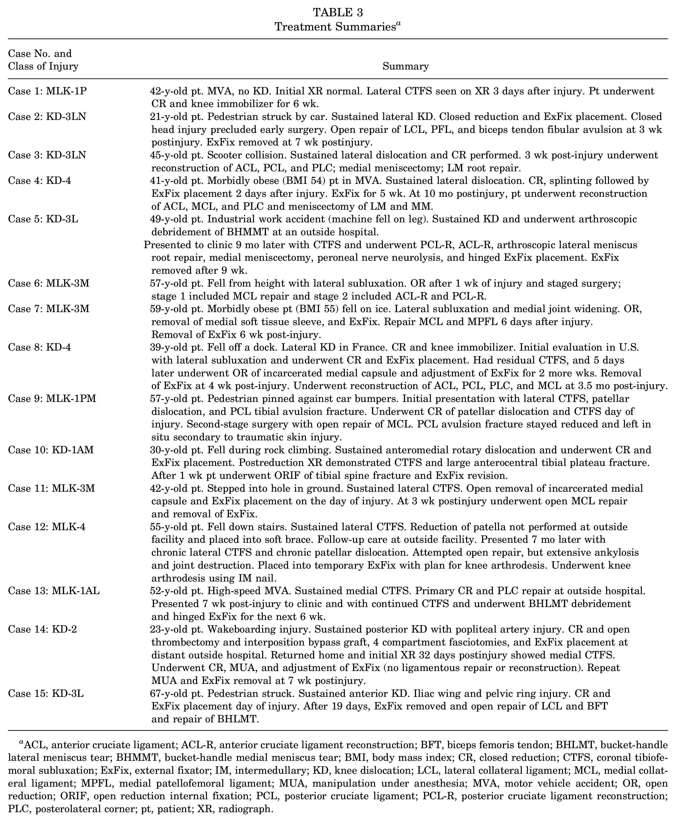
ACL, anterior cruciate ligament; ACL-R, anterior cruciate ligament reconstruction; BFT, biceps femoris tendon; BHLMT, bucket-handle lateral meniscus tear; BHMMT, bucket-handle medial meniscus tear; BMI, body mass index; CR, closed reduction; CTFS, coronal tibiofemoral subluxation; ExFix, external fixator; IM, intermedullary; KD, knee dislocation; LCL, lateral collateral ligament; MCL, medial collateral ligament; MPFL, medial patellofemoral ligament; MUA, manipulation under anesthesia; MVA, motor vehicle accident; OR, open reduction; ORIF, open reduction internal fixation; PCL, posterior cruciate ligament; PCL-R, posterior cruciate ligament reconstruction; PLC, posterolateral corner; pt, patient; XR, radiograph.

Hinged external fixation for case 5 due to chronic lateral uniplanar coronal tibiofemoral subluxation (UCTFS). The patient was evaluated with UCTFS knee 9 months after initial trauma. (A) Preoperative anteroposterior (AP) radiograph demonstrating lateral tibiofemoral subluxation. (B) Postoperative AP radiograph demonstrating posterior cruciate ligament reconstruction, anterior cruciate ligament reconstruction, and hinged external fixator placement. Hinged external fixation was placed to control coronal plane subluxation while allowing early postoperative knee range of motion.
The use of external fixation in these multiple ligament injured knees needs to be carefully monitored in the follow-up period, to avoid distraction and resultant subluxation of the joint, which was seen in 2 of these cases.
A mechanical blockage to reduction was seen in several of these cases, with sources as described in the Injury Patterns and Associated Injuries section as well as Table 3. Awareness of these potential causes, which may not always be apparent on a radiograph, CT scan, or even MRI scan, if there is metal artifact from hardware or the external fixator, is important for surgical planning.
One patient underwent closed reduction of subluxation and use of a knee immobilizer. While no mechanical blockage to reduction was seen, it was estimated that the laxity from a partial collateral ligament injury in addition to the bicruciate injury resulted in subluxation. The duration from injury to closed reduction and knee immobilizer placement was 3 days, and the patient was kept in the knee immobilizer for 6 weeks. Confirmation of reduction was monitored during follow-up visits through serial radiographs.
Discussion
This study was undertaken due to our experience of being confronted with these cases of UCTFS for which the literature has little description or guidance for management and treatment of the injury. 3 From this collection of cases, we identified several origins to the source of the subluxation. Among the 12 patients with lateral UCTFS, a mechanical blockage to reduction was found in 8 cases (Table 2). These included medial sleeve incarceration in 4 patients, patellar dislocation in 2 patients, a medial meniscus bucket-handle tear, a displaced tibial spine fracture, a PCL tibial avulsion, and a partial collateral ligament tear. Three patients in this group experienced 2 sources of mechanical block for UCTFS; however, a mechanical reason for lateral UCTFS could not be identified in 4 patients, suggesting that generalized laxity due to ligamentous tear (partial or complete) might have contributed, which was additive to their underlying cruciate ligament injury.
In the medial UCTFS group, all 3 patients exhibited a mechanical reason for subluxation, with 2 patients having a bucket-handle lateral meniscus tear and 1 patient being maintained in a subluxed position by the external fixator. In contrast, in the lateral UCTFS group, a mechanical reason for subluxation was indicated in 7 of 12 patients.
Tibiofemoral joint subluxation can result in short-term and long-term consequences that will impact treatment and prognosis, including difficulty obtaining or maintaining reduction, knee instability, arthrofibrosis, and severe osteoarthritis.1,8,14 While a missed KD is rare, a pure uniplanar tibiofemoral subluxation could be overlooked, especially in the setting of a patient with multiple traumatic injuries, where more urgent intervention may be necessary or if follow-up imaging is not obtained. 6 This is most clearly demonstrated by the case of chronic subluxation (case 12) that resulted in knee ankylosis and articular cartilage destruction necessitating knee arthrodesis. It was believed that the chronic patellar dislocation, medial ligamentous incompetency, and extensive soft tissue release to align the tibiofemoral joint would put a hinged total knee arthroplasty at high risk of failure in this rare clinical scenario in this 55-year-old patient.
The causes of unreducible KDs have been discussed in the literature.
Most commonly, the medial femoral condyle becomes “button-holed” through the anteromedial joint capsule, or a torn MCL becomes entrapped in the medial tibiofemoral compartment. While an attempted closed reduction is typically the initial step to address this problem, it often necessitates an open reduction approach.16,17 Rupture and entrapment of medial soft tissue structures in the medial joint space are considered common reasons for medial joint space widening and were seen in 4 of 15 of our cases; however, the spectrum of origins of UCTFS is lacking in the literature.
Our study identified several cause of UCTFS, including medial and lateral meniscus bucket-handle tears, medial sleeve incarceration, patellar dislocation, displaced tibial spine fracture, PCL tibial avulsion, and partial collateral ligament tear. Surgeons should be mindful that multiple mechanisms of mechanical blockage can also be simultaneous. Additionally, it should be noted that iatrogenic tibiofemoral subluxation could occur if the external fixator is misplaced or is maintaining traction and subluxation. No identifiable mechanical reason for lateral UCTFS was seen in 4 patients, and we theorize that it could potentially be because of the additive laxity due to partial or complete collateral ligamentous tear in association with bicruciate injury.
The management of UCTFS is rarely discussed in the literature. The only case report addressing a chronic medial UCTFS utilized a hinged external fixator to address the condition. 3 This approach offers the stability of multiplanar fixation to assist in obtaining and maintain reduction, especially in the coronal plane, and can allow for initiation of early range of motion.
Our approach to UCTFS depends on several factors, including the time from injury to diagnosis, the reducibility of the subluxated knee, the stability of the reduction, the pattern of ligamentous injury and associated injuries, and the presence of mechanical blockage. All patients with evidence of KD, subluxation, or MLKIs—based on the mechanism of injury and physical examination—undergo radiographic imaging and MRI. A CT scan is indicated if the patient presents with a complex fracture/dislocation pattern. The treatment options included closed reduction of subluxation, immobilization with a brace versus uniplanar or hinged external fixator placement, and ligamentous repair or reconstruction. We recommend biplanar radiographs, in addition to a physical examination, especially during hospitalization, while a MRI scan (with an MRI-compatible external fixator, if present) is being obtained and the treatment strategy is being finalized, to ensure the knee remains in a reduced position.
Closed reduction and knee immobilization were applied in 1 case in our series involving a patient with polytrauma injury. This individual was diagnosed with lateral UCTFS 3 days after the initial injury. Opting for this conservative plan was influenced by the necessity to address other injuries and the observed stability of the knee reduction during follow-up visits. As a result, acute surgical intervention to address knee ligamentous injuries was not required for this patient.
A uniplanar external fixator was applied in patients, with either KD or subluxation, whose knee was unstable after closed or open reduction or had a vascular injury that necessitated surgical intervention. In 4 of 9 of the patients in this group, the primary external fixator needed to be revised because of development of UCTFS while in the external fixator.
Hinged external fixation was utilized in 2 of 15 (13%) patients who presented in a chronic fashion. The usage of a hinged external fixator in the setting of KD/MLKI injuries has been controversial in the literature. The treatment of KD and MLKI is challenged by the dichotomous needs to achieve a stable knee while also preserving reasonable knee range of motion. 11 The hinged external fixator, originally developed for the treatment of elbow instability, aims to initiate early range of motion while preserving ligamentous repairs/reconstructions. Its application in KD is aimed at providing coronal stability while initiating knee sagittal movements to prevent knee arthrofibrosis, all while preserving the ligamentous repair/reconstruction in the early stages. A prospective randomized study on patients with KD conducted by Stannard et al, 18 comparing the use of hinged external fixation versus hinged brace after ligamentous repair/reconstruction, found fewer failed ligament reconstructions in the group with hinged external fixation with comparable knee range of motion. In our series, the indication for using an external fixator was patients with chronic presentation of UCTFS, with particular concern regarding knee arthrofibrosis after surgery.
Limitations
The limitations of this study include its retrospective nature, which relies on medical record information and may result in missing data for some patients. Additionally, some patients were referred to our practice in a subacute or chronic manner, leading to limited data on their initial intervention or encounter. Second, being a multicenter study aimed at providing a comprehensive presentation of this rare condition, it posed challenges in approaching the various pathologies. Third, outcome measures and follow-up data have not been presented in this study. However, the aim of this study was to illustrate the phenomenon of UCTFS, identify reasons for this condition, and demonstrate the variety of treatments that can be utilized to provide a stable knee while preserving knee range of motion.
Conclusion
Uniplanar coronal tibiofemoral dislocation or subluxation is a rare event that has not been well described in the literature. This case series demonstrated several factors that may contribute to its presentation. The majority of cases will have ≥1 source of mechanical blockage to reduction. Awareness and management of this condition early in the course of treatment can lead to obtaining and maintaining a more stable joint reduction, but UCTFS can be found at various time points from injury. Biplanar radiographic imaging serves as a screening tool to recognize potential subluxation in postoperative follow-up, while MRI (compatible with the external fixator if present) is essential for identifying intra-articular blockages that may contribute to the condition. External fixation has been frequently used in these cases in order to maintain reduction, and chronic UCTFS is a challenging clinical dilemma that may require multiplanar or hinged external fixation to assist in maintaining tibiofemoral reduction.
Footnotes
Final revision submitted September 24, 2024; accepted October 18, 2024.
One or more of the authors has declared the following potential conflict of interest or source of funding: G.A. has received fellowship support from Arthrex; stock or stock options from Cytonics and Sparta Biopharma; educational support from Evolution Surgical; other financial or material support from OrthoFix; consulting fees from RubiconMD, Bioventus, and Sideline Sports Doc; and research support from Smith & Nephew. M.J.A. has received consulting and speaking fees from Arthrex; consulting fees from DePuy Mitek, JRF Ortho, Medical Device Business Services, and BodyCad; research support from Orcosa; education payments from Suvon Surgical and Gotham Surgical Solutions & Devices; and hospitality payments from Horizon Therapeutics. M.J.M. has received speaking fees from DePuy Mitek and Smith & Nephew; nonconsulting fees from Synthes GmbH; and consulting fees from Smith & Nephew. AOSSM checks author disclosures against the Open Payments Database (OPD). AOSSM has not conducted an independent investigation on the OPD and disclaims any liability or responsibility relating thereto.
Ethical approval for this study was waived by Yale University (2000023454).