
Abstract
Background:
Common classification schemes, measurements, and surgical planning for trochlear dysplasia are predicated on 2-dimensional imaging views.
Purpose:
To investigate patellofemoral joint osseous anatomy using 3-dimensional (3D) printed models to describe osseous anatomic trochlear variations in patients with recurrent patellar dislocation.
Study Design:
Cross-sectional study; Level of evidence, 3.
Methods:
Computed tomography scans were obtained from 20 patients with recurrent patellar dislocation and 10 healthy control knees, and 3D prints generated from these computed tomography scans were studied with respect to mediolateral positioning of the proximal trochlear groove and groove obliquity as well as changes in the appearance, height, and orientation of the medial and lateral trochlear ridges. Each trochlea was centered with respect to a vertical line perpendicular to the distal femoral condyles and through the central intercondylar notch roof, with the 3D models resting on their posterior femoral condyles. A novel 3D measurement method was devised to assess groove obliquity, termed the entry point–transition point (EP-TP) angle. The EP was defined as the midpoint of the flattened region of the proximal trochlea where the lateral and medial ridges of the proximal trochlea meet, and the TP was the point along the trochlear groove at which the groove shape changes from an oblique orientation proximally to one more vertical distally. Measurements were obtained by 3 reviewers, and reliability analyses were performed.
Results:
With the dysplastic knees arranged according to flattening of the trochleas, increased obliquity of the trochlear grooves was observed, as reflected by increased EP-TP angles as well as more lateral patellar EPs into the proximal trochleas of these recurrent patellar dislocation knees when compared with the control knees. The degree of trochlear dysplasia (according to the Dejour classification and trochlear flatness in the frontal and axial planes) was associated with diminished prominence of the lateral trochlear convexity, increasingly lateralized proximal trochlear EPs, increased trochlear groove obliquity, lateral trochlear curvature, and progressive medial ridge deformity.
Conclusion:
The 3D reproductions enabled unique conceptualization of trochlear deformity associated with recurrent patellar dislocation.
Stability and function of the patellofemoral joint are directly related to its osseous anatomic and articular surface-related geometric properties. The morphology of the trochlear groove, in addition to static and dynamic stabilizers of the joint, is of interest in patellar tracking, instability, and dislocation. 1 Patellofemoral instability is most commonly observed during the first 30° of knee flexion, as the patella fails to engage fully with the trochlear groove. 7,8
The geometry of the trochlear groove is complex, as its shape and cross-sectional anatomy change along its length. The trochlear groove is studied using axial radiograph views introduced by Merchant to visualize patellofemoral contact areas at 30° and 45° of knee flexion. 1 However, axial radiography is limited by the impaired visibility of the transepicondylar and posterior condylar axes such that measurements must be made with respect to the geometry of the groove alone, with the knee flexed and the patella well beyond its point of maximal instability in early flexion. 1
Classification schemes of trochlear dysplasia have been devised by the appearance of the patellofemoral joint on conventional radiographs. The Dejour classification is predicated on the overlapped appearance of the lateral and medial trochlear facets and the degree of proximal flatness or convexity of the trochlear groove as defined on lateral, transverse, and axial knee images. 3,4 Advanced imaging—computed tomography (CT) and magnetic resonance imaging—particularly through the early degrees of knee flexion more completely captures articular dysplasia than do radiographs. 10,11 These radiographs still depict and evaluate anatomy nonorthogonally to the trochlear groove in the transverse plane, while patellar tracking follows an oblique path in the setting of trochlear dysplasia such that standard CT images cannot fully capture the complexity of the dysplastic patellar tracking path and the “global picture” of trochlear dysplasia within 2 dimensions. 9
Three-dimensional (3D) imaging and printing have numerous applications within orthopaedic surgery, from preoperative planning for primary and revision arthroplasties using 3D printed models to assessments of anatomy and bony defects, the development of patient-specific instrumentation and implants, and educational aides for patients and surgical trainees. 6,12 The 3D imaging and models provide a more comprehensive understanding of complex dysplastic anatomy and have the potential to improve diagnostic understanding and surgical decision making, adding to the already popular Dejour classification.
The purpose of this study was to investigate patellofemoral joint osseous anatomy using 3D printed models to describe osseous anatomic trochlear variations in patients with recurrent patellar dislocation relative to the trochlear morphology seen in individuals with no history of patellar instability or dislocation. We hypothesized that trochlear morphology would differ significantly between the control and recurrent patellar dislocation cohorts, as represented using 3D prints, and that unique morphologic features would be appreciated that are not appreciable on conventional 2-dimensional (2D) imaging. Secondarily, we hypothesized that measurements of proximal trochlear morphology in 3 dimensions would differ significantly between the recurrent patellar dislocation and control cohorts and that these measurements would not be appreciable on standard 2D tomographic or radiographic imaging.
Methods
Institutional review board approval was waived for this study. The 3D prints were generated from patients with a history of recurrent patellar dislocation seen and treated by the senior author (J.P.F.) between January 2020 and November 2021. The sample originally included all patients with a history of recurrent atraumatic patellar dislocation for whom a CT scan of the knee was obtained. Exclusion criteria were congenital deformity and a history of trauma to the knee or arthritic disease, leaving 20 patients with recurrent patellar dislocation (15 female, 5 male) for study inclusion. Medical records were evaluated with respect to the following parameters: history of patellar dislocation, excessive lateral patellar translation and/or apprehension on clinical examination, presence of J sign, Insall-Salvati ratio, Caton-Deschamps (CD) ratio, tibial tubercle–trochlear groove (TT-TG) distance, femoral sulcus angle, trochlear depth, lateral trochlear inclination (LTI), and trochlear appearance on radiographic views (anteroposterior, axial, and lateral). These measurements were obtained using axial 2D CT images.
In addition, 3D prints were generated from 10 female knees with no history of patellar dislocation whose whole body CT scans were included in the New Mexico Decedent Image Database. This data set is composed of whole body CT scans from >15,000 New Mexicans who died between 2010 and 2017, with associated metadata on 60 variables, such as demography and cause of death. These control knees were selected with respect to age matching and atraumatic causes of death not involving damage to the knee joint.
Bilateral knees were segmented and printed for each control, yielding a total of 20 knees. The CD ratio, TT-TG distance, femoral sulcus angle, and LTI measurements were obtained and compared with those of the patellar dislocation cohort.
CT Images and 3D Printing
All CT examinations were performed on either a 128-slice (Revolution EVO; GE) or a 64-slice (Optima CT660 and LightSpeed VCT; GE) multidetector CT scanner with 0.625-mm slice thickness, 120-kVp tube voltage, and spiral pitch ratios of approximately 0.5. Tube current values ranged from 100 to 150 mA. Contrast material was not administered with these acquisitions. Soft tissue reconstruction algorithm images of 0.625- or 1.25-mm slice thickness on CT were applied. The field of view was adjusted to the patient’s physique and bone window kernel, and 3D reformatted images were chosen for image reconstruction.
The CT DICOM image files (Digital Imaging and Communication in Medicine) were loaded onto a medical visualization application (Intuition; TeraRecon) or Simpleware ScanIP (Synopsys) for rendering of a 3D volumetric data set. Segmentation of the distal femurs and patellae was performed via thresholding, edge detection, and manual segmentation. Briefly, image processing was conducted via automated and manual segmentation using thresholding for bone, with refinement of structures using manual segmentation. Smoothing was kept to a minimum to remove noise while preserving osseous details. Stepwise recursive Gaussian smoothing was then applied. The entirety of the distal femoral metaphysis and femoral condyles was included, in addition to the entirety of the patella. Stereolithography files (STL) were generated from each segmented data set. The surface model files generated in the Intuition platform were submitted to a commercial 3D printing service (Mediprint). ScanIP-generated models were oriented for printing in Cura (Ultimaker).
The Intuition-based models were printed 100% to scale in the thermoplastic material acrylic styrene acrylonitrile using a high-resolution material extrusion printer (Stratasys F370). ScanIP-generated models were printed in the thermoplastic material polylactic acid with water-soluble polyvinyl alcohol supports using a high-resolution dual-extrusion printer (Ultimaker S5). Models were printed at a layer height of 0.005 inches (0.127 mm) with a low-infill triangular lattice center. All printing supports were removed from the models, leaving only the bony region of interest.
Evaluation of 3D Morphology
The 3D printed models were independently assessed by an orthopaedic surgeon with 40 years of subspecialized experience in patellofemoral joint pathology (J.P.F.), a fourth-year orthopaedic surgery resident (C.A.S.), and a senior medical student (K.E.Y.). The morphology of the femoral trochleas was visually assessed, analyzed, and categorized. To standardize observations, the trochlea was described relative to the face of an analog clock and centered with respect to a vertical line perpendicular to the distal femoral condyles and through the central intercondylar notch roof. Severity of dysplasia was assessed with respect to proximal trochlear flatness when viewed in the frontal and axial planes and Dejour classification with the 3D prints resting on their posterior femoral condyles. Discrepancies in the categorization of the 3D prints were addressed as a group and by reorganization of the models by each reviewer according to a particular trend of interest. Only observations for which a group consensus was reached are described in the present investigation. Two additional reviewers, a fourth-year orthopaedic surgery resident (C.A.S.) and a pediatric orthopaedic surgeon with 47 years of experience (D.R.C.), were presented with the models and asked to focus on a specific feature of interest (eg, trochlear groove orientation) while arranging the models in a progression from least to most dysplastic (ie, more severely) to establish external consistency.
To quantitatively evaluate and compare trochlear groove curvilinearity between models in the control and recurrent dislocation cohorts, a novel 3D measurement method was devised for use on the 3D printed models. This measurement was termed the entry point–transition point (EP-TP) angle, in which the EP was defined as the midpoint of the flattened region of the proximal trochlea that first engages with the patella in early knee flexion where the lateral and medial ridges of the proximal trochlea meet. The TP was defined as the point along the trochlear groove at which the direction of patellar tracking changes course from an oblique orientation proximally to more vertical toward the intercondylar notch distally. All EP-TP angle measurements were obtained with the models resting on their posterior femoral condyles. Measurements were obtained by 3 reviewers (K.E.Y., B.B., J.P.F.), and inter- and intrarater reliability analyses were performed. For the interrater reliability, 2 of the reviewers (K.E.Y., B.B.) performed the measurements at 2 distinct time points 4 weeks apart. Finally, the anteroposterior height of the femoral condyles was measured using a metric ruler as the models lay on their posterior femoral condyles. Normalization was not performed.
Statistical Analysis
Statistical analyses were performed in Prism 8 (GraphPad Software). Two-way unpaired Student t tests were performed between the control and recurrent patellar dislocation cohorts with respect to CD ratio, TT-TG distance, LTI, and femoral sulcus angles, as measured on conventional 2D CT scans. Two-way unpaired Student t tests were also used to compare EP-TP angles, as measured on the 3D prints, between cohorts. Inter- and intrareliability analyses were performed with respect to EP-TP angles for each cohort using intraclass correlation coefficients (ICCs). Descriptive statistics are reported as mean and standard error. P < .05 was considered significant.
Results
The mean age of patients whose models were included was 19.2 years (range, 15-43 years). The mean age of controls whose models were included was 19.7 years (range, 15-25 years). Statistically significant differences were observed in CD ratio, LTI, and TT-TG distance between the control and recurrent patellar dislocation cohorts (P = .011, P = .0011, and P = .01, respectively). All measurements in the control cohort were within normal limits, defined as a CD ratio between 0.8 and 1.2, LTI >11°, and a TT-TG distance <15 mm. Specifically, these patients had a mean CD ratio of 1.029 (SE = 0.05), 21.8° LTI (SE = 1.4°), and 10.2-mm TT-TG (SE = 1.3), as compared with a mean CD ratio of 1.327 (SE = 0.09), 9.9° LTI (SE = 1.3°), and 17.3-mm TT-TG (SE = 1.5) in the patellar dislocation cohort.
In the control knees, it was observed that the EP for patellar engagement with the trochlea lay at the most proximal and central aspect of the trochlear groove. The trochlear groove was observed to lie close to midline with respect to the center of the femoral shaft under normal conditions (Figure 1, A-C). Were the distal femur and trochlea oriented and described relative to the face of an analog clock and centered with respect to a vertical line perpendicular to the distal femoral condyles and through the central intercondylar notch roof, this patellar EP into the proximal trochlea would be located at approximately 12 o’clock in normal knees. This clockface analogy is rooted in the clinical manifestation of patellar position with progressive knee extension, resulting in J signs of varying degrees of severity. In our cohort of controls, the patellar EP varied slightly from the 12-o’clock position but did not quite reach 1 o’clock (left knee) or 11 o’clock (right knee). By contrast, in our 3D printed knees of recurrent patellar dislocation, the trochlear grooves curved from lateral to medial from the proximal lateral aspects of the trochleas and back laterally toward the distal ends of the trochleas. The curved trochlear groove was traced for each knee from the most proximal aspect of the trochlea to the intercondylar notch (Figure 1D).

(A) Normal trochlear morphology, as represented on a 3D printed model from a knee with no history of patellar dislocation. The point at which the patella likely first engages with the proximal trochlea—the trochlear groove entry point—is midline relative to the femoral shaft. Anterior view of the osseous morphology of the trochlea. (B) Inferior view of the trochlea with the distal femur resting on the posterior femoral condyles. (C) Superior view of the trochlea. (D) The curvilinear path of the trochlear groove in a dysplastic trochlea varies among individuals and is readily appreciable on 3D printed models of the patellofemoral joint. This path is demonstrated by the curved course of the black wire that extends from the proximal to distal trochlea. 3D, 3-dimensional.
The proximal groove EPs in our recurrent patellar dislocation cases varied from mildly to severely lateralized as compared with normal trochleas, resulting in distinctly curvilinear trochlear grooves (Figures 2 –4). In left knee models, the EP migrated to the 2- to 3-o’clock position with progressive dysplasia (Figure 2), whereas in right knee models, this EP was observed at the 9- to 10-o’clock position with increasing dysplasia severity. Less dysplastic trochleas resembled inverted parabolas when viewed from a frontal perspective, while severely dysplastic trochleas possessed highly oblique and lateral entry paths where the medial and lateral trochlear convexities converge proximally. A novel 3D measurement method, the EP-TP angle, was devised to evaluate differences in curvilinearity between models and cohorts. The EP-TP angle was found to vary significantly between the control and recurrent dislocation cohorts by 2-tailed unpaired Student t test, P < .0001, with greater angles (ie, more obtuse) that approached 180° in the control cohort relative to the recurrent dislocation cohort, which correlated with the observation that the trochlear groove has a less curvilinear appearance in control knees. The EP-TP measurements demonstrated good interrater reliability (mean ± SE; ICC, 0.77 ± 0.09) and intrarater reliability (ICC, 0.85 ± 0.08).

Trochlear groove entry point lateralization with increasing degrees of dysplasia, from left, as shown on left knee models. The entry point into the trochlea lateralizes, and the trochlear groove becomes more curved and oblique. The path of the trochlear groove as it starts from the entry point is delineated by the red line in each model. As the entry point into the trochlea becomes increasingly lateralized, the medial trochlear ridge appears increasingly angulated (blue line). With greater extents of lateral trochlear curvature, the medial ridges appear increasingly distorted and blend into the plane of the trochlear sulcus proximally, yielding prominent apices to the proximal trochlear groove, which are commonly referred to “supratrochlear spurs” based on their appearance on 2-dimensional radiographs.

Entry point–transition point (EP-TP) angle as shown in an age-matched control knee model (left) and a recurrent patellar dislocation knee model (right). EP was defined as the midpoint of the flattened region of the proximal trochlea that first engages with the patella in early knee flexion where the lateral and medial ridges of the proximal trochlea meet. The transition point was defined as the point along the trochlear groove at which the direction of the trochlear groove changes from an oblique orientation proximally to more vertical toward the intercondylar notch distally. An EP-TP angle of approximately 141.3° was measured in the control knee, as compared with an EP-TP angle of 120.1° in the dislocator knee.

Two models show right knee trochleas classified as Dejour type C (left) and B (right) dysplasia. The medial trochlear ridge courses obliquely toward the lateral aspect of the femur in both knees, though disparate extents of proximal trochlea and lateral condyle flattening are observed, which were worse in the Dejour B trochlea.
With increasing lateralization of the proximal trochlear EP, the lateral trochlear ridge appeared progressively flatter, forming a shallow depression of variable width (Figure 5), which is consistent with the Dejour A and B classifications. Therefore, as the knees became increasingly dysplastic, as a function of 3D trochlear groove curvilinearity as described earlier, greater lateralization and flattening of the proximal trochlear EPs were observed.

The “height” of the lateral and medial trochlear facets was observed to vary with increasing degrees of dysplasia, from left, as illustrated by the same left knee models shown in Figure 2 from the anteroposterior view. The models are viewed from an axial vantage point as they rest on their posterior femoral condyles, which provide a convenient and well-accepted plane of reference. Increased proximal trochlear flattening on the anteroposterior view was observed to correlate with more lateral trochlear entry points, increasing lateral curvature, and loss of lateral trochlear prominence from the axial perspective.
The morphology of the proximal midline trochlear groove that normally lies between the medial and lateral trochlear ridges changes with increasing lateralization of the EP, causing the proximal trochlea to appear increasingly parabolic and curved. With greater dysplasia, the medial ridge becomes increasingly oblique at the proximal trochlear sulcus such that the progressively flat proximal trochlea is accompanied by a prominent apex to the proximal trochlear groove as the lateral trochlear ridge is obliterated (Figure 6).

These 3-dimensional printed models show severely distorted morphology of the superior trochlea. These morphologies vary from wide parabolic configurations to narrow V-shaped peaks. The entry point into the trochlear groove is denoted in red. One can readily appreciate the severe curvatures of these dysplastic trochleas from patients with recurrent patellar dislocation. These models also demonstrate that the entry point in trochlear dysplasia is oblique, progresses from the lateral side, and may not be definable with traditional 2-dimensional images that are sectioned transversely and sagittally.
We observed variations in the morphologies of the distorted proximal medial ridges along the superomedial borders of trochleas of recurrent patellar dislocation cases. These distorted medial ridges seem to define the trochlear groove paths. The proximal extent of this ridge forms what is referred to as a supratrochlear “spur” on conventional imaging. With increasing severity of dysplasia, as reflected by increased trochlear groove curvilinearity and proximal trochlear flatness, the angle between the superior and distal components of the medial ridge becomes increasingly obtuse, causing the medial ridge to adopt an oblique appearance with an angle that approaches 180° (Figure 7). The morphologies of medial trochlear convexities varied with respect to their heights orthogonal to the trochlea, length in the anteroposterior plane, and curvature. A spectrum of medial ridge prominences was confirmed, ranging from elevated, highly convex ridges on the medial aspects (accompanied by significant depressions on the superomedial undersurfaces of those ridges) to more flattened, less apparent medial convexities bordering shallow and oblique trochlear grooves (Figure 2). Medial ridge prominence varied with greater lateral curvature and obliquity of the trochlea. Linear obliquity of the medial trochlear ridge was more common in trochleas with more lateral EPs and less lateral condylar prominences.
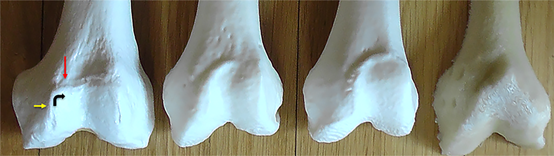
Medial trochlear ridge morphology changes with progressive dysplasia, from left. The medial ridge is composed of a superior aspect perpendicular to the long axis of the femur (red arrow) and a distal projection parallel to the long axis of the femur (yellow arrow). With increasing severity of dysplasia, this angle between the superior and distal components of the medial ridge “flattens” and becomes increasingly obtuse, causing the medial ridge to adopt an oblique appearance with a medial ridge angle that approaches 180°.
When the models were viewed axially as they rested on their posterior femoral condyles—comparable to the standard 2D posterior condyle reference plane used in CT imaging—the angles between the medial and lateral femoral condyles were progressively shallower with greater degrees of dysplasia (Figure 4). The anteroposterior height of the lateral femoral condyle decreased with respect to the medial femoral condyle height as the extent of dysplasia increased such that the appearance transitioned from that of Dejour type C dysplasia, with a prominent lateral convexity and a hypoplastic medial condyle, to that of 2 condyles similar in prominence and morphology (Figure 5). Greater trochlear flattening proximally was observed and the EPs for patellas increasingly lateralized as the condyles became more equal in size from this inferior perspective. The angle between the medial and lateral trochlear ridges, which form the borders of the trochlear groove and act as buttresses for the patella as it traverses the groove, may be readily seen, measured, and compared along the trochlea on 3D prints. This angle of the proximal trochlea formed by the intersection of the medial and lateral trochlear convexities is distinct from the femoral sulcus angle.
Discussion
In this descriptive study, we identified a unique set of trends in dysplastic patellofemoral trochlear osseous anatomy in patients with recurrent patellar dislocation. Manipulation of these 3D models enables unique conceptualization of patellar tracking paths, which are delineated by the curvature and obliquity of the medial trochlear ridge and may manifest as a J sign in recurrent patellar dislocation. As the extent of trochlear dysplasia increased—according to increased trochlear groove curvilinearity and proximal trochlear flatness described for the first time herein—3D printed reproductions showed increasingly lateral and distal positioning of patellar EPs into their trochleas, accentuated lateral curvature of the trochlear grooves, and relative hypertrophy of the medial trochlear ridges with encroachment toward the proximal aspect of the trochlear sulcus, yielding prominent proximal medial ridges commonly identified as supratrochlear spurs on conventional imaging.
The distinct descriptions of proximal trochlear morphology described herein introduce a new paradigm of trochlear dysplasia conceptualization in the analysis of recurrent patellar dislocation. Fritz et al 5 compared the study of 50 3D printed models of the distal femur with conventional radiographs and CT scans, 28 of which represented patients with trochlear dysplasia. The authors observed that the trochlear groove courses midline in controls, while the course of patellar tracking appears more curved among dysplastic cases. 5 Loudon 8 documented that the patellar tracks in a lateral-medial-lateral course in the arc from tibiofemoral extension to flexion, with little lateral or medial translation during flexion in normal knees, such that the patella remains centered on the trochlea. This lateralization of the patella begins around 30° of flexion, which is maintained for the remaining arc of knee flexion in what has been termed a C-curve pattern. 8 The shape and orientation of the trochlear groove 2 described by Loudon resemble the path of patellar tracking described herein. This curvilinearity of the trochlear groove is reflected by the significant difference in the EP-TP angle, which was larger and reflected less curvilinear grooves in the control cohort relative to the smaller EP-TP angles and more curvilinear grooves in the recurrent dislocator cohort.
Conventional imaging leaves much to conjecture and extrapolation in surgical planning for trochlear dysplasia, as this complex problem exists and must be understood in 3 dimensions. The Dejour classes A and B, with their shallow to flat proximal trochleas defined by transverse tomographic images, partially capture the 3D trochlear distortions that involve medial and lateral trochlear convexities obliquely converging along variably curved grooves with variably lateralized EPs. We observed that the trochlear EP of the patella was more midline and that the proximal trochlea appeared less flat when the axial view of the femoral condyles was consistent with a Dejour C classification, suggesting that the Dejour C classification is less deformed than Dejour A/B when considering 3D curvature and obliquity.
The supratrochlear spur seen on 2D imaging is actually the proximal medial trochlear ridge described in detail herein. Fritz et al 5 analyzed 3D printed models of the distal femur and observed a “prominent crest along the superomedial aspect of the trochlea,” but they did not describe variations in the morphology of the medial ridge. With increasing severity of dysplasia, this medial trochlear ridge appeared increasingly oblique relative to the long axis of the femur, and the angle between the superior and distal components of the medial trochlear ridge became increasingly flattened. Our 3D prints illuminated that the shallowness of proximal trochleas, as depicted by proximal transverse nonorthogonal radiographic images, does not sufficiently define their true 3D contours, which impact patellar tracking. The origin of the trochlear groove proximally is more lateral in severely dysplastic knees such that 2D axial images are of limited value, considering that lateral trochlear entry planes for patellar engagement are far from transverse.
Surgical Planning Using 3D Images and Prints
The 3D prints afforded greater understanding and discrimination among patterns of trochlear morphology between recurrent patellar dislocation cases and controls. The enhanced understanding of trochlear curvature and obliquity in 3 dimensions has important implications for surgical design, in which the result must be stable entry and maintenance of a patella within a variably curved trochlea. Proximal flatness will likely be treated differently in the patient with recurrent patellar dislocation with a minimally curved trochlea as compared with the patient with a curved and/or oblique trochlea and lateral patellar EP. The 3D imaging and printing are likely best reserved for patients with more challenging and complicated manifestations of recurrent patellar dislocation for use as a complementary tool to optimize surgical planning.
The senior author (J.P.F.) has found that 3D understanding of trochlear curvature improves decision making, particularly regarding when to add tibial tubercle transfer medially, anteromedially, or distally or trochleoplasty. For example, surgical decision making was altered for 2 patients with patellar dislocation using 2D measurements and impressions as compared with those based on 3D printed reconstructions. Patient A demonstrated a CD ratio of 1.2, 17-mm TT-TG, 8° LTI, small J sign, and Dejour B pattern and was treated successfully with medial patellofemoral ligament reconstruction alone. Patient B, with similar 2D measurements (CD, 1.0; TT-TG, 14 mm; LTI, 8°; prominent J sign; and Dejour B), was treated by anteromedial tibial tubercle osteotomy and medial patellofemoral ligament reconstruction owing to the 3D appreciation of greater trochlear curvilinearity in addition to a lateral EP.
Three-dimensional visual analysis of patient B’s curvature pattern suggested the value and logic of adding an anteromedial tibial tubercle osteotomy to advance the patella into the longer, curved, convex trochlea. Both might be considered for trochleoplasty using 2D criteria, but 3D evaluation suggested that these surgical procedures would suffice in providing permanent patellar stability. Advantages of 3D reconstructions include appreciation of extreme curvature; the extent, relevance, and location of a prominent medial ridge; the proximal patellar EP and its relationship to the distal trochlear center; and full understanding of the multiplanar curvilinear tracking path that the patella must follow with progressive knee flexion.
Limitations
Our study comments on femoral trochlear morphology alone. The observations described herein were made entirely from static models of the femoral trochlea in a single degree of flexion rather than dynamic tracking. Although these descriptions of variations in trochlear morphology expand on current methods of trochlear dysplasia conceptualization and were made with the kinematic factors associated with patellar tracking in mind, additional dynamic analyses of patellofemoral motion and characterization of the course of patellar engagement with these variably shaped trochleas must be conducted and elucidated. Furthermore, the patients with recurrent patellar dislocation whose prints were included represent a single surgeon’s practice over a short time frame. As our observations primarily involve descriptive characterizations of osseous trochlear morphology, formal reliability analyses were not performed. Despite this, consensus was obtained for the trends described, and reviewers not involved in the original characterizations were able to reproduce the ordering of models according to specific trends with 80% accuracy. In addition, LTI measurements are conventionally performed on magnetic resonance images rather than CT scans. Although LTI measurements were included and compared between cohorts to verify a discrepancy in a 2D parameter commonly used to evaluate trochlear dysplasia, they did not constitute a primary focus of the present investigation. Last, the anteroposterior height measurement of the femoral condyles (Figure 4) was not normalized for femurs of different sizes. Research into how these measurements can best be normalized using 3D prints is ongoing.
Conclusion
In this study, critical evaluation of 3D reproductions enabled unique conceptualization of trochlear deformity associated with recurrent patellar dislocation. As the severity of trochlear dysplasia increased, 3D printed reproductions showed (1) increasingly lateral and distal positioning of patellar EPs into their trochleas, (2) accentuated lateral curvature of the trochlear grooves, and (3) relative hypertrophy of the medial trochlear ridges with encroachment toward the proximal aspect of the trochlear sulcus, yielding prominent proximal medial ridges. These findings establish a rationale for selectively using 3D models in the study of trochlear dysplasia to improve the visualization and characterization of patients with recurrent patellar dislocation.
Footnotes
Acknowledgment
The authors appreciate the contributions of David Diduch, MD, and Miho Tanaka, MD, for providing 3D models and Daniel Wiznia, MD, for providing guidance regarding CT segmentation. The authors also thank Lisa Lattanza, MD, chair of the Yale School of Medicine Department of Orthopaedics and Rehabilitation, without whose support these efforts would not have been possible.
Final revision submitted August 3, 2022; accepted September 8, 2022.
One or more of the authors has declared the following potential conflict of interest or source of funding: R.K. has received hospitality payments from GE Healthcare. J.P.F. has received grant support from Encore Medical and hospitality payments from Smith & Nephew. AOSSM checks author disclosures against the Open Payments Database (OPD). AOSSM has not conducted an independent investigation on the OPD and disclaims any liability or responsibility relating thereto.
Ethical approval for this study was waived by Yale University (No. 2000032369).