
Abstract
Background:
Pulling the long head of the biceps tendon into the joint at arthroscopy is a common method for evaluation of tendinopathic lesions. However, the rate of missed diagnoses when using this technique is reported to be as high as 30% to 50%.
Hypothesis:
Tendon excursion achieved using a standard arthroscopic probe does not allow adequate visualization of extra-articular sites of predilection of tendinopathy.
Study Design:
Descriptive laboratory study.
Methods:
Seven forequarter amputation cadaveric specimens were evaluated. The biceps tendon was tagged to mark the intra-articular length and the maximum excursions achieved using a probe and a grasper in both beach-chair and lateral positions. Statistical analyses were performed using analysis of variance to compare means.
Results:
The mean intra-articular and extra-articular lengths of the tendons were 23.9 and 82.3 mm, respectively. The length of tendon that could be visualized by pulling it into the joint with a probe through the anterior midglenoid portal was not significantly different when using either lateral decubitus (mean ± SD, 29.9 ± 3.89 mm; 95% CI, 25.7-34 mm) or beach-chair positions (32.7 ± 4.23 mm; 95% CI, 28.6-36.8 mm). The maximum length of the overall tendon visualized in any specimen using a standard technique was 37 mm. Although there was a trend to greater excursion using a grasper through the same portal, this was not statistically significant. However, using a grasper through the anterosuperior portal gave a significantly greater mean excursion than any other technique (46.7 ± 4.31 mm; 95% CI, 42.6-50.8 mm), but this still failed to allow evaluation of Denard zone C.
Conclusion:
Pulling the tendon into the joint with a probe via an anterior portal does not allow visualization of distal sites of predilection of pathology. Surgeons should be aware that this technique is inadequate and can result in missed diagnoses.
Clinical Relevance:
This study demonstrates that glenohumeral arthroscopy does not allow visualization of common areas of pathology of the long head of the biceps tendon.
Abnormalities of the long head of the biceps (LHB) tendon are a frequent cause of anterior shoulder pain. However, it is well recognized that physical examination is unreliable for diagnosis. 10 The literature reports low sensitivities and specificities for common clinical tests as well as imaging modalities, including ultrasound and magnetic resonance imaging (MRI). 1 –3,13 These figures are determined by comparison with shoulder arthroscopy. 1 –3,10,13,14
To visualize the extra-articular portion of the tendon, it is standard practice to pull the tendon into the joint through an anterior midglenoid portal while viewing through a posterior portal. Several technical reports have described how elevation, abduction, and rotation of the arm as well as elbow flexion can reduce tension in the LHB tendon, potentially allowing 3 to 5 cm of tendon to be pulled into the joint. 4,8 Despite this, Murthi et al 16 reported that approximately 50% of macroscopic tendon lesions were missed at arthroscopy because these abnormalities were located beyond the limit of visualization afforded by tendon excursion alone.
It is clear from other studies that pathology distally in the groove is common. 16 Refior and Sowa 20 reported in a histological study that the most common site of degenerative change in the LHB was at the most distal part of the intertubercular groove. The predilection to abnormalities in this location was attributed to it being a vascular watershed area and therefore more susceptible to degenerative change. 6,19 In addition, this area is the narrowest part of the groove, and the potential for frictional damage is high and may be further exacerbated by degenerative change in the groove itself. 20 Moon et al 15 reported outcomes from a series of patients who underwent rotator cuff repair followed by open subpectoral tenodesis. These authors categorized the LHB into the 3 zones described by Denard et al 7 (zone A, the proximal 2.5 cm of the tendon; zone B, between 2.5 and 5.6 cm; and zone C, distal to 5.6 cm) and reported that the prevalence of tears was 100% in zone B and 77.8% in zone C and that degenerative changes were observed for all cases in zones A and B and in more than 80% of cases in zone C.
The aim of this study was to determine the length of the LHB tendon that can be pulled into the joint during glenohumeral arthroscopy. We hypothesized that the amount of excursion achieved would be less than that described in early technical reports and would not allow visualization of areas of predilection of pathology. This article therefore seeks to challenge the established doctrine that conventional shoulder arthroscopy is an adequate gold standard for the diagnosis of LHB pathology.
Methods
The study protocol was awarded health research ethics board approval. A sample size calculation determined that 7 specimens were required (μ1, 30 mm; μ2, 40 mm; Σ, 5 mm; α, 0.05; power, 0.95). Ten fresh-frozen forequarter cadaveric specimens were evaluated; 3 were excluded as the LHB tendon was not intact. The mean age of the remaining 7 specimens was 74 years (range, 44-96 years). All specimens were female; 4 limbs were right-sided and 3 were left-sided. Each specimen was thawed for a minimum of 24 hours prior to assessment. Arthroscopy was performed by 2 fellowship-trained attending orthopaedic shoulder surgeons (A.S., J.O.). Each shoulder underwent arthroscopy in both lateral decubitus and beach-chair positions.
Standard glenohumeral arthroscopy was performed using a 30° arthroscope while viewing through a posterior portal. A spinal needle was then used to localize the position of the anterior midglenoid portal, which was placed in the rotator interval just above the upper edge of the subscapularis tendon at the midpoint of the visible part of the tendon. Diagnostic arthroscopy was performed to identify any pre-existing intra-articular pathology. In particular, careful evaluation of the biceps pulley was performed to exclude any specimens in which subluxation of the tendon may have affected measurements of excursion. An 18-gauge spinal needle was then passed from the anterolateral edge of the acromion to pierce the most distal part of the LHB tendon visible (Figure 1). A 3-0 monofilament suture was shuttled through the needle and subsequently retrieved and knotted.

A spinal needle inserted from the anterolateral edge of the acromion piercing the long head of the biceps tendon at the most distal aspect visualized.
In the lateral decubitus position, the arm was maintained in the standard position for glenohumeral arthroscopy, which was 45° of abduction, 15° of forward flexion, and 7 kg of traction. In the beach-chair position, excursion of the tendon was facilitated by 90° of elbow flexion and positioning the arm at 30° of elevation, 40° of abduction, and neutral rotation. The tendon was then pulled maximally into the joint using an arthroscopy probe inserted via a midglenoid anterior portal, and the process was repeated to tag the maximum length of the tendon that could be visualized (Figure 2).

A suture being placed while the long head of the biceps tendon is being maximally pulled into the joint using an arthroscopy hook after having already placed the first suture.
The tendon was then pulled into the joint with a grasper through the same portal, and a suture was placed to mark the additional visualized length (Figure 3). Tendon excursion was also assessed by using a grasper to pull the tendon into the joint through the anterosuperior rotator interval portal. This portal was located immediately anterior to the leading edge of supraspinatus (Figure 4) such that the tendon could be grasped right at the entrance to the pulley and tagged again at maximum excursion.

Additional suture placement with spinal needle while maximally pulling the tendon into the joint using an arthroscopic grasper.

The anterosuperior portal is placed in the rotator interval immediately anterior to the leading edge of the supraspinatus, allowing the tendon to be grasped at the entrance to the biceps pulley.
After the arthroscopic part of the procedure was completed, the LHB tendon was retrieved via an open deltopectoral approach. The tendon was sharply detached from its proximal attachment using a No. 15 scalpel while preserving maximum length. Digital calipers (model 88N6260; Marathon Ltd) were then used to record measurements of length. These measurements included the full length of the tendon from its origin to the musculotendinous junction, the length of the extra-articular portion of the tendon (calculated by deducting the intra-articular resting length from the total length), and the lengths that could be visualized by pulling the tendon into the joint during arthroscopy in both lateral decubitus and beach-chair positions. To minimize objectivity in assessment of the location of the musculotendinous junction, it was defined as the point at which muscle fibers became visible on the anterior surface of the tendon, as seen in Figure 5.
Determination of the location of the musculotendinous junction by appearance of muscle fibers on the anterior surface of the tendon.
Statistical analyses were performed using analysis of variance (GraphPad Statistical Software) to compare mean tendon excursion data.
Results
Partial tears or degenerative fraying of both the supraspinatus (n = 5) and subscapularis (n = 4) were common in the study population, but the biceps pulley was intact in all specimens and none needed to be excluded for LHB subluxation. The mean length of the tendon measured from its origin to the musculotendinous junction was 106 mm (range, 94-125 mm). The mean length of the intra-articular portion of the tendon was 23.9 mm (range, 22-26 mm). The mean length of the extra-articular portion was 82.3 mm (range, 72-99 mm).
A summary of the LHB excursion data along with statistical analysis is presented in Table 1. The mean overall length of tendon that could be visualized by pulling it into the joint with a probe through the anterior midglenoid portal was not significantly different when using either lateral decubitus (mean ± SD, 29.9 ± 3.89 mm; 95% CI, 25.7-34 mm) or beach-chair positions (32.7 ± 4.23 mm; 95% CI, 28.6-36.8 mm). The maximum length of the overall tendon visualized in any specimen using a standard technique was 37 mm. In both beach-chair and lateral positions, there was a trend toward greater excursion using a grasper through the same portal, but this was not statistically significant. However, using a grasper through the anterosuperior portal gave a significantly greater mean excursion than any other technique (46.7 ± 4.31 mm; 95% CI, 42.6-50.8 mm) but this still failed to allow evaluation of Denard zone C.
Summary of LHB Excursion and Visualization Data a

a LHB, long head of the biceps tendon.
Discussion
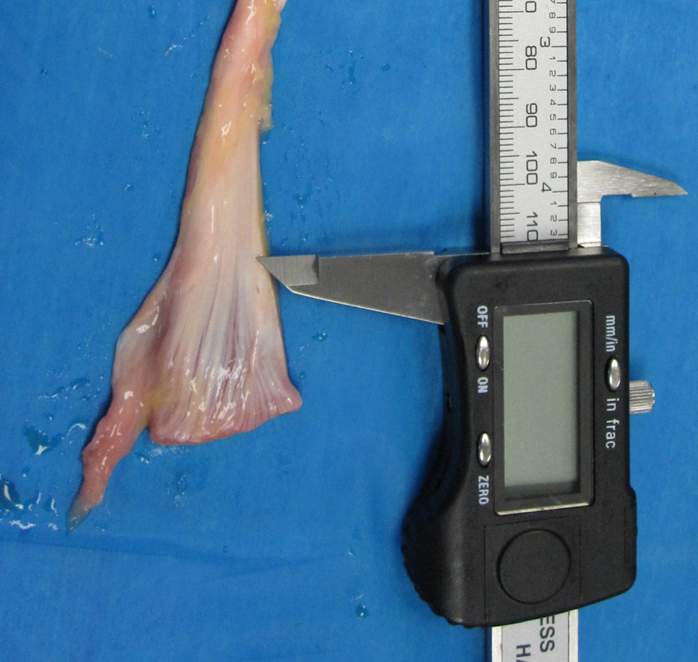
Pulling the LHB into the joint during glenohumeral arthroscopy using a hook/probe is widely considered to be the gold standard for the diagnosis of tendon lesions. 1 –3,10,13,14 This study demonstrates that by using such a technique, only a mean of 30 to 33 mm (28%-32%) of tendon can be seen, which is at the lower end of previously described technical reports using this technique. 4,8 When these data are further analyzed, it is apparent that the additional excursion with this technique is only 6 mm (lateral decubitus position range, 2-12 mm) and 9 mm (beach-chair position range, 2-14 mm) of the extra-articular portion of the tendon. It is therefore apparent that despite meticulous attempts to standardize portal placement in some cases, tendon excursion remained very limited. Potential reasons for this include differing degrees of tethering by vinculae, scarring of the biceps tendon, and variations in overall anatomy between patients. 12 Furthermore, these data are consistent with work by Gilmer et al 11 who reported that only 5 to 28 mm of tendon could be visualized at the time of arthroscopy. However, in that study, in contrast to the authors’ normal practice, a grasper was used instead of a hook. Our study demonstrates that using a grasper permits visualization of a significantly greater proportion of the LHB when compared with using a probe. This is because a grasper can be brought further into the joint in any direction while still capturing the tendon, whereas a probe is required to be pulled inferiorly to maintain tension on the tendon. In particular, we found that using a grasper via an anterosuperior rotator interval portal provided a much greater excursion than any other method studied. This was greater than using a grasper via an anterior midglenoid portal because from that position, excursion tends to be limited by tension in the portion of the LHB between the grasper and the biceps anchor. The use of the anterosuperior portal allows the tendon to be grasped more distally before it is pulled into the joint and therefore allows greater excursion before the proximal portion of the tendon restricts motion. However, using a standard arthroscopic grasper causes considerable damage to the tendon and is therefore not recommended (Figure 6). This is an important limitation of previous studies as not only is this technique not used in clinical practice but it also overestimates the amount of tendon that is normally visualized. 11,22

Damage to the long head of the biceps tendon sustained while using a standard arthroscopic grasper to pull the tendon into the joint.
To our knowledge, there is only 1 published report of an atraumatic grasper technique. Parada et al 18 described using an Allis clamp and rotating the tendon around its closed jaws. Although they reported no iatrogenic injuries, they did not report the length of tendon that could be evaluated using their technique; however, data from the current study suggest that even when using a standard grasper, a large proportion of the tendon is still not visualized.
In our best-case scenario, by using a hook alone, a maximum of only 14 mm (17%) of the extra-articular portion of the tendon was visualized when using a standard technique. When considering that Wafae et al 23 demonstrated that the bicipital groove is approximately 8 cm in length, it is apparent that the distal part of the tendon is not adequately evaluated with this technique. Inability to visualize this region is an important cause of missed diagnoses, which are reported to occur at a rate of 30% to 50%. 9,11,16,22 This also implies that published figures for sensitivities and specificities of common physical examination tests and imaging modalities based on arthroscopy as a gold standard are invalid. 1 –3,10,13 It would therefore be appropriate for further research to revisit these values based on open exploration to obtain valid data.
The results of this study concur with the work of Festa et al, 9 who also showed that arthroscopy in the beach-chair position only allows visualization of a short length of the extra-articular portion of the tendon. Our study demonstrates that in the lateral decubitus position, this distance is significantly less. However, despite this, we do not advocate one position over the other. This is because the amount of additional excursion achieved in a beach-chair position, though statistically significant (mean difference, 2.86 mm; P = .0327), is not likely to be clinically important as known sites of predilection of pathology are much more distal. This also means that removing the arm from traction to allow optimum positioning for greater tendon excursion in a lateral decubitus position is unlikely to reduce the rate of missed diagnoses.
Several recent studies have demonstrated that arthroscopy alone misses 30% to 50% of LHB lesions compared with open evaluation. 11,22 This adds to the increasing evidence that a “normal” standard arthroscopic examination of the LHB by pulling it into the joint with a hook and viewing with a 30° arthroscope is inadequate to exclude pathology and should no longer be relied upon to assess LHB pathology.
Previous authors have described attempts to improve visualization by using a 70° arthroscope but how well this improves the ability to evaluate the extra-articular portion of the tendon is not reported. 4 Bhatia et al 5 achieved excellent visualization of the extra-articular portion of the tendon by direct arthroscopy of the bicipital groove using a 4-mm, 30° arthroscope inserted through the Neviaser portal. However, this has not gained popularity, perhaps because of the risk of injury to the suprascapular nerve and supraspinatus tendon and a lack of knowledge of the limitations of traditional arthroscopic methods.
An alternative approach is open exploration, and historically, this has been a popular option. 17 However, with the widespread use of arthroscopy for assessment of the LHB, this has fallen out of favor. Given the findings of this study and the other evidence considered herewith, it may be that there is a role for open exploration when LHB pathology is suspected but arthroscopy is “normal.” 18 A concern with such an approach would be that open surgery risks instability if subsequent tenodesis is not indicated. However, it is interesting to note that Neer 17 and others 16,21 have reported that division of the transverse humeral ligament alone is a safe procedure and does not risk tendon subluxation.
Limitations
The main limitation of this study is that it represents an evaluation of a small number of cadaveric specimens. It is possible that in vivo, tendon excursion may be different. However, the risk of this was minimized by using fully thawed, fresh-frozen specimens with intact distal extremities. Because of the small number of specimens available, anatomic variation due to sex differences was minimized by using female cadavers only. However, excursion data may be different in male specimens, and this is not evaluated in this study. It is also important to note that this study was not designed to determine the minimum amount of tendon that needs to be visualized to avoid missed diagnoses. This is not currently known and is a topic for further study.
Conclusion
The results of this study challenge the accepted doctrine that pulling the LHB into the joint during shoulder arthroscopy is adequate for the diagnosis of pathology of the long head of biceps tendon. This technique fails to fully visualize Denard zones B or C, which are areas of predilection of tendon pathology. This results in a reported rate of missed diagnoses in 30% to 50% of patients and potentially results in suboptimal outcomes after shoulder surgery. 11,16,22 Furthermore, data regarding sensitivities and specificities of common clinical tests reported in the literature have been based on this technique. This raises the concern that these values are invalid as they are based on a technique that is associated with a high false negative rate.
Footnotes
One or more of the authors has declared the following potential conflict of interest or source of funding: This work was supported by The Pan Am Foundation.