
Abstract
Sexually transmitted diseases are a major public health problem both in developing and in developed countries, and especially with the co-synergy with HIV infection, there is an increasing need to have a proper understanding of the clinicodemographic patterns of sexually transmitted infections (STIs) for planning and implementing control strategies. Worldwide, there is an increased preponderance of viral STIs. Increasing incidence and altered clinical presentation of viral STIs in patients with HIV pose a diagnostic challenge; thereby, we studied the demographic profile of HIV-seropositive patients and compared clinical manifestations of viral STIs in HIV-seropositive patients to those in seronegative individuals. Twenty-seven HIV-seropositive patients with viral STI (herpes/molluscum/warts) and same number of age-, sex-, and STI-matched seronegative patients were studied for variability in clinical profile. There were significant differences in the demographic factors (education, income, and migration) and sexual practices (number of contacts and source of infection) in the 2 groups. Lesional symptoms, increased extent of lesions, and resistance to treatment were significantly more common in HIV-seropositive patients.
Introduction
The explosive spread of HIV and the AIDS epidemic have progressively increased the attention toward the study of factors associated with increased transmission of this virus. 1 According to The Joint United Nations Programme on HIV/AIDS (UNAIDS) Gap report 2016, the prevalence of HIV in India is 0.3%, but due to its high population, India has the third highest number of HIV-infected patients, with 21 million people living with HIV (36.7 million people worldwide) and 86 000 new HIV infections every year (UNAIDS data 2015). Worldwide, approximately 85% of HIV transmission cases occur through sexual transmission. Sexually transmitted diseases (STDs) constitute a major public health problem for both developing and developed countries. Evidence suggests role for both ulcerative and nonulcerative STDs in augmenting the transmission of HIV and accelerating the progression of HIV to AIDS by decreasing CD4 counts via a variety of biological mechanisms (risk estimates ranging from 2.0 to 23.5). 1,2 This mutual interaction between HIV and STD can profoundly affect the transmission dynamics of HIV. 3 –8 These STDs present with atypical clinical manifestations in seropositive patients with extensive, persistent involvement, altered morphology, and coexistence of multiple sexually transmitted infections (STIs). 1 –3,5,6,8 In countries like India, where there is high prevalence of STIs, targeted STD detection and treatment especially in seropositive patients may have a central role in reducing HIV burden as suggested by randomized controlled intervention trials. 2,5,7,9 The current program, National AIDS Control Programme-IV (NACP-IV), 2012 to 2017, aims to reduce annual new HIV infections by 50% through targeted interventions for key affected groups with high risk of transmission (patients with STI). 9 There is a general consensus that the bacterial STDs (chancroid, donovanosis, and gonorrhea) are declining possibly because of the widespread use of effective antimicrobials, while the prevalence of viral STDs is increasing. 10 –15 As the most common causative agent of genital ulcers worldwide, herpes simplex virus (HSV) has emerged as an important cofactor affecting HIV transmission dynamics, 11,13,15,16 contributing to two-third of the total STI case burden. 12,14 Of these 3 STIs (herpes, warts, and molluscum), herpes is found to be the commonest STI associated with HIV infection. 12,14 Moreover, since viral STIs are persistent and recurrent, they are associated with a significant reduction in quality of life scores, with the greatest impact seen in patients coinfected with HIV. 17 To our knowledge, there is paucity of studies focusing on the clinical profile of viral STDs in seropositive patients. The purpose of our study was to obtain relevant epidemiological data on various viral STDs in seropositive patients and study the clinical differences in their presentation in seropositive patients when compared to seronegative patients.
Materials and Methods
The study was conducted in department of dermatology of a tertiary hospital in Northern India, in collaboration with department of microbiology. All the patients attending STD clinic over a period of 2 years (target population) were counseled and tested (pretest and posttest) at Integrated Counseling Testing Centre for HIV (National AIDS Control Organisation [NACO] Strategy III using 3 different rapid antibody tests), VDRL, and hepatitis B. Appropriate laboratory investigations were done to confirm the STI. HIV-seropositive patients already on antiretroviral therapy, other immunosuppressive therapies, pregnant females, and psychologically unstable patients were excluded from the study. All the patients were explained the nature of the study, and written informed consent was obtained. Based on the clinical findings, relevant investigations, for example, Giemsa, Gram, Tzanck, dark-ground illumination, wet mount, tissue biopsy, KOH, acetowhitening, culture, serology (Venereal Disease Research Laboratory [VDRL], Trephonema Pallidum Hemagglutti [TPHA], and Herpes Simplex Virus [HSV]), and so on, were done wherever required and feasible to confirm the STI.
After clinical and laboratory investigations, 60 patients (seropositive and having viral STI) were recruited as patient population. Of these, 32 consented to follow-up and chose to enter the study (study entrants) out of which 5 patients were lost to follow-up. Case analysis was restricted to a final 27 study participants (cases) who were allocated on the basis of their age (<20 years, 20-40 years, and >40 years), sex (male/female), and STI (herpes, warts, and molluscum contagiosum). Group B (controls) consisted of age- and sex-matched HIV-seronegative patients with same viral STI as patients of group A (STI matched). A preformed questionnaire including the demographic details of the patient and the sexual partner and history of similar or different STIs was filled for both cases and controls. Detailed information was obtained regarding sexual orientation (behavior and practices) and addictions. The clinical morphology of lesions; extent, course, and duration of lesions; and any associated features (pain, dysuria, dyspareunia, bleeding, discharge, constitutional or systemic complaints) were also recorded. Photographic records were maintained for both the groups. Specific treatment for the STI diagnosed (according to Centers for Disease Control and Prevention guidelines) was given to the patients and the partners with appropriate counseling, and patients were followed till clinical cure and further up to 1 year for any recurrences, complications, or residual complaints. Relevant statistical analyses (χ2 and Fisher exact t test) were carried out at the end of the study period to compare the clinical profile of viral STIs in seropositive versus seronegative patients. A P value of <.05 was considered significant. Ethical clearance was obtained from the ethical committee of Lok Nayak Hospital before the commencement of the study.
Results
Most of the patients presenting to STD clinic were in the sexually active age-group with male predominance, having education up to primary level, unskilled labourers, belonging to lower socioeconomic status, and residing in middle-class residential quarters. The differences in the demographic profile between the 2 groups are highlighted in Table 1.
Differences in Demographic Detail, Sexual Behavior, and Practices of the Patients in Both Groups.

Abbreviations: CSW, commercial sex worker; STD, sexually transmitted disease; STI, sexually transmitted infection.
There was significant difference in the income, socioeconomic status, residential area, and occupation between the 2 groups, with higher prevalence of poverty, illiteracy, and history of migration in the seropositive group. Regarding sexual behavior and practices, most of the patients were heterosexual in both the groups, and 22.2% of patients were bisexual in the seropositive group. Interestingly, number of pre- or extramarital contacts in the past 6 months, unprotected sex with commercial sex worker (CSW), and peroral contact were significantly commoner in the HIV-seropositive patients. The most common mode of acquiring HIV was sexual contact (96.3%), although 3 patients had history of intravenous abuse also. The differences in the sexual profiles of both the groups are highlighted in Table 1.
The 3 viral STDs studied were herpes, molluscum, and genital warts. Of 27 patients presenting to the clinic, 13 (48.5%) patients presented with raw areas over genitalia, while 14 (51.85%) patients had complaints of growth over the genitalia. Genital herpes was the most common viral STI (48.15%) followed by genital molluscum (37%), while genital warts were least prevalent (29.63%). Four (14.8%) patients had simultaneous existence of 2 STDs with combination of molluscum with warts in 2 patients and single patient of herpes with molluscum and wart each, compared to none in the seronegative group. None of the patients tested positive for hepatitis B. The oral involvement and cutaneous lesions were also significantly commoner in the HIV-seropositive group, with candidiasis being the commonest oral finding (14.8%), while xerosis (37.03%) was the commonest cutaneous manifestation.
As regards the site of involvement, in male patients, prepuce (80%) was the commonest site in herpes patients, while in genital warts and molluscum patients, the most common sites were glans penis (50%) and shaft of the penis (75%), respectively. In female patients, labia majora (85.7%) was the commonest site of involvement for all the 3 viral STIs. Although the genital lesions were more symptomatic (burning sensation, pain, dyspareunia) in the seropositive group (77.8%) when compared to the seronegative group (51.8%), the difference was not significant (P > .05). Significant difference existed in the extent of extragenital, periurethral, and perianal involvement (erythema/erosion/growth/discharge). Inguinal lymphadenopathy was also significantly commoner in seropositive group.
For herpes patients, clinically significant difference was noted in the extent of lesions, recurrence rate, and longer healing time in the seropositive patients (Figure 1). With warts and molluscum, clinically significant difference was seen only for extent (size, number, and extragenital involvement) of lesions. Atypical manifestations such as giant lesions were seen in 4 (28.57%) patients (Figure 2), ulceration with bleeding in 2 (14.29%), while destructive lesions and scarring in another 2 patients in the seropositive group. One of the patients (giant wart) showed dysplastic changes in histopathology. The variability in the clinical profile of these STDs is depicted in Table 2.

Extensive, necrotic, and deep ulcerations of herpes in seropositive patients.
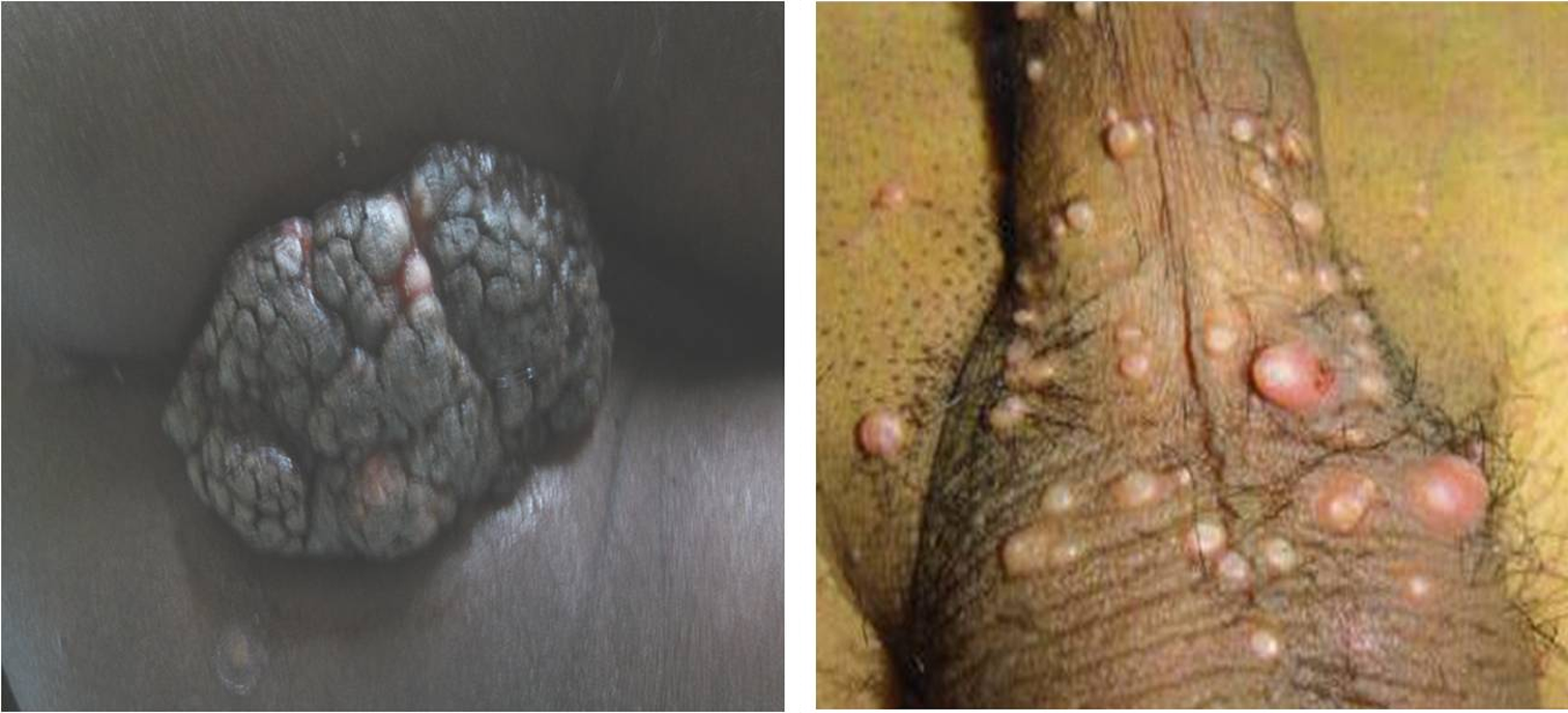
Extensive, numerous, and larger (giant forms) lesions of genital warts and molluscum seen in seropositive patients.
Variability in the Clinical Profile of Sexually Transmitted Infections in Both Groups.

Discussion
Our study highlights the variability in clinical presentations of viral STIs in seropositive patients and focuses on the differences with respect to HIV-seronegative patients.
Demographic Profile of Patients Presenting to STD Clinic
The majority of the patients were in the sexually active age-group (20-40 years), with males (74.1%) predominating, which is explainable by sociocultural inhibitions and gender bias, lower awareness, reluctance about seeking care or consultation in gyne clinics rather than STD clinics, and asymptomatic nature of infections in females. 3,14,15,18 –21 Half (55%) of the seropositive patients had migrated, and 18.5% patients had separated from their partners (either divorced or widow) comparing well with the findings of Rodrigues and Bachmann et al. 3,20 Most of the seropositive male patients (two-thirds) gave history of contact with CSW and also multiple sexual contacts (more than 6) in the past 6 months. This is because CSWs still serve as source of infection pool for HIV. 3,12,16,21 The majority (85.2%) of the HIV-seropositive patients were either unemployed or unskilled workers, belonging to lower socioeconomic strata and having multiple addictions, for example, smoking, alcoholism, and tobacco chewing in concordance with studies in the literature. 19 –21 Although unprotected sexual contact was commoner in the seropositive group, the difference was not significant when compared to seronegative patients. This is because lack of condom use facilitates transmission of both HIV and other STIs. 10,16,21 Sexual mode of transmission was the cause of HIV in 96.3% patients, which is the commonest mode of HIV transmission in developing nations such as India. 16,19 Heterosexual contact was the commonest type of sexual contact (78%) followed by bisexual contact which compares well with the other studies. 3,14,16,19,21 There was no significant difference in the knowledge and awareness regarding HIV infection, as there is in general lack of awareness regarding HIV and protective role of condoms. 3,13,21 Importantly 3 of the 27 seropositive patients had history of intravenous drug abuse, which is increasingly becoming an important risk factor for HIV transmission (risk factor = 0.3) 3,10,18
Morphological Differences in the Clinical Presentation of STIs in the 2 Groups
The 3 viral STDs studied were herpes, molluscum, and genital warts. Genital herpes was the most common viral STI confirmed in 13 (48.15%) patients followed by genital molluscum seen in 10 (37%) patients, while genital warts were present in 8 (29.63%) of the 27 patients. In our study, herpes was the most common viral STI (14 patients) which goes well with the studies that show increasing prevalence of herpes in HIV-seropositive patients. 10,11,13,16,22 In fact, Herpes Simplex Virus 2 (HSV2) infection is associated with an approximately 5 times increase in probability of HIV infection 16 and chronic HSV2 ulcers of more than 1-month duration are an AIDS-defining illness. In our study, we however found genital molluscum to be commoner than condylomata acuminate. This contrasts with earlier studies where condyloma acuminata is more prevalent. 10,14,19 However, this may be explainable by regional differences in the prevalence of STIs and matches the findings in a study by Sharma et al where molluscum was commoner than warts. 10,11,15 There were 4 (14.81%) seropositive patients who presented with multiple STDs, which is because of the increased susceptibility due to the destruction of mucosal barrier and lowered immunity. 8,19
Ulcerative Viral STI: Genital Herpes
In our study, the lesions of herpes were larger, extensive, deeper, and more necrotic in the seropositive group when compared to the seronegative group as supported by earlier studies 6,8,23,24 (Figure 1). There have been numerous case reports highlighting the atypical presentation of herpes in seropositive patients such as chronic perianal ulcerative lesions, exuberant, hypertrophic, condyloma-like, tumor-like nodules, and vegetative plaque-type lesions. 25 –28 The hypertrophic form may in fact mimic early squamous cell carcinoma and may be mistreated. 29 The herpetic ulcers in seropositive patients were significantly less responsive to treatment and had more frequent recurrences than seronegative patients. Increased risk of acyclovir resistance in seropositive patients has also been well reported in the literature. 6,8,12,27,30 –32 This has clinical implications as recent studies have shown that the risk of seroconversion in HIV-discordant couples is more in patients with genital herpes, thereby stressing the need for effective alternative treatment modalities such as cidofovir, imiquimod, and foscarnet in seropositive patients. 7,16
Growths: Genital Warts
Genital warts occur with increased frequency in seropositive patients, especially in men who have sex with men (MSMs) and with decreasing CD4 counts. 33 –36 In our study, the size and extent of lesions including perianal (50%) and extragenital (37.5%) involvement were significantly commoner in the seropositive patients (Figure 2). The correlation of human papilloma virus (HPV) and HIV infection has been reviewed extensively, and numerous case reports exist in the literature depicting giant form of condyloma acuminata, perianal involvement, widespread lesions, and severe painful ulcerations to be more frequent in the seropositive group. 35 –38 In our study, one-fourth of patients with genital warts were resistant to conventional treatment. This is similar to a study by De Panfilis et al where the warts in seropositive patients were persistent (treatment resistant) and had more frequent relapses. 39 Thus, the presence of atypical warts or multiple relapses should be a driving reason for HIV testing. 37 There is an increased risk of dysplasia in the seropositive patients, as seen in one of our patients. 33,34,36,40 Anderson et al showed that the condyloma in seropositive patients (34 patients) were shown to be harboring squamous cell neoplasia more often than seronegative patients, with endoanal location being a risk factor for recurrence. 40 Since the risk of premalignant and malignant sequelae is high in seropositive patients, they require more intensive screening such as proctoscopy and anoscopy.
Molluscum Contagiosum
In our study, the lesions of molluscum were significantly more extensive and larger in size in seropositive patients when compared to seronegative patients. This is similar to the findings of numerous studies that described atypical aspects of molluscum (morphology, growth pattern, and size) occurring in nearly one-third of seropositive patients. 41 –44 Numerous cases of seropositive patients presenting with disseminated involvement 42 –45 and atypical forms such as nodular, furuncle, or horn like, resembling basal cell carcinoma, cryptococcosis, or panniculitis and giant forms, have been reported. 43 –48 They may also present as disfiguring agminate lesions, sebaceous nevus of Jadassohn, ecthyma, or giant condylomata acuminate. 42 The lesions are persistent and tend to be resistant to conventional therapy and recur rapidly even after surgical excision. 41,45,46,49,50
Conclusion
The persistent and recurrent nature of viral infections is responsible for their increasing trend in the current STI scenario and also enhanced susceptibility of an individual to acquire or transmit HIV through sexual contact (epidemiologic synergy). Our study highlights the epidemiological profile of seropositive patients with STIs finding possible high risk factors that might serve as potential targets in the prevention of spread of this epidemic. The study reinforces the variability in the clinical manifestations of viral STIs in seropositive patients, the lesions being more extensive, atypical, and destructive and treatment resistant. More focused screening and interventions directed at early diagnosis (clinical variability serving as a sign of immunodeficiency) and appropriate management of these viral STIs and therapeutic measures (eg, anti-HSV therapy in seropositive patients with herpes or vaccine-based prevention strategies in HPV) may prove to decrease the risk of HIV transmission and play a key role in the HIV prevention program in an endemic country like India.
Limitations of the Study
All the STDs were not studied, the focus was only on viral STDs, and the number of study patients was too small to infer definite conclusions.
Footnotes
Declaration of Conflicting Interests
The author(s) declared no potential conflicts of interest with respect to the research, authorship, and/or publication of this article.
Funding
The author(s) received no financial support for the research, authorship, and/or publication of this article.