
Abstract
Aim:
This study aims to assess how well light-cured resin cements and the corresponding try-in pastes match in terms of color.
Materials and Methods:
A1 color blocks containing three different types of ceramic materials were used to generate 120 specimens (5 × 5 mm2). These specimens were split into two thickness-based groups, and the groups were then randomly allocated to receive various kinds of resin cement. Composite substrate blocks with A3 color and dimensions of 4 mm thickness were used to simulate dentin substrates. Color measurements were taken before and after the application of try-in pastes and resin cements using a spectrophotometer. The obtained results were analyzed using the CIELab and CIEDE2000 frameworks, and statistical analysis was performed using a three-way ANOVA test and paired sample t-test with a significance level of α = 0.05.
Results:
There were no significant interactions between material, thickness, and cement in relation to color in both evaluation systems (p > .05). However, there were significant differences in the a* values (green-red axis) between the try-in pastes and resin cements (p < .05), while no significant differences were observed in the L* (lightness) and b* (blue-yellow axis) values (p > .05).
Conclusion:
The try-in pastes utilized for pre-cementation evaluation exhibited favorable color compatibility with the corresponding resin cements.
List of Abbreviations
computer-aided design and computer-aided manufacturing
lithium disilicate glass ceramics
zirconia-reinforced lithium silicate ceramics
lithium disilicate strengthened lithium aluminosilicate glass-ceramic
high translucency
Calibra Veneer Esthetic Resin Cement
Nexus NX3 resin cement
Introduction
Restorations that satisfy aesthetic standards can be made with new ceramic materials that are prepared using technologic devices. Innovative technologies are being used to obtain materials with better esthetic, biological, and mechanical properties. 1 Feldspathic ceramics; leucite reinforced ceramics; lithium disilicate reinforced ceramics; lithium disilicate reinforced ceramics reinforced with zirconia; glass infiltrated ceramics; polycrystalline ceramics (alumina based and zirconia based); hybrid ceramics; resin nanoceramics; zirconia-silica ceramics added to the resin matrix were produced by using computer-aided design and computer-aided manufacturing (CAD-CAM). 2 Lithium disilicate glass ceramics (LDGC) are frequently preferred in the production of laminate veneers in prosthetic dentistry due to their excellent esthetic characteristics. 3 LDGC contains 57% to 80% SiO2, 11% to 19% Li2O, 0% to 13% K2O, 0% to 11% P2O5, 0% to 8% ZrO2, 0% to 5% Al2O3, and 0% to 5% MgO. 4 LDGCs are reinforced with a durable material such as zirconia in order to expand the usage areas and to enable them to be used in extreme pressure areas.5,6 In the light of these developments, zirconia-reinforced lithium silicate ceramics (ZLS) have taken their place in the markets as an esthetic, durable, and applicable material. 6 ZLS contains 56% to 64% SiO2, 5% to 21% Li2O, 1% to 4% K2O, 3% to 8% P2O5, 8% to 12% ZrO2, 0% to 4% CeO2, and 0% to 6% pigments. 7 Glass-ceramics are polycrystalline materials composed of partially crystallized glass, mainly determined by precipitated crystals. 8 Lithium disilicate strengthened lithium aluminosilicate glass-ceramic (LAS), another type of glass ceramic used in clinical applications, offers a low and negative thermal expansion coefficient, high optical properties, and high chemical resistance.8,9 LAS contains <70% SiO2, ~11% Li2O, ~11% Al2O3, <3% K2O, ~2% Na2O, <8% P2O5, <0.5%ZrO2, <2% CaO, <9% other oxides. 10
Restorations with less thickness can be produced with newly produced high-resistance ceramics. For this reason, the literature recommends using porcelain laminate veneers that range in thickness from 0.5 to 0.7 mm in order to provide sufficient mechanical performance.11–15 Resin cements containing bonding agents, such as acids, primers, and different colors in the kit, are widely used in the cementation of ceramics. The presence of diverse color choices holds crucial significance in attaining the utmost visual appeal of the dental restoration. Research on color restoration indicates that a number of factors are important in defining a restoration’s ultimate color. The tooth’s thickness and color, the ceramic’s light-transmitting properties, the fire process, the staining method, the surface treatment techniques, and the thickness, color, and transmittance of the resin cements employed are some of these variables.16,17
For the success of adhesive cementation, which is irreversible and highly technically sensitive, the clinician can experiment with try-in pastes of the same color of the resin cements to see the ultimate color of the restoration.18,19 The companies’ try-in pastes consist of glycerin that dissolves in water and has been enhanced with coloring additives and mineral ingredients. 19 When applied before cementation, the clinician can evaluate the substrate’s color, the ceramic’s translucency, and the cement’s hue. 19 The controversy lies in the information regarding whether try-in pastes can accurately represent the ultimate color of corresponding cements, taking into account factors, such as brand variations in applications, the thickness of ceramic, and the effects of aging. 20 Thus, the color match can be clearly adjusted by experimenting before cementation.
The study’s objective is to evaluate how well the corresponding try-in pastes and light-cured resin cements match in color. According to the null hypothesis, there would be no difference in the color match between try-in pastes and resin cements based on the type, thickness, or substance of cement utilized.
Materials and Methods
The power level of the study was identified using G*Power software (version 3.0.10), provided by Heinrich Heine University in Düsseldorf, Germany. The analysis revealed that a minimum of 10 specimens was needed to achieve the desired power level of 80, with an α level of 0.05. In total, the study included 120 specimens, with 10 specimens in each group.
Preparation of Ceramic Specimens
Within the scope of the study, the following three different types of ceramics were preferred: LDGC (IPS e.max CAD; Ivoclar Vivadent AG, Schaan, Liechtenstein); ZLS (Celtra Duo; Dentsply Sirona, DeguDent GmbH, Hanau-Wolfgang, Germany); and LAS (Straumann Nice; Straumann, Freiburg, Germany). A total of 120 specimens, 40 from each ceramic type, were prepared in the study.
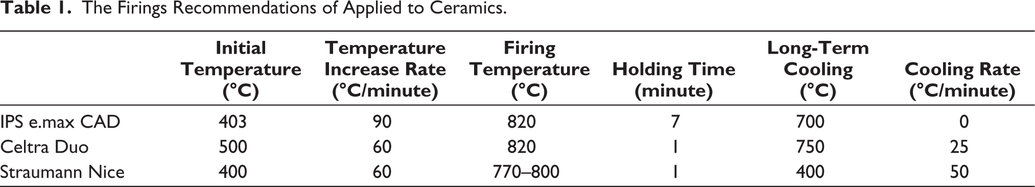
Each material was produced by high translucency (HT) A1 color blocks supplied by the manufacturers. The specimens were prepared in a square shape of 5 × 5 mm2 and in two separate thicknesses of 0.5 mm and 0.6 mm, which could simulate the minimal laminate veneer thickness in clinical use. The specimens were subjected to crystallization firing and glazing according to the manufacturer’s guidelines, following the process outlined in Table 1. Subsequently, the specimens underwent a 10-minute ultrasonic cleaning process with distilled water. Each specimen’s adhesion surface was then polished by using 600, 800, 1,000, and 1,200 grit abrasive paper with a consistent cooling effect from water. The specimens were then placed in distilled water at 4°C for 24 hours.
The Firings Recommendations of Applied to Ceramics.
Substrate Fabrication
To ensure consistency and simulate the dentin substrates, substrate blocks were created from A3 color composite (Polofil NHT; VOCO, Cuxhaven, Germany) with identical dimensions and a thickness of 4 mm before measuring the color of the specimens. 21 In the production of the composite blocks, a metal mold with the same length and width dimensions as the specimens was used. Composite resin was placed in the middle of the metal mold and polymerized with a light device (VALO Grand; Ultradent, South Jordan, Utah, USA) for 20 seconds. After the polymerization, the composite blocks were removed from their slots, and edge leveling and polishing processes were applied. All composite resin specimens were subjected to continuous water cooling and polished using abrasive paper with grit sizes of 600, 800, 1,000, and 1,200. After being created, 120 composite blocks were kept for 24 hours at 4 °C in distilled water.
Try-in Paste Application
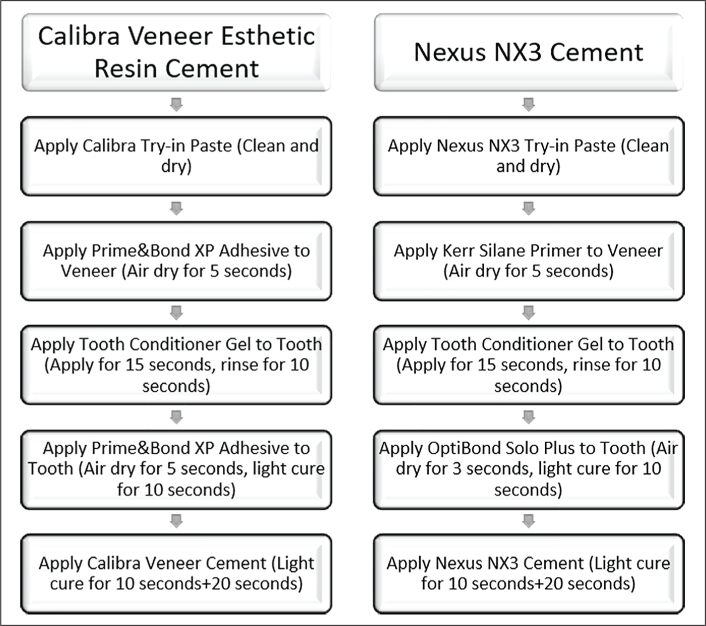
Using basic randomization, specimens with two distinct thicknesses from each ceramic group were split into two groups (n = 10), numbered, and cementation procedures were started with 2 different light-cure resin cements (C, Calibra Veneer Esthetic Resin Cement; Dentsply Sirona, Konstanz, Germany) (N, Nexus NX3; Kerr, Orange, CA, USA). To provide standardization in the cementation process, the closest shades of both resin cements (Calibra-Translucence, Nexus NX3-Clear) were selected. For each specimen, a try-in paste of the selected color of the resin cements was applied on the cementation surface of the materials, measured with a digital micrometer at a thickness of 100 ± 1 µm, 22 and then placed on the composite blocks. A force of 200 g was exerted on the upper surface of the specimens for a duration of 10 seconds, following which any excess paste was eliminated using a brush.
Spectrophotometric Analysis and Color Measurement
The color measurements of the specimens were measured with a spectrophotometer (Spectroshade Micro, MHT, Verona, Italy) on a gray background.
23
The illuminator was positioned with respect to the specimen’s horizontal plane at a 45-degree angle. This setup gave the specimen an optical configuration with 45 degrees of illumination and 0 degrees of observation. The reflectance spectra between 380 and 780 nm at a wavelength interval of 2 nm were measured with the spectrophotometer and then converted to CIE L*a*b* values (2 degrees observer and illuminant D65).
24
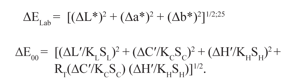
Measurements were repeated three times in the same specimen, and the mean L*, a*, and b* values were recorded as the initial measurement values (Lt*, at*, and bt*). Following their separation from the composite blocks, the ceramic specimens’ try-in pastes were cleaned with an air-water spray and allowed to dry. The same process was done for the composite blocks, and after making sure that there were no try-in paste residues, resin cement cementation was started. The cementation process was applied to both the ceramic specimens and the composite blocks, as illustrated in Figure 1, according to the contents of the resin cement sets and the recommended application procedure. To complete the resin cementation procedure, a digital micrometer-measured thickness of 100 ± 1 µm of resin cement was applied to the inside surfaces of the ceramic specimens and set on the composite blocks. The top surface of the ceramic veneers was then subjected to 200 g of pressure. After removing the excess materials from the edges, polymerization was achieved from the composite block to the ceramic specimens and from the ceramic specimens to the composite block with a light device (VALO Grand) for 20 seconds. The same spectrophotometer, same gray background, and identical optical setup were used for the color measurement. Measurements were repeated three times in the same specimen, and the mean L*, a*, and b* values were recorded as definitive values (Lr*, ar*, br*). Calculations were made using the CIELab and CIEDE2000 color systems to determine the mean color difference values for try-in paste and resin cement. The specified formulas were used for calculations:
The Diagram Showing Application Protocols of Resin Cements.
In this equation, ∆L0, ∆C0, and ∆H0 represent variations in lightness, chroma, and hue, respectively. Additionally, SL, SC, and SH are the weighting functions assigned to the lightness, chroma, and hue components, while KL, KC, and KH serve as parametric factors that necessitate adjustment in accordance with specific viewing parameters. It is significant to note that, for the sake of the current study, the values of these parameters have been set to 1. 26
Statistical Analysis
Statistical data analysis was conducted using a software program specialized in statistical analysis (IBM SPSS Statistics, v20.0; IBM, Armonk, New York, USA). Statistical analysis of the study groups and subgroups was carried out in accordance with the 3 × 2 × 2 factorial design. There, consequently, statistical analyses for the color difference assessments were conducted using a three-way ANOVA. To investigate the alterations in L, a, and b values, a paired sample t-test was employed. The Tukey multiple comparison test was also utilized to compare the mean values between groups (α = 0.05).
Results
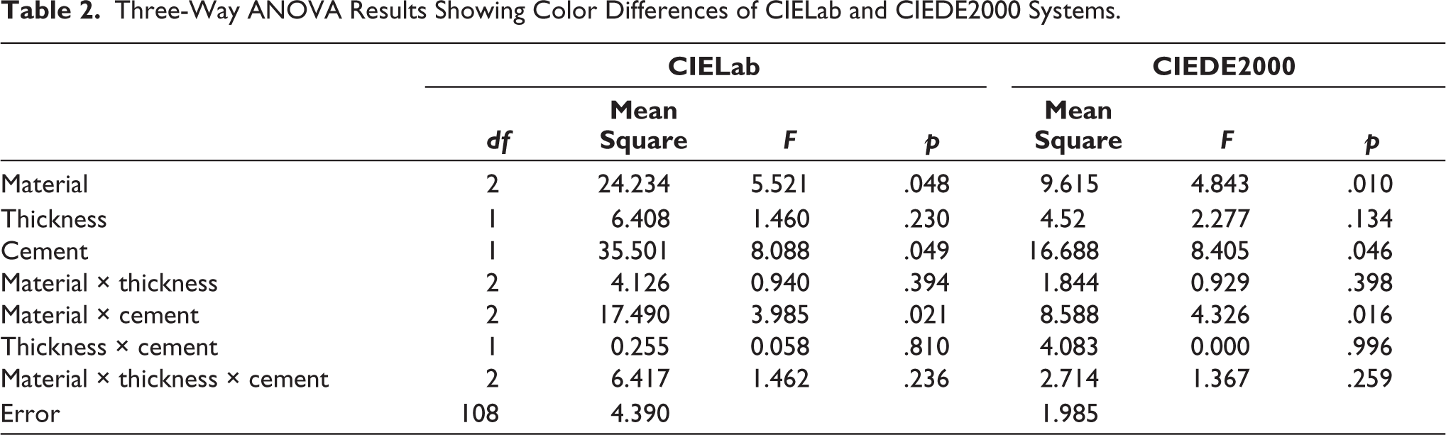
After analyzing the CIELab test outcomes, it was determined that there were no significant variances in the material × thickness × cement, material × thickness, and thickness × cement interactions (p > .05), as indicated by the results of the three-way ANOVA test. However, significant differences (p < .05) were found in the material × cement interaction (Table 2). Significant differences were observed between the LAS (3.69 ± 3.22) and other ceramics (p = .048) based on the findings of the Tukey multiple comparison test. N resin cement (3.36 ±2.8) had higher mean ΔE values (p = .049) than C resin cement (2.28 ± 1.43). However, no significant differences were found between the thicknesses (p = .23) (Table 3 and 4).
Three-Way ANOVA Results Showing Color Differences of CIELab and CIEDE2000 Systems.
The Least Squares Mean and Standard Deviation (SD) for the Color Differences of the Groups in the CIELab System.
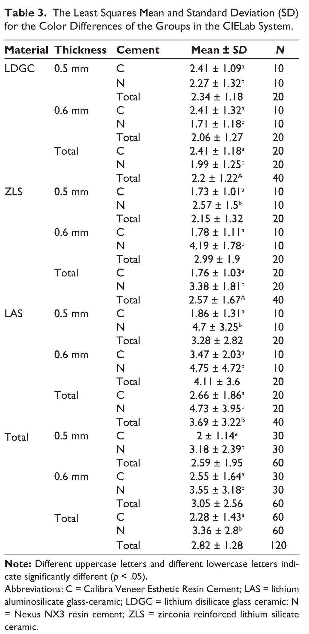
Abbreviations: C = Calibra Veneer Esthetic Resin Cement; LAS = lithium aluminosilicate glass-ceramic; LDGC = lithium disilicate glass ceramic; N = Nexus NX3 resin cement; ZLS = zirconia reinforced lithium silicate ceramic.
When analyzing the CIEDE2000 test results, no significant changes were identified in the material × thickness × cement, material × thickness, or thickness × cement interactions (p > .05), according to the results of the three-way ANOVA test. However, significant differences (p < .05) were found in the material × cement interaction (Table 2). The LAS specimens showed the highest ΔE values (2.54 ± 2.15) (p = .01), while the LDGC specimens showed the lowest ΔE values (1.58 ± 0.82) (Tukey multiple comparison test results, p = .01); C resin cement (1.62 ± 1.01) showed lower ΔE values than N resin cement (2.37 ± 1.85) (p = .046). However, no significant differences were found between the thicknesses (p = .13) (Tables 3 and 4).
The Least Squares Mean and Standard Deviation (SD) for the Color Differences of the Groups in the CIEDE2000 System.
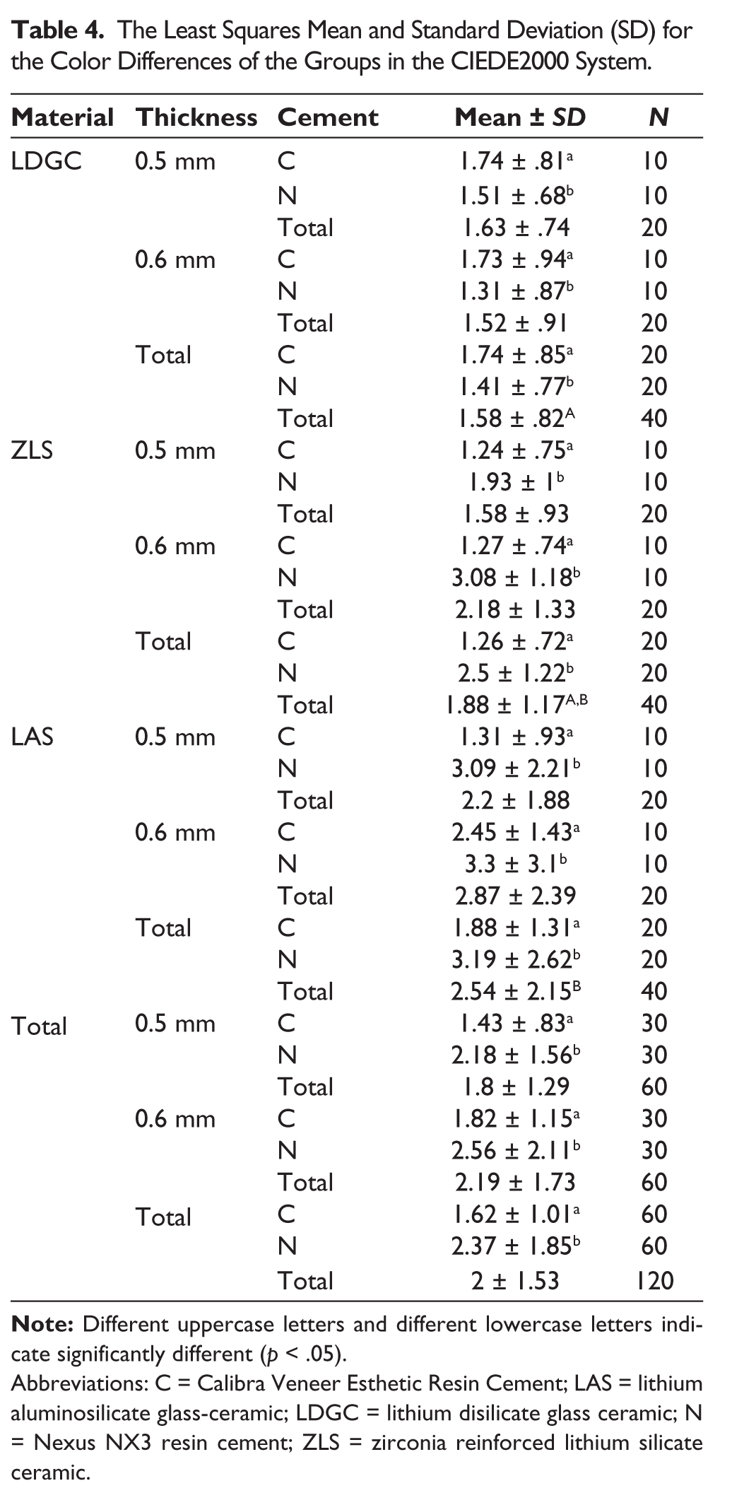
Abbreviations: C = Calibra Veneer Esthetic Resin Cement; LAS = lithium aluminosilicate glass-ceramic; LDGC = lithium disilicate glass ceramic; N = Nexus NX3 resin cement; ZLS = zirconia reinforced lithium silicate ceramic.
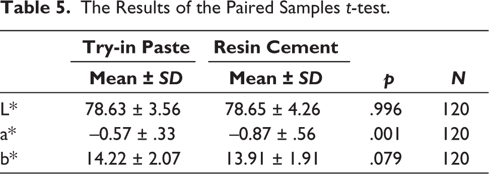
Significant disparities were identified between try-in paste and resin cement when evaluating the results of the paired sample t test based on the a* values (p = .001). The a* value of resin cement (–0.87 ± 0.56) exhibited inferior outcomes compared to the a* value of try-in paste (–0.57 ± 0.33). Nevertheless, there were no distinctions observed between try-in paste and resin cement concerning the L* and b* values (p > .05) (Table 5).
The Results of the Paired Samples t-test.
Discussion
The null hypothesis was partially accepted despite the differences in the materials, resin cements, and their interactions; there were no variations in thickness-dependent differences in thicknesses and interactions.
The perceptible and acceptable ΔE values (ΔELab) were 1.2 and 2.7 for the CIELab system, corresponding CIEDE2000 (ΔE00) values were 0.8 and 1.8, respectively. 27 According to the CIELab system, the findings of the current study showed that LDGC and ZLS specimens exhibited clinically acceptable values, while LAS specimens exhibited clinically unacceptable values. According to the CIEDE2000 system, only LDGC was found to exhibit mean color difference values at clinically acceptable levels. These results reveal the differences between both color systems.
It is advised to employ try-in pastes that match the color of the resin cements prior to cementation in order to confirm the ultimate color and state of the restorations.28,29 A very slight color change occurred during the polymerization of the resin cement, and there were notable variances between the three distinct try-in pastes and the matching resin cements, according to a study analyzing veneer porcelain restorations manufactured at a thickness of one millimeter. 30 The usage of thicknesses smaller than 1 mm may be the reason for the lack of variances observed in the color matching of try-in pastes and resin cements in this investigation. Chen et al 31 found that although the color of the try-in pastes and the corresponding resin cements had a high level of matching in LDGCs, there were significant differences in all shades applied in ZLSs. The compatibility of the try-in pastes and the corresponding resin cements of the ZLS specimens with other ceramics in the present study, according to both color evaluation systems, may be due to the use of only 1 shade of resin cements. In a study by Paken et al 32 aquagel was placed between zirconia-reinforced ceramic specimens and tooth-colored substrate. In the other groups, try-in paste and resin cement were placed between the ceramic specimens and tooth-colored substrate material samples, respectively. Try-in pastes applied to ZLS specimens, and the corresponding resin showed significant differences in all shades. In this investigation, try-in pastes had lower ΔE00 values compared to resin cements. The reason for the current outcome is that mixing the base and catalyst pastes causes the resin cement to become darker. Studies that show no difference was seen between the try-in paste and matching cements in terms of color perception are similar to the findings of the current investigation.33,34
Research35–37 investigating the impact of ceramic thickness on the compatibility of the try-in paste and its corresponding resin cement has shown that the thickness of the ceramic material has a noticeable influence on this compatibility. However, the ΔE values between the try-in pastes and resin cements have been found to increase with decreasing ceramic thickness, suggesting a decline in compatibility. Gomes et al 38 used 0.5 mm and 0.8 mm thick feldspathic ceramic veneers and reported that ceramic thicknesses affect the ultimate color of the restoration and that using thin ceramics has a significant effect on the result. Ayata et al 39 reported that the thickness of the veneer material used was more effective on the color masking of the final restoration than the tooth color and the translucency value of the material. If a laminate restoration of 0.5 mm or thinner is planned, it is very important to consider the type of resin cement, tooth color, and ceramic material. In a different research, monolithic zirconia specimens were utilized with varying thicknesses: 0.5 mm, 1 mm, 1.5 mm, and 2 mm. A combination of opaque and transparent cement hues was used to cement these specimens. The results of the investigation showed that the material’s thickness affects the final hue. Furthermore, the specimens showing the biggest degree of alteration were those with a thickness of 0.5 mm. 40 Kürklü et al 41 reported that no color changes were found in the 0.5 mm and 1 mm laminate veneers using transparent resin color. The fact that no significant differences were found between the 0.5 mm and 0.6 mm thickness values and the statistical interactions between the parameters did not show significance in the present study also supports Kürklü et al. 41 This result shows that thickness has no effect on try-in paste and resin cement color matching, contrary to the studies in the literature.
According to Munsell color evaluation system coordinates, 42 both try-in paste and resin cements had color parameters on the grayish axis based on L* values and on the yellowish axis based on b* values. Based on a* values, statistical differences were found between the try-in paste and the corresponding resin cement, while the resin cement had greener axis color characteristics than the try-in paste.
The ultimate color of translucent restorations, particularly veneers, may be negatively impacted by the hues of resin cements.43,44 The kind, color, and thickness of the resin cement as well as the shade of the ceramic were what ultimately defined the veneer restorations’ optical color. 45 Silva et al 46 reported that cement color can reduce the effect of veneer translucency on the ultimate color of the veneer, while some cement colors can make the effect of veneer translucency selection more prominent. Both the try-in paste and the resin cement had their thicknesses normalized to 100 ± 1 µm22 and maintained constant because the purpose of this study was to assess how closely the colors of the two materials matched. Since the colors of the resin cement corresponding to the try-in paste were already used as the color, this parameter was also eliminated.
The fact that the current study only used one color for the try-in pastes and resin cement was one of its limitations. Another restriction was the assessment of just two distinct thicknesses. Another drawback of the study was that it was conducted in vitro, which meant that the oral environment could not be perfectly replicated. Investigating different thickness and color options in future studies may lead to different results.
Conclusions
The in vitro study’s results led to the following deductions being made:
Color match was found between the try-in pastes and their corresponding resin cements according to both color evaluation systems. LAS had the highest ΔE value for both color systems. C resin cement showed lower ΔE values than N resin cement in both color systems. Thicknesses (0.5 mm and 0.6 mm) did not have any effect on the color match between the resin cements and try-in pastes.
Footnotes
Acknowledgements
This study was supported by the Department of Scientific Research Projects (TSA-2022-10239) Atatürk University, Erzurum, Turkey.
Authors’ Contribution
Conceived and designed the experiments: AO. Performed the experiments: GE, OG, and FK. Analyzed the data: AO. Wrote the paper: AO, GE, OG, and FK.
Data Availability Statement
The data that support the findings of this study are available on request from the corresponding author.
Declaration of Conflicting Interests
The authors declared no potential conflicts of interest with respect to the research, authorship, and/or publication of this article.
Ethical Approval
Ethics committee approval is not required for this in-vitro material study.
Funding
The authors disclosed receipt of the following financial support for the research, authorship, and/or publication of this article: This study was supported by Department of Scientific Research Projects (TSA-2022-10239) Atatürk University, Erzurum, Turkey.
Informed Consent
Not applicable