
Abstract
Aims:
To evaluate the color changes that occur after accelerated aging in feldspathic ceramic crowns cemented with three different dual-cured resin cements.
Materials and Methods:
For each of the A2-colored RelyX U200, G-CEM LinkForce, and Panavia V5 cement groups, 45 dies from A2-colored zirconia blocks and 45 crowns from CEREC blocks were prepared. Color measurements after 24 h of cementation (T1) and after cycles of aging of 1750 (T2), 3500 (T3), and 7000 (T4) in the thermal cycle device were made using SpectroShade Micro device. The coordinates of the color were used L*, a*, and b* as base and the color change was calculated with ∆E00 in determining the color. One-way analysis of variance test was used to compare the times in terms of ∆L*, ∆a*, and ∆b* values.
Results:
It was found that ∆L* value decreased significantly in period of the T2, T3, and T4 times compared to T1 in all groups (P < .05), whereas the change between period of T2, T3, and T4 times was not significant (P > .05). The ∆a* value increased significantly in the period of T3 and T4 times compared to T1 in the only G-CEM LinkForce group, whereas the ∆b* value increased significantly in the period of T4 time compared to T1 in the only Panavia V5 group. The changes in ∆E00 values, which were observed in all period of times, were found to be between 0.43 and 1.04, 0.43 and 1.43, and 0.40 and 0.97 in RelyX U200, G-CEM LinkForce, and Panavia V5 groups, respectively.
Conclusion:
After accelerated aging, it was found that the color of all cements became darker and the G-CEM LinkForce group turned red and the Panavia V5 group turned yellow. However, it was found that the color changes that occurred were within clinically acceptable visible levels.
Keywords
Introduction
Resin cements are frequently used in cementation of full ceramic restorations due to their superior mechanical properties, support for ceramic, low solubility in the oral environment, and sufficient esthetics.1,2 Resin cements can mask the color underneath as well as change the color of the restoration in full ceramic restorations, which depend on three main factors: final colors of the tooth/substructure, the type of ceramic/resin cement, and the thickness of the ceramic/resin cement.3,4 Therefore, controlling and balancing the opacity and color parameters play a key role in achieving good esthetic results.3,5
It is reported that the most important problem that can be encountered in resin cements is exposure to internal and external coloring.1,2 While environmental conditions, ultraviolet rays, humidity, heat, and food pigments are called external discoloration agents, the formation of oxygen by-products related to the chemistry of the material is the internal color change. 6 Oxidation of reactive groups in amine accelerators and inhibitors causes a more yellow color change in chemically activated systems such as dual-cured and autocured resin cements.2,6,7 It is reported that this structural change in chemical composition is very difficult to control. 2
While visual color measurement methods used in dentistry are not preferred very often due to the presence of the subjective definitions, instrumental color measurement methods are more preferred due to the fact that the factors that can affect color determination are at minimum level, they contain numerical values, and their repeatability and reliability are high.8,9 Digital camera and image systems, colorimeters, spectroradiometers, and spectrophotometers are one of the most frequently used instrumental color measurement methods nowadays. 10 It was reported that the measurements obtained in a study, in which spectrophotometers named VITA Easyshade and SpectroShade Micro were compared, were extremely reliable and spectrophotometers could be used clinically to determine the changes in tooth color before and after treatment. 11
Although, in literature, there are many studies that evaluated the color stability of different resin cements in different full-ceramic restorations,2,12–15 no research has been found to evaluate RelyX U200, G-CEM LinkForce, and Panavia V5 in feldspathic ceramic crowns. At this juncture, it was aimed to evaluate the color changes, occurring after accelerated aging in feldspathic ceramic crowns cemented using RelyX U200, G-CEM LinkForce, and Panavia V5, by SpectroShade Micro in this study. In our null hypothesis, there is no significant difference in terms of color change between RelyX U200, G-CEM LinkForce, and Panavia V5 resin cements before and after aging.
Materıals and Methods
Study Groups
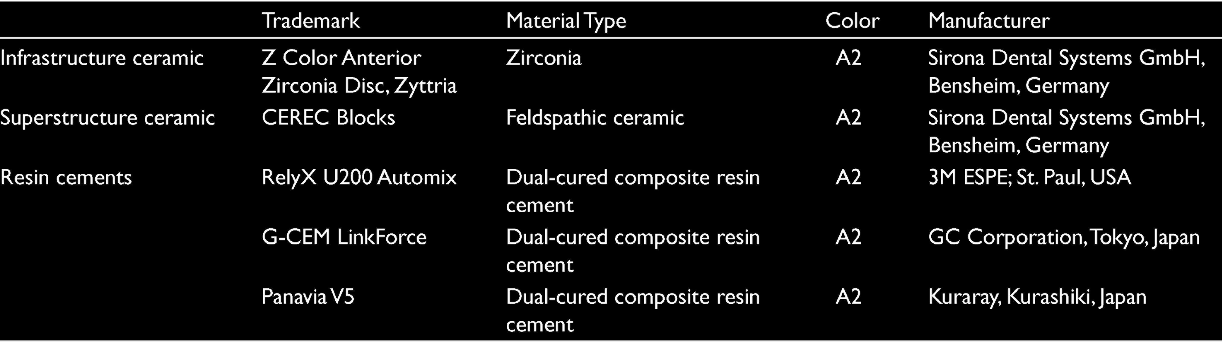
For RelyX U200 (3M ESPE; St. Paul, USA), G-CEM LinkForce (GC Corporation, Tokyo, Japan), and Panavia V5 (Kuraray, Kurashiki, Japan), 12 groups were found, with four subgroups for each group, according to times of aging in our study in which discoloration occurred after 24 h of cementation in dual-cured cements (T1) and 1750 (T2), 3500 (T3), and 7000 (T4) cycles of aging in the thermal cycle device (n = 15). Ceramic samples and resin cements used in the study are shown in Table 1.
Ceramic Samples and Resin Cements Used in the Study
Sample Preparation
Preparation of samples: Forty-five dies from zirconia blocks in A2 color and 45 crowns samples from CEREC blocks were prepared for each of the three different dual-cured cement groups in A2 color.
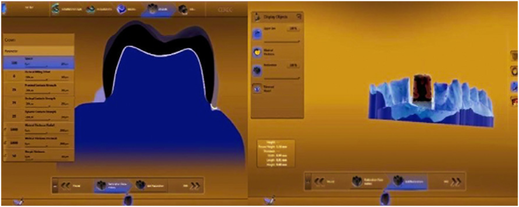
The model obtained from zirconia die designed with solid modeling in Solidworks program (Dassault Systems, Waltham, MA, USA) was scanned with a 3Shape device, and then their samples were produced in milling device (Coritec 250i, İmes-İcore GmbH, Eiterfeld, Germany). The zirconia die produced were placed by scraping in the plaster model missing tooth with number 11 and fixed with wax (Figure 1). Afterward, digital models were obtained after scanning with an intraoral scanning device (CEREC AC Omnicam, Sirona Dental Systems GmbH, Bensheim, Germany; Figure 2). Each side of the restoration is designed to be 1 mm thick and the cement thickness to be 100 μm to provide all crowns on these digital models with standard features (Figure 3).
Fixing the die with a wax in a plaster model for digital impression
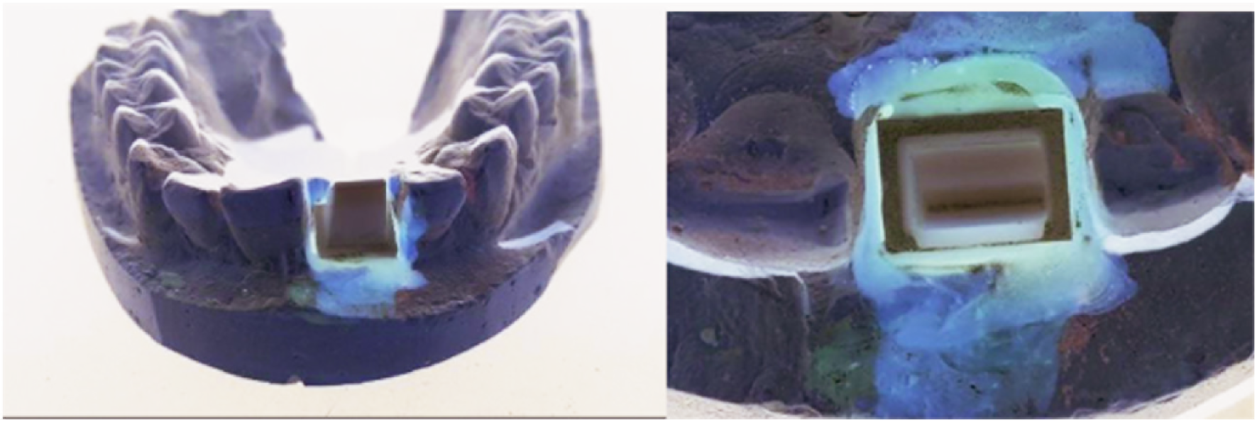
Obtaining the digital model
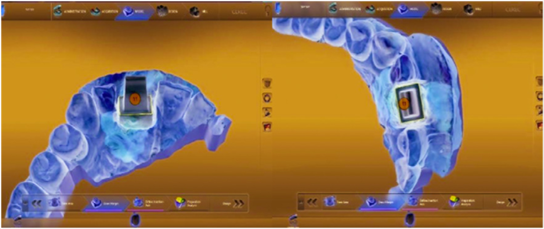
Creating digital design
The production of the restorations, considering their design was completed, was made using CEREC blocks in the two-stage milling mode in the scraping device (CEREC MCXL, Sirona Dental Systems GmbH, Bensheim, Germany). The produced feldspathic ceramic crowns are cleaned after keeping them for 120 s in ultrasonic cleaner and dried with air–water spray (Ultrasonic Cleaning Device, Guangdong GT Ultrasonic Industrial Co. Ltd., Shenzhen, China). Glazed material consisting of powder and liquid (VITA Akzent Plus, VİTA Zahnfabrik H. Rauter GmbH and Co. KG, Bad Säckingen, Germany) was mixed and applied to the surface using a brush.
Cementation Processes
Cementation processes in RelyX U200, G-CEM LinkForce, and Panavia V5 cement groups were performed in the direction of the manufacturer’s recommendations. In all groups, the surface of the zirconia die was blasted with aluminum oxide particles at a distance of approximately 10 mm for 60 s, then cleaned with 98% ethyl alcohol and dried with air without water and oil. The inner surfaces of the feldspathic crowns were washed with water for 30 s and dried with air without water and oil after acidifying them with 9% hydrofluoric acid in a gel form for 60 s. The application was repeated when the ground–glass appearance was not seen on the surface of the samples.
The individual primer of each cement group was applied to both zirconia die and feldspathic ceramic crowns for 5 s and dried by spraying the air without water and oil to evaporate the dissolvent. The resin cement was mixed with a plastic spatula and applied using a brush to the inner surface of the crown, and then placed on the zirconia die with finger pressure; after prepolymerization was performed for 3 s with a light device (Elipar Freelight 2 LED Curing Light, 3M ESPE, St. Paul, USA), the spillover redundancy was cleaned with the help of a probe. Afterward, they were repolymerized for 20 s from the mesial, distal, buccal, and lingual surfaces.
Color Measurements
SpectroShade Micro for color measurements made on a gray neutral background at T1, T2, T3, and T4 times (MHT, Verona, Italy) was used. After the SpectroShade Micro device was calibrated using the white and green calibration tiles made by the manufacturer, color measurements were made three times from each sample and the average of three was used.
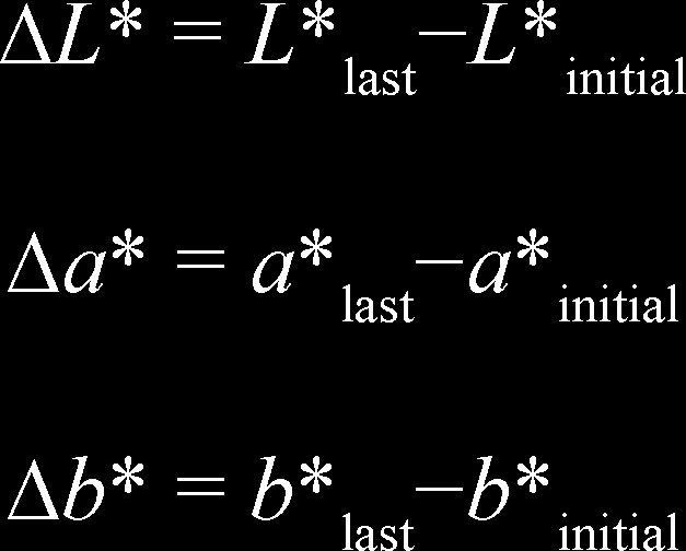
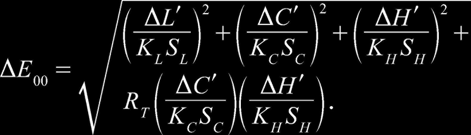
Discolorations in four different time periods were evaluated on the basis of the whole crown. In determining of color, a CIEDE 2000 (ΔE00) system expressing the coordinates of the color with the symbols L*, a*, and b* was used as base. The L* coordinate in the system describes the brightness of the color, the a* the amount of red–green color, and the b* the amount of the blue–yellow color. While the L* coordinate is located on the vertical axis containing values from 0 (absolute black) to 100 (absolute white), a* and b* coordinates rotate around the axes around L* and take values between −128 and 127. When the color a* takes positive value, it becomes red, when it takes negative value, it becomes green. When b* takes positive value, it becomes yellow, when it takes negative value, it becomes blue. ∆E00 values obtained as a result of the color measurement made using the CIEDE 2000 system indicate the degree to which the color can be perceived by the human eye. ∆E00 < 0.81 describes as clinically imperceptible discoloration. ∆E00 ≤ 1.77 describes as clinically acceptable visible discoloration.
16
Discolorations were calculated using the following formulations:
Statistical Analysis
Descriptive statistics for the features emphasized have been expressed as mean, standard deviation, and minimum and maximum values. One-way analysis of variance variance analysis method was used in the analysis of the results, and Tukey HSD test according to the homogeneity of the values. Statistical significance level was taken as 5% in the calculations and SPSS for windows version 23 (IBM Corp., Armonk, NY, USA) statistics package program was used for calculations.
Results
The changes observed in ΔL*, Δa*, and Δb* values in the period of T1, T2, T3, and T4 times of RelyX U200, G-CEM LinkForce, and Panavia V5 groups are shown in Table 2. In all groups, there was a significant decrease in ΔL* value as against T1 in the period of T2, T3, and T4 times. It was found that the change between the period of T2, T3, and T4 times was not significant. It was observed that there was a significant increase in Δa* value in the period of T3 and T4 times to that in T1 in the only G-CEM LinkForce group; while the change between the period of T2, T3, and T4 times and the period of T1 and T2 times was not significant. On the other hand, a significant increase in the Δb* value was found in the only Panavia V5 group to that in T4 with regard to the period of T1 and T2 times. The change between the period of T1, T2 and T3, and T3 and T4 times was not found to be significant.
∆L*, ∆a*, and ∆b* Values of RelyX U200, G-CEM LinkForce, and Panavia V5 Groups, Respectively, After 24 h of Cementation and After 1750, 3500, and 7000 Cycles of Aging
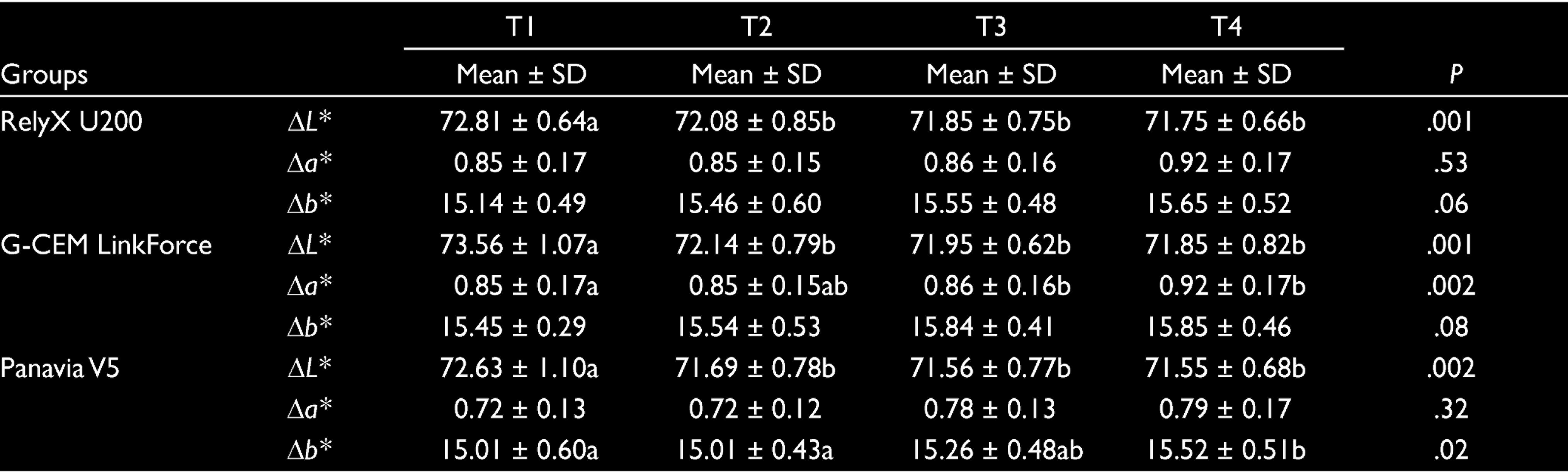
T1, after 24 h of cementation; T2, 1750 cycles after aging; T3, after 3500 cycles of aging; T4, 7000 cycles after aging.
The changes and their comparisons observed in ∆E00 values of RelyX U200, G-CEM LinkForce, and Panavia V5 groups in the period of T1–T2, T1–T3, T1–T4, T2–T3, T2–T4, and T3–T4 times were shown in Table 3. As seen in the table, it was found that the ∆E00 values of the period of only T2–T3 and T3–T4 times values in the RelyX U200 Automix group were lower than T1–T2, T1–T3, and T1–T4. It was found that ∆E00 values of the period of T2–T3, T2–T4, and T3–T4 times in G-CEM LinkForce group were lower than the values of T1–T2, T1–T3, and T1–T4. The ∆E00 value of the period of T3–T4 times was found to be lower than those of T1–T2, T1–T3, and T1–T4, whereas the ∆E00 value of the period of T2–T3 and T2–T4 times was found to be lower than the value of T1–T4 in the Panavia V5 group.
The Values After 24 h of Cementation of RelyX U200, G-CEM LinkForce, and Panavia V5 Cements and the ∆E Value After 1750, 3500, and 7000 Cycles After Aging
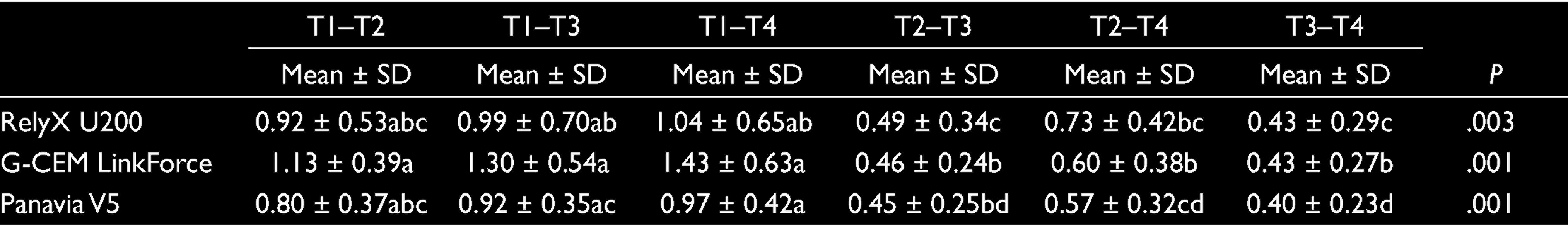
T1, after 24 h of cementation; T2, 1750 cycles after aging; T3, after 3500 cycles of aging; T4, 7000 cycles after aging.
Discussion
In our study, in which the discoloration in three different resin cements after accelerated aging in feldspathic ceramic crowns was evaluated using SpectroShade Micro, it was observed that the color of all cements became darker after aging and the G-CEM LinkForce group turned red and the Panavia V5 group turned yellow. Besides, it was found that clinically acceptable visible discoloration occurred in all groups after accelerated aging. According to some authors, it has been reported that the yellowing of a material over time may be related to camphoroquinone in its chemical structure. 17 Another explanation for the yellowing tendency has been reported that the Bis-GMA-based material may be exposed to ultraviolet light and heat. 18 Almeida et al. stated in their study that the reddening of the dual-cured cement they used was due to the presence of unreacted camphoroquinone and oxidized amine molecules. 19
Corrosion of metal-supported ceramic systems, which have been used for a long time as their mechanical properties are good, bad optical properties, and metal reflections at the gingival border have led to the preference of full-ceramic systems. 20 It is reported that the optical properties of full-ceramic restorations are close to that of the natural tooth, their biological compatibility with oral tissues is better than those produced by traditional methods, and their thermal expansion coefficient and thermal conductivity are close to that of dental tissue.3,13
The use of Computer Aided Design (CAD)/Computer Aided Manufacturing (CAM) systems has become popular in the construction of full-ceramic restorations. It has been reported that the internal and marginal compatibility of restorations produced using CAD/CAM are more compatible than other production methods. 21 Besides, it is stated that because the blocks produced industrially for use in CAD/CAM systems can be produced more homogeneously and with less defectives, restorations produced with CAD/CAM systems are more preferred than other restorative options. 22 Therefore, the crown restorations were produced by a CAD/CAM system using CEREC Block CAD/CAM material in our research.
It has been stated that zirconia, another dental material used in our study, has a high mechanical strength, esthetics, and biocompatibility.23,24 Using zirconia ceramics has provided multimember restorations to be made in full-ceramic restorations and increased the success rate. 24 Also, Hamm et al. evaluated the appropriate models for CAD/CAM in vitro studies by investigating different model materials regarding suitability for intraoral scanners and dimensional stability. They reported that polyurethane and the typodont model did not meet the requirement of dimensional stability, whereas scans of CoCrMo and ZI showed the best precision. 25 Because of these properties, zirconia was used instead of natural teeth and metal abutments in our research. In this way, the possibility of different optical and microstructural properties of the substructure samples obtained from natural teeth and the possibility of corrosion of metal abutments over time have been eliminated. 20
One of the most widely used aging techniques to simulate the effect of the intraoral environment on restorative materials is the exposure of restorative materials to cyclic thermal stresses. 26 What should be the bath temperature, keeping time, and number of cycles used in thermal aging technique is still being discussed.21,22 Although International Organization for Standardization recommended in the 5 to 55°C for a 20-s, 500-cycle aging procedure, some researchers prefer their own parameters instead of following this standard.26,27 In our study, 28 the protocol which Addison et al. assume that 3500 thermal cycles correspond to approximately one clinical year has been taken as a reference. At this juncture, samples subjected to aging of 7000 thermal cycles were assumed to stimulate the two-year intraoral environment. In addition, it was found that bath temperatures are 5 to 55°C, bath transfer time is 10 s, and keeping time in bath is 25 s.
Nowadays, light-cured or dual-cured cements are used in the cementation of full-ceramic restorations because they are more esthetic. 29 It has been stated that insufficient light polymerization in light-cured cements, which are frequently preferred in esthetic areas due to their color stability, causes increased solubility and gaps in marginal areas, secondary caries and pulpal reactions.12,29 Therefore, dual-cured cements have been developed to provide a complete polymerization of the chemically treated component in deep areas where light is cut down.7,12 It is reported that the tertiary amine, which acts as an initiator in its composition for the chemical polymerization reaction, causes discoloration in dual-cured cements.12,29 An amine reacts with a benzoyl peroxide, often aromatic or other amine, such as komforoquinone. The two molecules in the polymerization reaction between amine and benzoyl peroxide cannot be at the desired level because they cannot be in close contact with each other. This situation causes color instability in unreacted benzoyl peroxide molecules. 12 In our study, it was aimed to evaluate the discolorations after accelerated aging in three different cement groups with Panavia V5 without amine group, RelyX U200 without benzoyl peroxide, and G-CEM LinkForce containing benzoyl peroxide.
The American Dental Association recommends the use of the CIEDE 2000 color differential system for the evaluation of color differences. 30 Not only discoloration in our study was analyzed at ΔE00 level but also evaluations were made in terms of ΔL*, Δa*, and Δb*. Then, after 24 h of cementation at ΔL* value in the RelyX U200 group, it was observed that there was a significant decrease in aging at 1750, 3500, and 7000 cycles in thermal cycle device. It was found that the change observed for ΔE00 value in the period of all times is between 0.43 and 1.04. In comparison with the findings of our study, discoloration after cementation performed using RelyX U200 in monolithic zirconia ceramic discs with 0.6 mm and 1 mm thick were, respectively, 5.64 and 5.06 for ΔE00 value in the study, which was evaluated using spectrophotometer (model CM-2600d) before and after cementation, whereas it was observed that the decrease in L*, a*, and b* values was greater. 31 In the study in which discoloration after cementation performed using RelyX U200 on IPS e.max Press ceramic discs was evaluated using spectrophotometer (Color-Eye 7000A) after cementation and after 100 h of accelerated aging, it was found that the change in the ΔE00 value was 2.76 and the observed that the decrease in ΔL*, Δa*, and Δb* values were 1.04, 0.63, and 1.69, respectively. 32 The differences in the findings are thought to be due to the different ceramic discs used, different aging procedures, and color measurement conditions and equipment.
In the literature, there are a limited number of studies in which discolorations after cementation using G-CEM LinkForce and Panavia V5 in different full-ceramics are evaluated. Atay et al. 33 evaluated the discoloration, which after cementation performed using G-CEM LinkForce and Panavia V5 on IPS e.max press ceramic discs, using a spectrophotometer (VITA Easyshade) after cementation and 5000 and 10000 cycles after aging in thermal cycle device.
It was found that change observed for ΔE00 value in the G-CEM LinkForce group was 1.69 and 2.17, whereas there was significant decrease in L* and a* values and a significant increase was occurred in the b* value. It was found that changes observed for ΔE00 value in the Panavia V5 group were 1.55 and 2.47, whereas there was significant decrease in L* and a* values and significant increase was occurred in the b* value. In our study, there was, after the 24 h of cementation in L* value in the G-CEM LinkForce group, a significant decrease in 1750, 3500, and 7000 of cycles aging in thermal cycle device, whereas there was a significant increase in a* value in 3500 and 7000 of cycles aging in comparison with later 24 h than cementation. It was found that the change observed for ΔE00 value in the period of all times was between 0.43 and 1.04. After the 24 h of cementation in the Panavia V5 group, a significant increase was observed in 7000 cycles of aging in the thermal cycle device and it was found that the change observed in ΔE00 value in the period of all times was between 0.40 and 0.97. It is thought that the minimal differences between studies may be due to the ceramic discs used, different aging procedures, and the color measurement device.
In the literature, the studies in which the discoloration observed in the cements after accelerated aging were evaluated, and it was observed that all the faces of the cements that were bonded to different ceramic discs were directly exposed to the aging environment, except one coated face. Yet, cements in the oral environment are in direct contact with the external environment only from their marginal areas. Therefore, the cements are exposed to excessive external factors and hydrolytic deterioration occurs due to excessive water absorption in aging procedures. 19 In our study, samples were produced as crowns, and the cements cemented on zirconia core were exposed to aging environment only from their marginal areas to eliminate these negativities and to make them more suitable for clinical conditions. Therefore, the main limitations of this study are that different cement types were not evaluated in different ceramic samples and that it was an in vitro study. Thus, it is recommended to conduct in vivo studies on different ceramic samples, where different cement types are evaluated in the long term.
Within the limitations of this study, the following conclusions can be drawn.
After accelerated aging, it was found that the color of all cements became darker and the G-CEM LinkForce group turned red and the Panavia V5 group turned yellow.
24 h from cementation in all cement groups and the discoloration in the thermal cycle device after 1750, 3500, and 7000 cycles of aging were found to be clinically acceptable visible levels.
Footnotes
Acknowledgements
The authors wish to thank Dr Naci Murat for statistical analysis.
Declaration of Conflicting Interests
The authors declared no potential conflicts of interest with respect to the research, authorship, and/or publication of this article.
Ethics Approval
Ethics committee approval is not required for this study.
Funding
This research was supported by the Scientific Research Projects Division of Van Yuzuncu Yil University with project number TDH-2018-7009.
Patient Declaration of Consent
The authors declare that they have no conflict of interest.