
Abstract
Aim:
An assessment and comparison of three retreatment files was conducted to determine their effectiveness in the retrieval of gutta-percha.
Materials and Method:
In the investigation, 60 extracted single-rooted human teeth having straight canals and complete apices were used. The ProTaper Universal file system was used to prepare teeth and obturated using gutta-percha with sealant AH Plus utilizing down pack and backfill obturation technique. Removal was performed with ProTaper, D-RaCe files, MTwo, and R-Endo retreatment files. Using a stopwatch, the time needed to take out the GP was also calculated in seconds. We used ANOVA along with the post hoc Tukey’s test to analyze the quantity of obturating material in apical, middle, and coronal third following decoronation and longitudinal splitting.
Results:
The average percentage of gutta-percha points removed across root levels varied significantly among the ProTaper, D-RaCe MTwo, and R-Endo groups. With the exception of the ProTaper group, when the middle level displayed the most removal, the coronal level showed much higher removal than the mid-root level, while the apical level often exhibited the highest removal.
Conclusion:
ProTaper retreatment files outperformed MTwo, D-RaCe, and R-Endo among the four groups in terms of removing root canal filling material. However, none of the experimental groups removed gutta-percha completely from root canals.
Introduction
Successful outcomes in root canal procedures are through proper cleaning of the canal and the root canal system sealed completely. 1 With newer technologies developed to its aid, its success rate has been estimated to vary between 50% and 90% when properly conducted. 2
The use of novel materials and techniques in root canal therapy has demonstrated significant success, particularly in terms of enhancing tooth functionality and preservation. The recurrence of root canal infection can prevent the desired outcome of primary root canal therapy. 3 Failed endodontic cases are primarily due to inadequate root canal preparation and filling. 4 In root canal retreatment, by taking out the filler material and so getting rid of the related microorganisms, the infectious process is treated. 5
Non-surgical retreatments are the most conservative methods. 6 The success rate of appropriately executed retreatment, accompanied by suitable measures to control and prevent endodontic re-infection, varies between 62% and 85%. 7 The most popular filler material for root canals is gutta-percha, and its complete removal is critical to reviving entry to the apical foramen for successful retreatment procedures. 8
Numerous methods are employed to retrieve remaining filler materials from root canal space with the use of endodontic hand files, rotary instruments, heated instruments, and solvents. 9 Rotary nickel-titanium (Ni-Ti) instruments are the less time-consuming method to remove filling materials from root canal space effectively. 10
The primary rationale behind this finding is that the generation of frictional heat and softening of the gutta-percha during rotary instrumentation would help in easier removal of the obturating material. 11
Thus, the objective of this study is to evaluate and compare the efficiency of Ni-Ti rotary instruments, that is, MTwo retreatment, D-RaCe, ProTaper retreatment, and R-Endo retreatment files in the elimination of gutta-percha.
Materials and Method
Upon clearance from the ethical committee (with reference letter number SBBDC/2020/152118-A dated 22.12.2020), the study was conducted. Patient consent was not required as it was an in vitro study. Sixty human single-rooted teeth that were extracted having straight canals and completely developed apices were chosen for the investigation. Ultrasonic scalers were employed to eliminate calculus and residual soft tissue from the root surfaces. To ensure proper disinfection, teeth were immersed in NaOCl at 3% concentration. Subsequently, the teeth were kept until they were needed in distilled water.
Preparation of the Specimen
Access opening was done using an endoaccess bur (Dentsply) and high-speed airotor (NSK, Japan). Then, Gates-Glidden drills in sizes 1, 2, and 3 (Mani, Japan) were used to execute the coronal third preparation with a micromotor (API, India), and the working length was adjusted to be 1.0 mm less after measuring the length of the root canal using an endobloc (Dentsply). By inserting a 10 K-file (Mani, Japan) into the root canal, apical patency was evaluated. The crown-down approach was employed with X-SmartTM Endodontic Motor (Dentsply Maillefer) up to F2 by means of the ProTaper universal file system from Dentsply Maillefer. Upon each file change, a standard irrigation protocol was followed. The irrigation solution used was 5 mL of 3% NaOCl (Chemident, India). A further 5 minutes was spent rinsing with 17% EDTA (Anabond Desmear, India) and then 3% NaOCl (Chemident, India), and paper points were used to dry the canals after being cleansed with saline water for a minute.
Canal Filling
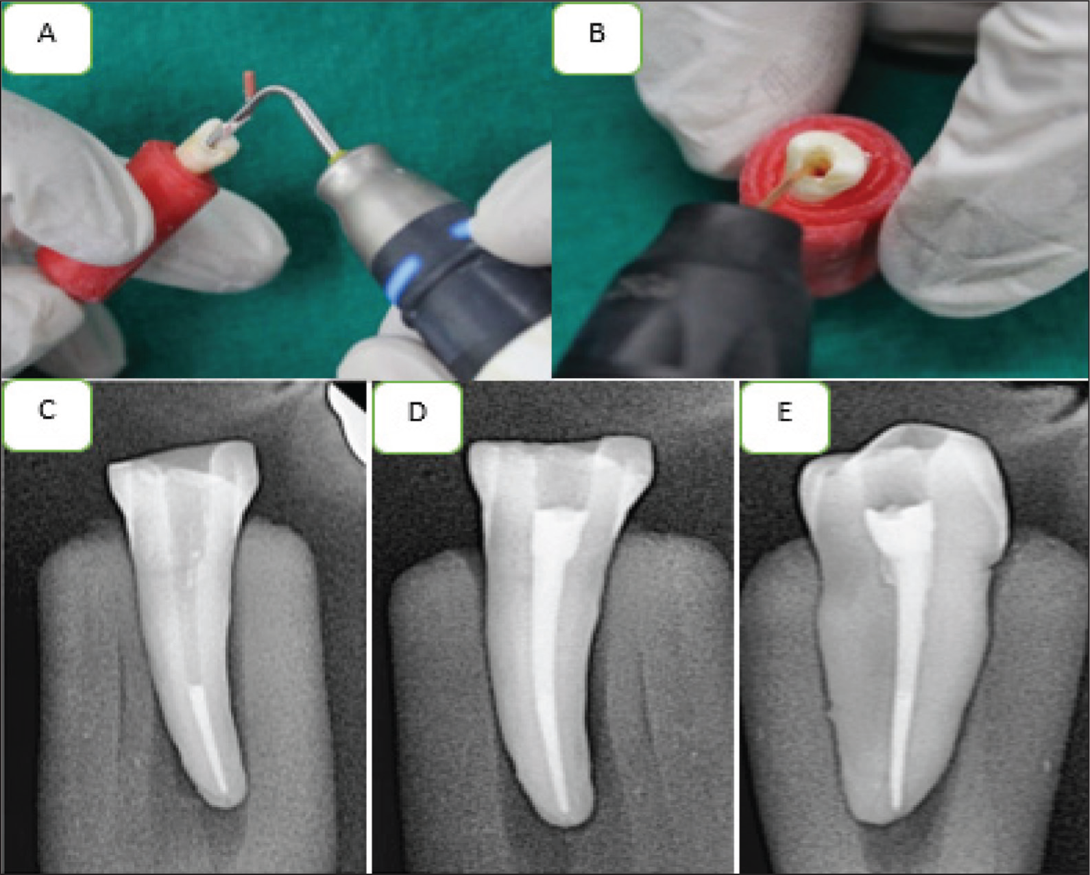
Sealant AH Plus, a resin-based product (Dentsply Maillefer) using gutta-percha points, was inserted into the canal. Gutta-percha points (Dentsply Maillefer, USA) were used to seal the root canals, and an apical plug was made with Fast Pack (Orikam Healthcare, India). Radiographs were performed to verify the apical plug formation. The remaining canal was sealed off using thermally plasticized gutta-percha pellets (Orikam Healthcare, India) using the Fast Fill obturation system (Orikam Healthcare, India). Using radiographs collected at various angles, the obturation was assessed. The specimens were kept in storage for 2 weeks at 100% humidity and 37°C after placing temporary restoration (Figure 1).
(A) Down Pack. (B) Backfill Obturation. (C) Apical Plug. (D and E) Bucco-Lingual and Mesio-distal View of Final Obturation.
Retreatment Procedures
In accordance with what instrument was applied to GP removal, the samples were sequentially separated into four groups. Group I: MTwo R (VDW, Germany); Group II: D-RaCe (FKG, Switzerland); Group III: ProTaper universal retreatment file (DENTSPLY, Switzerland); and Group IV: R-Endo (MICRO-MEGA, France).
Group A: MTwo Retreatment Files
Two retreatment files, 15/0.05 and 25/0.05, were employed in the removal of the obturating material in the following manner: R25 (Size 25 with a 0.05 taper) and R15 (Size 15 with a 0.05 taper). The suggested speed is 280 rpm. The maximum torque is 0.03 Ncm at 15/0.05 30 gcm. Maximum torque is 1 Ncm at 25/0.05 120 gcm. Once utilized in two root canals, the instruments were disposed of.
Group B: D-RaCe Retreatment Files
The D-RaCe set comprises two Ni-Ti instruments: DR1, which has a size of 30 and a taper of 0.10, and DR 2, which has a size of 25 and a taper of 0.04. DR1 was used in the coronal third, rotating at 1000 rpm and 1.5 Ncm of the desired torque, and DR 2 rotated at a recommended torque of 1 Ncm at 600 rpm in the middle and apical thirds. DR1 was discarded after being employed in three root canals, while DR2 was utilized only once before being discarded.
Group C: ProTaper Universal Retreatment Files
Featuring a convex triangular cross-sectional design, these are used to remove filling material. The coronal third is tapered with D1 (size 30, 0.09), the middle third is tapered with D2 (size 25, 0.08), and the apical third is tapered with D3 (size 20, 0.07). The lowest speed (500 rpm) is recommended. D1 is discarded after three root canals, while D2 and D3 are discarded after two root canals.
Group D: R-Endo Retreatment Files
They consist of five instruments: The sizes of the taper are 25, 0.04 for Rm, 25, 0.12 for Re, 25, 0.08 for R1, size 25, 0.06 for R2, and 25, 0.04 for R3. R1, R2, and R3 were utilized as follows, at rotational speeds of 300–400 rpm up to the working length, correspondingly for the coronal, middle, and apical third. R1 had been discarded after three root canals. The R2 and R3 were discarded after two root canals.
A stopwatch was utilized for recording the retreatment time (in seconds) for each group immediately after the application of each instrument.
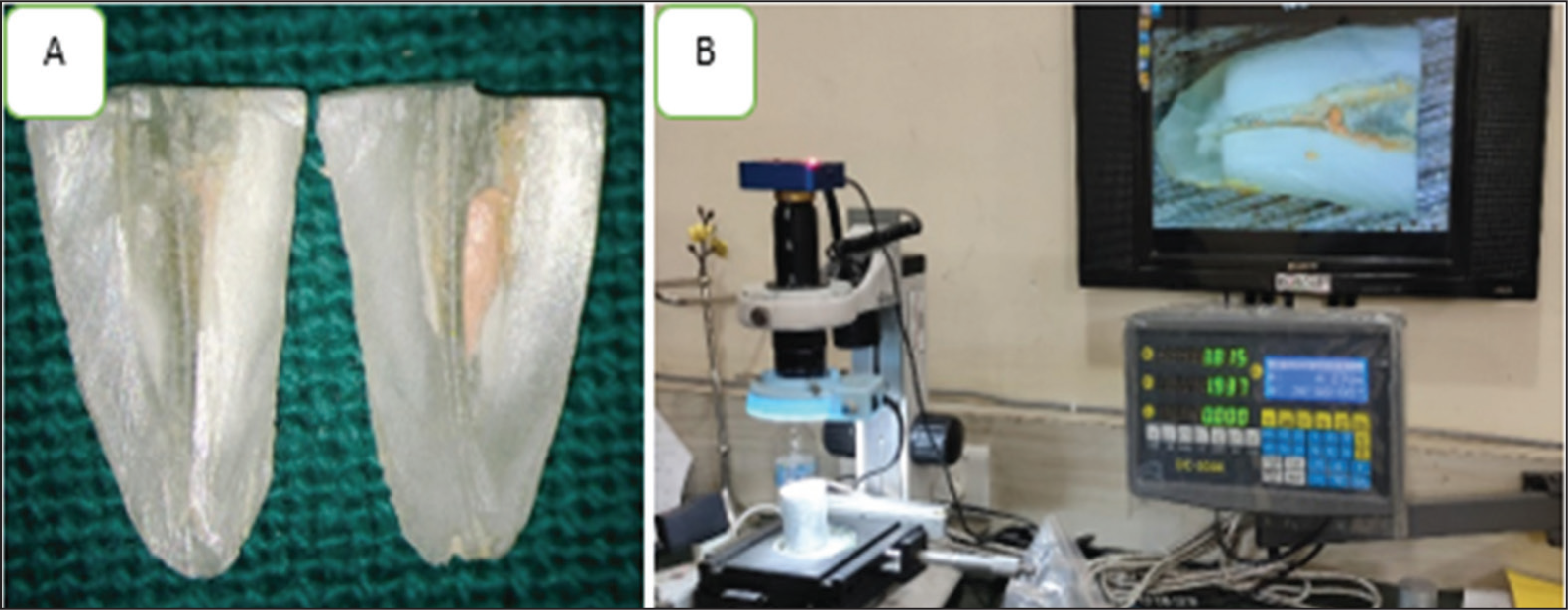
In all groups, between each instrument, samples received 2 ml of 3% NaOCl irrigation. Notably, 5 ml (17% EDTA) and 2 ml (3% NaOCl) served as the last resort of irrigation for 30 seconds. After that, a diamond disc was used to decoronate the roots to within 12 mm of the reference area. The roots were subsequently divided longitudinally with the aid of a chisel and mallet, and both root portions were captured on camera using a stereomicroscope set to 20× magnification (Bresser, Rhede, Germany). The root canals were split into three 4-mm sections using the Image J software. Consequently, root canals and regions with remaining filling material were demarcated, and their respective areas were measured in mm 2 (Figure 2).
(A) Photograph of LS Taken Under Stereomicroscope. (B) Magnification Under Stereomicroscope.
Evaluation Criteria
The Image J program (National Institutes of Health, version 1.53) was used to compute the area of the remnant gutta-percha and the overall area of the canal wall.
Using the formula, the proportion of the residual filling was determined:
Statistical Analysis
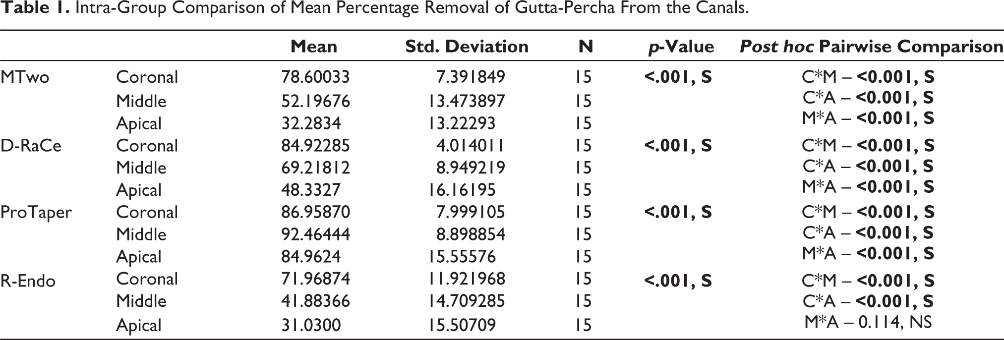
To conduct the statistical analysis, the SPSS software (IBM SPSS Statistics 19.0, IBM, Armonk, NY, USA) was used. The residual filler material was evaluated using a one-way analysis of variance (ANOVA). Additionally, a multiple comparison Tukey’s HSD test was performed with a significance of .05 (Table 1).
Intra-Group Comparison of Mean Percentage Removal of Gutta-Percha From the Canals.
Results
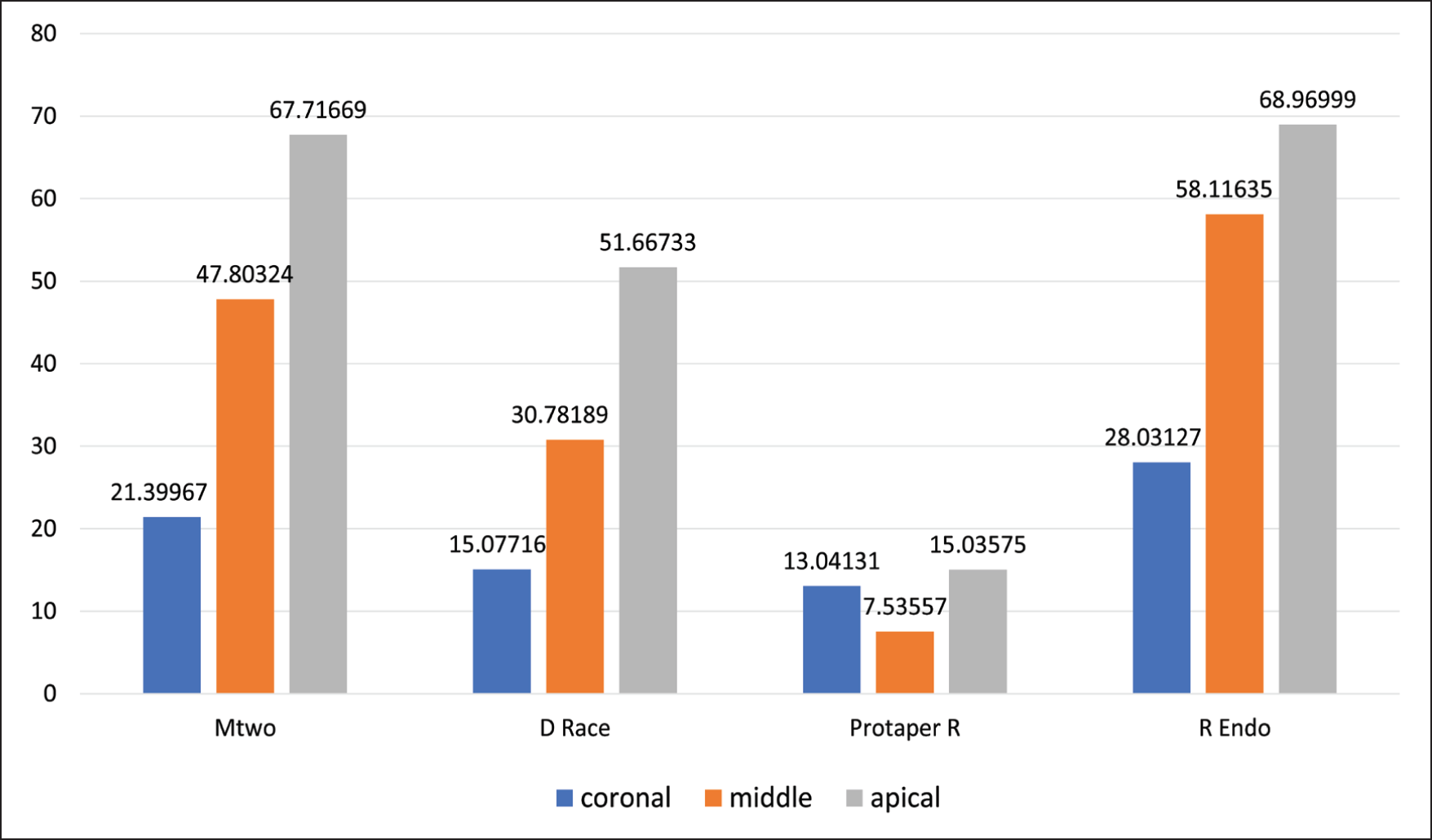
ProTaper universal retreatment file system exhibited the maximum percentage of GP removal (88%), followed by D-RaCe retreatment files (67%), MTwo retreatment files (54%), and R-Endo retreatment file systems (48%). For ProTaper, removal was highest in the middle third (92%), followed by the coronal third (87%) and the apical third (84%), whereas in MTwo, D-RaCe, and R-Endo, the removal was seen most in the coronal third (78%, 84%, and 71%), then the middle (52%, 69%, and 41%), and the apical (32%, 48%, and 31%), respectively. The qualitative analysis supported these findings and revealed that ProTaper universal retreatment has a higher mean percentage clearance of gutta-percha, followed by D-RaCe retreatment, MTwo retreatment, and R-Endo retreatment file systems. The differences were statistically significant between the groups, showing significant variances in variables that were measured (Figure 3).
Percentage of Residual Guttapercha on the Root Canal Walls After Retreatment.
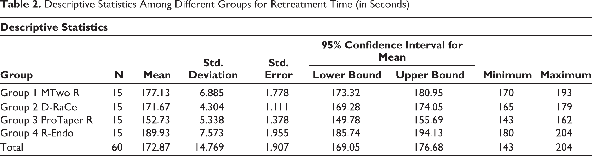
In this study, Table 2 illustrates the mean time in seconds required to remove obturating material for four distinct group systems (N = 15). It was found that group 4 has the highest value (189.93 + 7.57) for mean time in seconds required to remove filler material followed by group 1 (177.13 + 6.88) and group 2 (171.67 + 4.30). It was found that group 3 has the lowest value (152.73 + 5.33) for the mean time in seconds required to remove gutta-percha/sealer from the canals (Figure 4).
Descriptive Statistics Among Different Groups for Retreatment Time (in Seconds).
Time Taken to Remove Gutta-Percha From Canals in Four Groups.
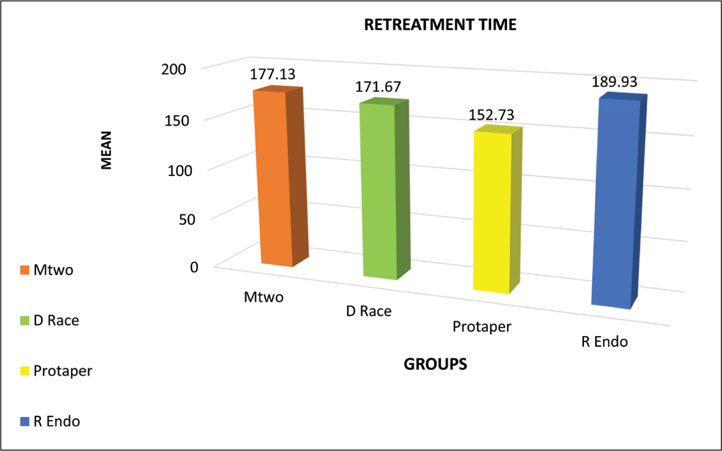
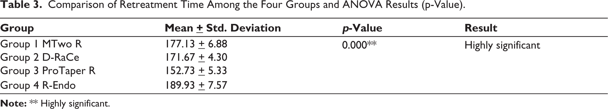
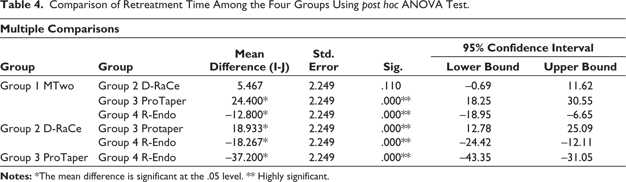
Table 3 illustrates that highly significant results were found for the mean difference between the four different group systems employing p-value ˂.05 for the one-way analysis of variance (ANOVA) test (Table 4).
Comparison of Retreatment Time Among the Four Groups and ANOVA Results (p-Value).
Comparison of Retreatment Time Among the Four Groups Using post hoc ANOVA Test.
Discussion
Even while initial endodontic therapy has a usually high success rate, mistakes can nevertheless happen due to procedural and nonprocedural errors. 9 Common factors associated with endodontic failures include persistent bacterial infection in the canal or periradicular area and preexisting periradicular rarefaction. Additional contributing factors encompass intricate root canal structure, separated instruments, mechanical perforations, root fractures, coronal leakage, the presence of periradicular lesions, and periodontal disease.9, 12
Nonsurgical retreatment is often considered the treatment of choice in the management of failed endodontic cases, with a success rate of 74%–98%. 8 The aim of nonsurgical endodontic retreatment is to extract the maximum amount of sealant and gutta-percha. The remnants of necrotic tissue and bacteria that may have contributed to the periapical irritation and pain will be recovered during the procedure. 13
Hand files as well as rotary files have been used to remove root canal filling material. The conventional file is associated with tediousness and time consumption for the operator, potentially leading to endodontic mishaps while removing well-condensed obturating material. In contrast, Ni-Ti files offer superelasticity, allowing for more centered canal preparations with reduced canal transportation. Moreover, the increased taper preparation facilitates effective irrigation. These files have greater cutting efficiency once engaged in a crown-down approach and a continuous reaming motion. As a result, it is possible to achieve root canal preparations that are rounder, with reduced straightening and minimal apical extrusion. However, despite the enhanced flexibility of Ni-Ti files, the risk of file separation remains a concern. 13
Although rotary devices facilitate or speed up the removal of root fillings, their usage should be cautious because more dentin was removed than with manual instruments. 14
The root canal sealer used in this investigation was AH Plus sealer. As AH Plus is a resin-based sealer, it has an edge in terms of biocompatibility and radiopacity. It is an epoxy resin with a non-toxic hardener. Radio-opacity is achieved through the inclusion of bismuth oxide, which also exhibits strong adhesive properties and undergoes slight contraction during the hardening process. Importantly, there is no release of toxic formaldehyde during the setting of these sealers. 15
According to a study done by Christopher et al., 16 the continuous-wave condensation approach led to an obturation with a higher density than that produced by cold lateral compaction. 16 This method was used to seal off canals. The quality and quantity of the obturation were judged with radiographs taken at different angulations. The filling material was then allowed to be set by storing the samples at 100% humidity for 2 weeks. After 2 weeks, all the samples were divided into four groups (n = 15) according to the four different types of Ni-Ti file systems used, namely, ProTaper universal retreatment files, D-RaCe files, MTwo files, and R-Endo retreatment file systems, for endodontic retreatment. 11
In the study conducted by Ersev et al., 17 radiographs were employed to assess the presence of remaining gutta-percha. However, it was noted that two-dimensional (2D) radiographs were insufficient for accurately representing the actual cleanliness of the canal and evaluating the effectiveness of retreatment procedures. 17 According to investigations by Kfir et al., 14 and Marques da Silva et al., 5 longitudinal splitting of roots for microscopic examination and photographic documentation was a preferable method. 14
Therefore, in this investigation, the roots were divided longitudinally, the amount of filling material that remained in the canal after the retreatment method was evaluated radiographically, and the leftover gutta-percha was examined using a stereomicroscope with the use of the Image J software.
No file system used in this investigation was able to fully remove the filler material from each of the 60 samples. However, the ProTaper universal retreatment file system showed the highest percentage of GP removal (88%), followed by D-RaCe retreatment (67%), MTwo retreatment (54%), and R-Endo retreatment file systems (48%). ProTaper removed more in the middle third (92%), after the coronal (87%) and the apical third (84%), whereas in MTwo, D-RaCe, and R-Endo, the removal was seen most in the coronal third (78%, 84%, and 71%), then the middle (52%, 69%, and 41%), and apical (32%, 48%, and 31%), respectively.
The importance of time in the root canal retreatment has several key factors, that is, efficiency and productivity, patient comfort, operational convenience, reducing chair side time, and good clinical outcomes. The disparity in retreatment time among the four groups yielded statistically significant results.
The results of this study were consistent with those of Marques da Silva 5 , Lalit et al., 10 Iriboz, 18 Joseph et al., 19 and Khedmat et al., 13 which show that ProTaper Universal retreatment file systems removed the most gutta-percha when compared with D-RaCe and MTwo systems. The distinctive flute design of ProTaper Universal Retreatment files is responsible for their exceptional performance. When removing root filling, these files efficiently cut through the superficial layer of dentin in addition to removing gutta-percha. The progressive tapers of D1, D2, and D3 files, which allow the sculpting of particular parts of a root canal with a single file, are additional characteristics. Without placing the instrument under undue strain in other areas, the variable tip diameter of the files enables accurate cutting action within the defined area of the canal. 10
Subsequently, a study done by Sunil et al. 15 claimed that the removal of gutta-percha from root canal walls using the MTwo R file system proved to be more successful than the R-Endo retreatment file system. The three design elements provide safe preparation, quick dentin removal, and effective shaping. 15 During instrumentation, M2 files exhibit an automatic cutting action as they progress apically. Additionally, they also perform lateral cutting. 15
Complete removal was not achieved in any of the groups. It is crucial to understand that this study was conducted in vitro, which limits the direct applicability of the results to clinical scenarios. Additionally, because the study only included teeth that have straight canals, it is impossible to immediately extend its findings to teeth exhibiting curved canals. To ascertain the effectiveness and if rotary Ni-Ti devices are safe and preserve native root canal morphology when used in the retreatment of teeth with intricate root canal anatomy in clinical conditions, more research is required.
Conclusion
The mean percentage reduction of GP at the coronal level among MTwo, D-RaCe, and R-Endo was discovered to be substantially greater than that in the middle third which is considerably greater than that at the apical region, while among the ProTaper group, the mean percentage removal of GP at the middle level was found significantly higher than the coronal level, which was much greater than the value at the apical level. ProTaper retreatment files performed better as compared with MTwo, D-RaCe, and R-Endo in the removal of root canal filling material.
Footnotes
Declaration of Conflicting Interests
The authors declared no potential conflicts of interest with respect to the research, authorship, and/or publication of this article.
Ethical Approval
Ethical committee approval vide letter no: SBBDC/2020/152118-A.
Funding
The authors received no financial support for the research, authorship, and/or publication of this article.
Informed Consent
Not required.