
Abstract
Aim:
The study’s objective is to assess the capacity of 17% EDTA (ethylene diamine tetraacetic acid) and 17% EDTA-S (sodium lauryl sulphate) on periodontally infected root surfaces as well as their capacity to remove the smear layer.
Materials and Methods:
Groups A (EDTA) and B (EDTA-S) were randomly selected from among 40 single-rooted teeth that had to be extracted due to the mobility. Sections of these teeth were subjected to a 3-minute “Active Burnishing Technique” treatment with 17% EDTA and 17% EDTA-S, followed by a 2-minute saline washing, and then investigated by using an electron microscope. The teeth’s root surface changes were evaluated using Sampaio’s Index, surface morphology, smear layer presence, dentinal tubule patency, and other factors. The results of this study were statistically examined using the paired “t” test. The significance threshold was established at P-value = .05.
Results:
The outcomes demonstrated that Group B benefited statistically from the elimination of the smear layer and the patency of the dentinal tubules (P < .01). In the EDTA and EDTA-S groups, there were 85.508.75 and 91.706.20 patent dentinal tubules revealed, respectively. The total surface area exposed by EDTA was 163.88 ± 24.68 and EDTA-S was 182.91 ± 28.16 with a P-value <.01.
Conclusion:
According to the study’s findings, as compared to EDTA, EDTA-S is more successful at exposing dentinal tubules and removing the smear layer, making it a useful root conditioning agent.
Introduction
Bacterial plaque is the main etiological component in periodontal disease. As a result, the tissues of the gingival region form inflammation lesions and the supporting periodontal tissues gradually start to degrade. 1
In order to create an environment that is conducive to epithelial and connective tissue cell adhesion and attachment, periodontal therapy aims to restore the health of the damaged periodontal tissue as well as damaged root surfaces. 2
Root surfaces that have periodontitis are over-mineralized and contaminated with harmful and other biologically relevant chemicals. Because of the development of a smear-like layer composed of organic and inorganic waste, the standard mechanical equipment used to treat periodontally damaged roots is unable to completely eradicate the infection. Chemical root conditioning has been proposed as a remedy for these mechanical therapies’ drawbacks. 3
Although root debridement and ultrasonic scaling are relatively common treatment methods in a clinical setting for physically cleaning the radicular surface during periodontal treatment, they are unable to completely decontaminate the dental hard tissues.3,4
Since the radicular surfaces of periodontally compromised teeth are severely contaminated with bacteria and their endotoxins, periodontal regenerative therapies may be less effective as a result of the delay in the formation of a new connective tissue link. Obtaining a flawless, even, and clean radicular surface during periodontal surgery is essential for facilitating creative connective tissue reattachment and accelerating the healing of the tissue.3-5
Several methods for conditioning root surfaces have been offered to achieve the goal of demineralization. In medicinal practice, chemicals including tetracycline, citric acid, phosphoric acid, and ethylene diamine tetraacetic acid (EDTA) have been utilized as conditioning agents. Since EDTA has a neutral pH and has been used as a root conditioner for a long time, it does not harm collagen fiber or periodontal tissues. Early cell migration and periodontal ligament attachment to the root surface are made feasible by exposing collagen fibers. To modify EDTA, 17% EDTA-S (sodium lauryl sulphate) is created by adding the sodium lauryl sulphate derivative Texapon to EDTA in a 1:1 ratio. 6
According to the current study’s research hypothesis, root surface modification, dentinal tubule patency maintenance, and smear layer removal may all benefit from the use of 17% EDTA-S. 3 We tried to examine the efficiency of 17% EDTA and 17% EDTA-S in eliminating smear layers because there is currently very little information about 17% EDTA-S. Dentinal tubule continuity, surface erosion, tooth material loss, along with the root surface modification index (RSMI) were compared at a magnification of 1500×.
Materials and Methods
In vitro study was conducted in Vishnu Dental College, Bhimavaram, Andhra Pradesh, India from June 2022 to December 2022. Forty single-rooted human teeth with advanced periodontitis were used. The teeth under consideration lacked erosions, cervical restorations, or caries. They were accordingly stored in regular saline and distilled water. A water-cooled high-speed bur was used to remove the root’s crown, healthy part, and two to three millimeters of its apical area after scaling and root planning. For the study, dentine specimens with a 5 mm × 5 mm dimension were prepared. Each specimen’s labial surface was employed for the investigation. Two groups were formed from all 40 roots. Fully erupted teeth, a lack of recent periodontal instrumentation or dental prophylaxis history, and periodontally diseased teeth with at least 60% attachment loss that were recommended for extraction were the inclusion criteria. Those teeth with proximal caries that reached the cementum and those with restorations that went past the CEJ were both disqualified from the research.
Objectives
Comparing the efficacy of smear layer removal was the study’s main goal. Finding the overall count of dentinal tubules in each sample, the ratio of exposed tubules to the overall count of tubules prevailing, the total surface area, dentin erosion, the loss of tooth substance, and the treatment of the root surface were the secondary objectives.
Sample Size in Each Group
40 (with justification)
Analysis
A priori: Compute required sample size
Tail (s)
= Two
Effect size
= 0.9316332
α err prob
= 0.05
Power (1-β err prob)
= 0.80
Allocation ratio N2/N1
= 1
Noncentrally parameter
= 2.9460829
Critical t
= 2.0243942
Sample size group 1
= 20
Sample size group 2
= 20
Total sample size
= 40
To achieve consistency, a standardization and calibration exercise was carried out before to the study’s start. Ten removed teeth were taken into consideration, five of which received 17% EDTA treatment, and the remaining five had 17% EDTA-S treatment. The effectiveness of the two approaches for removing the smear layer was tested on observers. The effectiveness of two compounds was evaluated, and the Cronbach’s alpha value―a gauge of consistency―was calculated and found to be 0.9. Between the two observers who evaluated the effectiveness of root conditioning, there was 90% agreement.
Group A: For a total of three minutes, cotton pellets soaked in a 17% EDTA (Meta Biomed MD Cleanser EDTA with pH 7, South Korea) solution were placed on the root specimens, and the solution was changed every 30 sec. Group B: For a total of 3 min, cotton pellets soaked in a 17% EDTA-S (Meta Biomed MD Cleanser EDTA with pH 7, South Korea) solution were placed on the root specimens, and the solution was changed every 30 sec. Comparing a neutral pH root conditioning agent (EDTA) to a low pH etching can improve healing.7,8
Using the “Active Burnishing Technique” and tweezers, saturated cotton pellets of the test solutions were rubbed onto the dentin samples for three minutes, changing the pellets every 30 sec to maintain a constant concentration. 8 The teeth were immediately placed in around 20 cc of distilled water after etching and gently swirled for 20 sec to cease the chemical reaction.
Spray Coated teeth were taken out of the desiccators jar and placed on the SEM stubs with the aid of a sputter coating apparatus. For the non-conducting sample to be observed under an electron microscope, sputtering imparts a conducting surface to it. All specimens were dehydrated using an ethanol gradient with 1% acetone as the last step. The roots of the teeth were placed in the center of the stubs and the labial side of the teeth faced the SEM’s beam after being dried for 30 min. The teeth were then mounted on the stubs with adhesive tape. Roots were scanned and inspected on the computer monitor with the SEM set to 1500×. Photographs of the representative areas were taken. The Paired t test was employed in every statistical analysis, with a significance level of P = .05. Sampaio’s Index, surface morphology, the existence of smear layers, the integrity of the dentinal tubules, and other variables were used to evaluate the teeth for root surface modification.
Statistical Analysis
The Statistical Package for Social Sciences (SPSS) version 20.0 program (IBM SPSS, IBM Corp., Armonk, NY, USA) was used to analyze the data. The RSMI was assessed using the Kruskal-Wallis H test. Using paired “t” test, RSMI was compared between the two groups. The significance threshold was established at P-value = .05.
Results
Forty periodontally ill teeth were removed and analyzed for the study. The following factors were examined using a scanning electron microscope after they were divided into two treatment groups (Table 1).
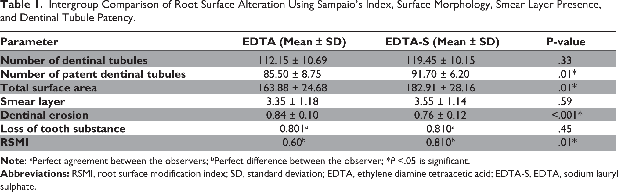
Intergroup Comparison of Root Surface Alteration Using Sampaio’s Index, Surface Morphology, Smear Layer Presence, and Dentinal Tubule Patency.
Total Surface Area
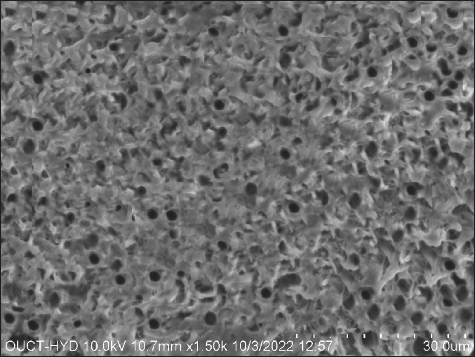
It had been noticed that change in root surface appearance at 1500× magnification was 163.88 ± 24.68 in Group A and 182.91 ± 28.16 in Group B (Figure 1). The research revealed that Group A had a surface that was more uneven than Group B with P-value = .01*.
Group A Shows Almost Complete Removal of the Smear Layer with Some Patent Dentinal Tubules.
Smear Layer
At 1500× in Group A, the mean ± SD of smear layer removed was 3.35 ± 1.18, and in Group B, it was 3.55 ± 1.14 (Figure 2). It was observed that Group B showed to have more efficiency in removal of smear layer than Group A, which was not statistically significant (P-value = .59).
Group B Shows Complete Removal of the Smear Layer with more Patent Dentinal Tubules Compared to Group A.

Number of Dentinal Tubules
At 1500×, it was seen that the number of dentinal tubules in Group A was 112.15 ± 10.69, and 119.45 ± 10.15 in Group A (Figure 1). The study showed that Group B showed to have more dentinal tubules as compared to Group A but was not statistically significant (P-value = .33).
Dentinal Tubules Patency
At 1500× in Group A, the mean ± SD of dentinal tubule patency was 85.50 ± 8.75, and the mean ± SD of dentinal tubule patency in Group B was 91.70 ± 6.20. Group B showed more patent dentinal tubules than Group A with P-value = .01*.
Dentinal Erosion
At 1500× in Group A, the mean ± SD of dentinal erosion occurred was 0.84 ± 0.10, and the mean ± SD of dentinal tubule patency in Group B was 0.76 ± 0.12. Group A showed more dentinal erosion than Group A with P-value < .001*.
Loss of Tooth Substance Index (LTSI)
In both groups, most of the specimens had corrugated surfaces with localized regions of cementum that had been partially removed, according to the data. There was no statistical difference between the two groups’ LTSI scores at 1500× (P = .45). As a result, both groups experience the same level of tooth material loss.
Root Surface Modification Index (RSMI)
At 1500× magnification, the RSMI scores did not indicate any statistical significance (P = .01) between Group A and Group B. At 1500× magnification, the RSMI scores of 2 in Group B showed that EDTA-S has superior root bio-modification characteristics.
Discussion
Root bio-modification chemicals have been utilized for many years to remove this smear layer before any regenerative operation. Acid etching is one of the methods for cleaning the root surface that is suggested.8,9
The root surface was detoxified utilizing a range of root bio-modification agents prior to periodontal regeneration. Additionally, it has been observed that neutral pH etching preserves the vitality of nearby tissues, whereas low pH etching results in the necrosis of the gingival tissue and nearby periodontal tissues 30 sec after the exposure.8-11
It is well known that EDTA-S removes smear layers more effectively than EDTA by itself. In the medical industry, soft soap is frequently used to remove incrustation in scaly skin conditions. Surface tension is reduced by the incorporation of mild soap, which acts as an active detergent. 12
Sixty single-rooted teeth were taken from the study by Kancharla et al. in 2019 due to periodontal disease. The teeth were given root conditioning agents treatment and examined under an electronic scanning microscope. The authors stated that the application of Carisolv as a root conditioning agent demonstrated favorable outcomes and could therefore be advised for use in vivo after evaluating the teeth for surface anatomy, the existence of smear-like film, the condition of dentinal tubules, and the loss of tooth material index. The findings of this investigation conflicted with those of our study, which found that the elimination of smear layers with EDTA and EDTA-S was similarly effective but not statistically significant. 13
Twenty single-rooted human teeth were classified into Group A and Group B for the study, which was carried out in 2012 by Nanda et al. Using an aggressive burnishing technique for 3 min, Group A dentin specimens received EDTA treatment, while Group B specimens received 10% tetracycline HCl solution treatment. After that, a scanning electron microscope was used to investigate the root surface samples. These findings were in line with the findings of the current investigation, which showed that 17% EDTA and 17% EDTA-S were effective at removing smear layers.14,15
Babgi et al. conducted research on 15 healthy single-rooted teeth in 2020. Four equal groups of scaled roots were created; the first group had no chemical treatment, while the other three received either HA, 17% EDTA, or 0.2% CHX gel as conditioning agents. Adjunctive therapies did not improve the GF attachment, and SRP is still the best way to restore cell adherence to the surface of the root and promote periodontal health. The results of the current experiment, which demonstrated the efficacy of 17% EDTA and 17% EDTA-S in eliminating smear layers, were consistent with these findings. 16
The application of chemo-mechanical therapy resulted in the elimination of the infected cementum along with the exposure of the unaffected cementum, as evidenced by the specimens treated with EDTA-S in the current experiment, which had a mosaic-like appearance showing the cementum. This was consistent with a prior investigation conducted by Grisi et al. 17
Dentinal tubule patency was seen in the EDTA-S group at a magnification of 1500×. The dentinal tubule integrity could be visible even at a magnification of 200×, according to an investigation conducted by Banerjee et al. 18 This was not observed in line with research by Grisi et al. 17 The dentinal tubules of the EDTA group were found to be less permeable than those of the EDTA-S group at a magnification of 1500×. Similar results were seen in the case of EDTA in a research investigation by Amaral et al., who studied the effects of the chemicals phosphoric acid, EDTA, tetracycline, and citric acid on the removal of minerals. 19
The diminution of tooth structure index was assessed across the four groups in the current study. How the dentist worked, how much power was used, and even how angular and sharp the curettes were can all be inferred from the total quantity of tooth material that was removed. 14
In the current study, the root surface post-modification was evaluated using the RSMI, which Sampaio JEC and colleagues proposed. This experiment’s usage of the RSMI at a magnification of 1500× revealed that EDTA-S had a significant root bio-modification property. It was determined that the root surface had a mean score of 2, meaning that the dentinal tubule gaps showed evidence of smear layer and that the root surface was free of smear layer. This is consistent with the outcomes of the research by Banerjee et al. 18
In a study published in 2022, Górski et al. examined the effects of root conditioning with 24% EDTA on the outcomes at 12 months following the use of the modified coronally advanced tunnel (MCAT) and subepithelial connective tissue graft (SCTG) to treat multiple gingival recessions (GR). When several RT1 and RT2 GR were treated with MCAT and SCTG, the use of 24% EDTA for root conditioning had no effect on the results at the 12-month mark. While the present investigation showed better smear layer removal, maintenance of dentinal tubules patency, and root surface modification. 6
The latter was shown to have a higher RSMI score when EDTA and EDTA-S were compared at 1500× magnification. When EDTA and EDTA-S were examined at 1500× magnification, it was found that EDTA-S had a higher RSMI score, indicating that its root bio-modification characteristics were superior.20,21
Generalizability and Limitations
Root bio-modification should be a part of periodontal therapy because it has been shown to improve the root surface’s capacity to receive new repopulating cells. In both the earlier experiments and the current investigation, the chemo-mechanical agent EDTA-S has shown to lower the amount of smear layer when combined with SRP. The research hypothesis is confirmed in light of the findings of the most recent investigation. The small sample size was one of the study’s drawbacks. In vivo research must be conducted in the direction of support findings because in vitro studies failed to yield the same outcomes in experimental settings.
Conclusion
It was revealed that the root bio-modification capabilities of 17% EDTA-S are superior to 17% EDTA in properties of smear layer removal, dentinal tubule patency, surface exposure, and root surface modification. Because root bio-modification has been shown to increase the root surface’s capacity to receive the newer replenishing cells, it needs to be incorporated for a significant step in the treatment of periodontal disease.
Footnotes
Declaration of Conflicting Interests
The authors declared no potential conflicts of interest with respect to the research, authorship, and/or publication of this article.
Ethical Approval
Ethical clearance were waived by the Institutional Review Board of Vishnu Dental College, as the leftover human specimen/remnant of Human teeth was collected for routine clinical care or analysis that would otherwise have been discarded. This is in accordance with the criteria for exemption from the Investigational Device Exemptions regulation at 21 CFR 812.2 (c) (3).
Funding
The authors received no financial support for the research, authorship and/or publication of this article.
Informed Consent
Informed consent was waived by the Institutional Review Board of Vishnu Dental College, as the leftover human specimen/remnant of Human teeth was collected for routine clinical care or analysis that would otherwise have been discarded. This is in accordance with the criteria for exemption from the Investigational Device Exemptions regulation at 21 CFR 812.2 (c) (3).