
Abstract
Aim:
The study aimed at evaluating the antibacterial activities of four adhesive systems against three bacterial species in two time periods.
Material and Method:
Four different antibacterial adhesive materials, a nonantibacterial adhesive (Clearfil SE Bond), and a vancomycin-impregnated antibiogram disc were used in the study. The antibacterial activities of Gluma 2 Bond, Clearfil SE Protect Bond (CPB) Primer, FL Bond II (FLB), and Peak Universal Bond (PUB) on Enterococcus faecalis, Streptococcus mutans, and Lactobacillus acidophilus were evaluated by the disc diffusion method. Antibiogram discs onto which the adhesive agent was dripped were placed on agar plates at intervals. Inhibition zone diameters around the discs, which were incubated, were measured. Statistical analysis was done with the one-way analysis of variance.
Results:
As a result of our study, it was concluded that the CPB Primer was the agent with the highest antibacterial activity, including the control group. One of the remarkable findings in our study was that CPB Primer showed the most significant effect against Streptococcus mutans among microorganisms. Although not statistically significant, the antibacterial effect of adhesive agents at the end of the 48th h was found to be higher than at the end of the 24th h (P > .05).
Conclusion:
The strong antibacterial activity of the CPB Primer on all three bacteria suggests that it is a preferable adhesive agent in deep dentinal caries as well as in initial caries lesions.
Introduction
The process of dental caries can be summarized as the decrease in the pH of the plaque with the flourishing of cariogenic microorganisms over the host defense mechanism, the subsequent dissolution of the crystal structure of the tooth, and the formation of cavitation. 1 It has been stated that cariogenic microorganisms can pass through the filling–tooth interface and survive in the microcavities of the dental tissue in teeth that have been restored by applying treatment procedures, and they can cause secondary caries and then pulp irritation as a result of microleakage. 2 Cavity disinfection methods have been developed to prevent possible problems, and among these methods, the use of antibacterial or disinfectant-containing adhesive materials has been increased because they are easy to apply in the clinic. 3
Gluma 2 Bond, one of the adhesive agents used in our study, is a light-cured, single-component adhesive containing 5% glutaraldehyde, 35% hydroxyethyl methacrylate (HEMA), and 60% water. The glutaraldehyde in its content provides antibacterial activity. This adhesive agent, which is considered to be noncytotoxic, is frequently preferred in dentin sensitivity treatments because it can desensitize dentin. 4
CPB Primer contains methacryloyloxy dodecyl pyridinium bromide (MDPB) as well as many monomers. The MDPB monomer in its content, which provides its antibacterial activity, has a bactericidal effect by breaking down the cell membranes of bacteria. This adhesive agent, which is generally preferred for cavity disinfection and sealing, has an indication for use in the treatment of hypersensitivity and exposed root surfaces. 5
Another bonding agent used in our study, FLB, is a self-etch adhesive resin that can be used with methacrylate-containing composites. The bond contains surface prereacted glass-ionomer/surface-treated glass-ionomer (S-PRG)-filler-based fluoroalumino silicate glass (FASG), urethane dimethacrylate (UDMA), TEGDMA, 2-HEMA, and an initiator. Its active ingredient, fluorine, plays a role in gaining its antibacterial activity. This bond, which can be recharged with fluorine ions in the environment, forms a strong and resistant structure against demineralization. 6
PUB is an adhesive resin that can be injected with a bottle or a single syringe. It can be polymerized with the highest intensity light devices, including LEDs. Chlorhexidine in its content provides not only antibacterial activity but also long-term bond strength. 7
Clearfil SE Bond (CB) is a 2-stage self-etch adhesive that can be used with all methacrylate-containing composites. It has an advanced marginal covering feature. However, its use is recommended in the treatment of hypersensitivity and/or exposed root surfaces. 8
Some materials, such as MDPB monomer, glutaraldehyde, chlorhexidine, and fluorine, are known as compounds that can increase antibacterial activity when added to dental adhesives. In our study, MDPB-containing CPB Primer, glutaraldehyde-containing Gluma 2 Bond, chlorhexidine-containing PUB, and fluorine-containing FLB adhesive agents, which have been shown in various studies to have antibacterial effects, were used. We wanted to determine the degree of antibacterial activity of adhesive agents with added antibacterial components. While doing this, we also wanted to compare adhesive systems with antibacterial components added to those without. Our aim was to determine whether the antibacterial activity belongs to the adhesive resin itself or to the added component. For this reason, we preferred to use CB adhesive resin, which is known to have no antibacterial properties, as the negative control group. We preferred to use antibiogram discs on which vancomycin was dripped as a positive control group to compare it with routinely used antibiotic preparations in order to benefit from its antibacterial effect.
In our study, the use of Streptococcus mutans, which is responsible for the initiation of dental caries, Lactobacillus acidophilus, which is considered effective in the progression and development of caries pathogenesis, and Enterococcus faecalis, which is considered the main cause of secondary infections, was preferred. Mutans with acidogenic, aciduric, and cariogenic properties are gram (+) cocci present in small amounts or absent in noncarious teeth. 9 Lactobacilli, which are frequently found in saliva, on the vestibular mucosa, and on the tooth surface, can increase in number with the increase in carious tooth tissue. 10 It is known that enterococci, which are facultative anaerobic gram (+) cocci, are the most common resistant bacteria in root canal fillings. 11
In this study, four different adhesive materials with antibacterial added (GB, CPB Primer, FLB, and PUB), a nonantibacterial (CB) adhesive system, and antibiogram discs with antibacterial effect vancomycin instilled, were examined at different times. The aim of the study was to evaluate the antimicrobial activities of Streptococcus mutans, Lactobacillus acidophilus, and Enterococcus faecalis bacterial strains by using the disc diffusion method. The null hypothesis of the study was that there would be no significant difference between the four adhesive systems in their antibacterial activity against Streptococcus mutans, Lactobacillus acidophilus, and Enterococcus faecalis bacterial strains at both 24 h and 48 h time periods.
Materials and Methods
Adhesive Agents Used in the Study
The adhesive agents used in the study are shown in Table 1.
Adhesive Agents Tested for Antibacterial Activity in the Study
Supply and Preparation of Microorganisms
Our study was carried out at Ankara University Faculty of Medicine, Department of Microbiology. The microorganisms used were obtained from Ankara Refik Saydam Hygiene Center. In this study, standard, lyophilized Enterococcus faecalis (ATCC 29212), Streptococcus mutans (ATCC 25175), and Lactobacillus acidophilus (RSKK 03037) strains were homogenized with a 5-mL physiological salt solution (PSS). E. faecalis and S. mutans strains were inoculated in brain–heart infusion (BHI) broth and L. acidophilus strain in MRS (Man, Ragosa, and Sharpe) broth. The cultures were incubated for 24 h to 48 h in an incubator (Heraeus, Germany) with CO2 at 37°C. After 48 h, the bacteria whose growth was controlled by Gram stain were passaged onto BHI agar and MRS broth agar solid media. Then, they were kept in an oven with CO2 at 37°C for 24 h. After the incubation, bacterial colonies that were found to be grown at sufficient levels were taken with sterile swabs, and bacterial suspensions were prepared in 3-mL PSS and 0.5 McFarland turbidity (1.5 × 10⁸ CFU/mL). Bacteria were spread on all surfaces of brain–heart infusion agar (BHIA) and MRS broth agar solid plates with sterile swabs from these solutions mixed with a vortex device (Biosan SIA, Latvia).
Preparation of Samples
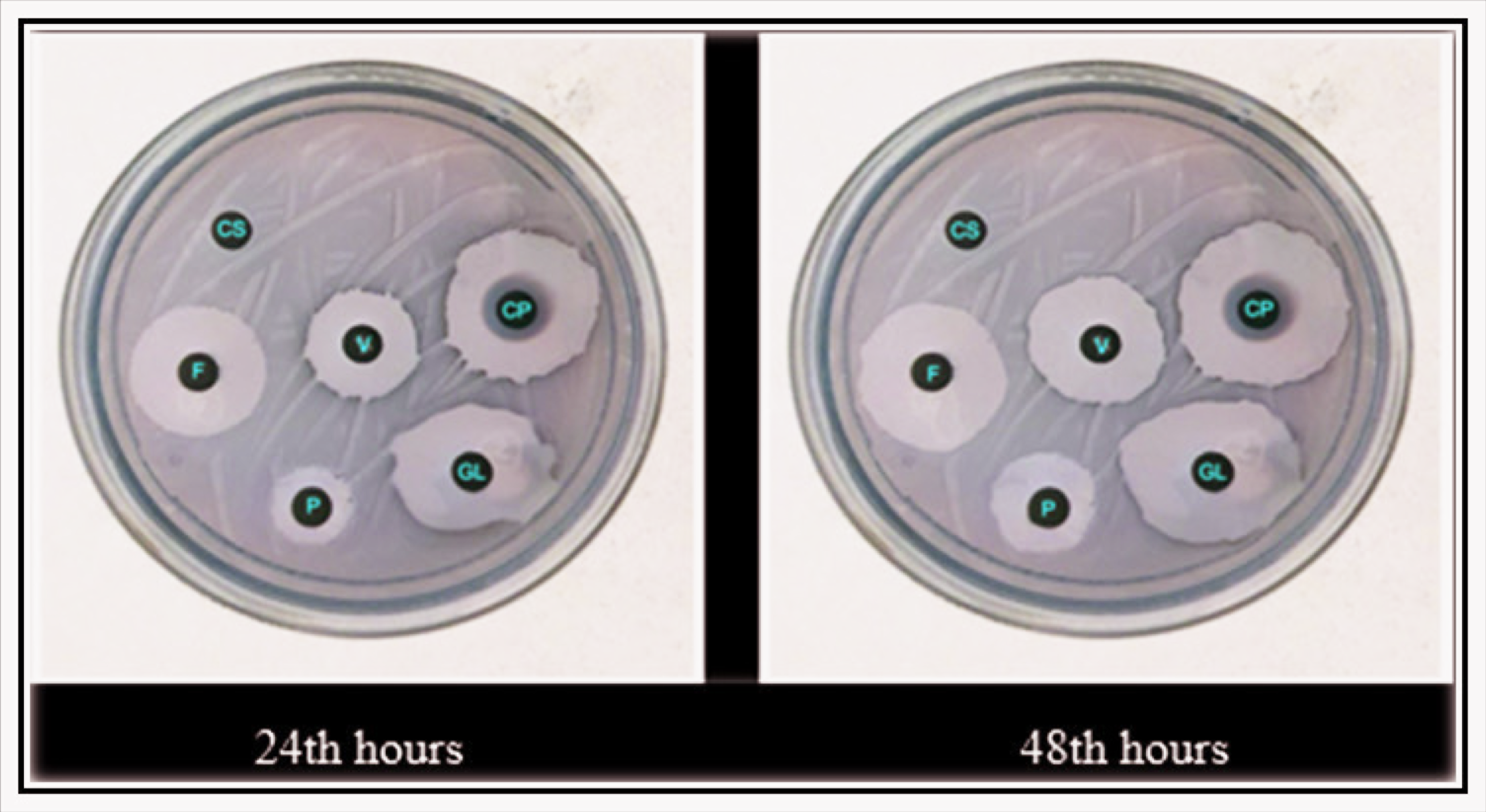
Preprepared standard, 5 mm diameter sterile and blank antibiogram discs were placed in sterile wells containing 30 µL of adhesive agents for 10 s and then placed on the plates. Five antibiogram discs impregnated with different adhesive agents and one antibiogram disc with 30-mg vancomycin instilled were used as the positive control group in each petri dish. Ten petri dishes were formed from each petri dish containing six discs impregnated with the adhesive and control group. Discs were placed in 10 separate petri dishes for each bacterium. A total of 180 adhesive and control group impregnated antibiogram discs were used in 30 separate petri dishes. After the procedures were completed, they were kept in a CO2 oven for 24 h and 48 h at 37°C for the growth of bacteria on the plaques.
Measuring Zone Diameters
After controlling the growth of the plaques removed from the oven, the inhibition diameters around the discs were evaluated at 24 h and 48 h by measuring with a millimetric inhibition zone scale. Measurements were made from the two outermost points of the inhibition ring formed around the disc. Inhibition zone diameters were recorded in millimeters by two different physicians in order to evaluate the results effectively.
Statistical analysis
In our study, the compatibility of continuous variables with the assumption of normality distribution was evaluated with the Kolmogorov–Smirnov test, and homogeneity was evaluated with the Levene tests. The sample size calculation was done according to the paper published by Noordzij M et al., 12 using G*Power software (version 3.0.9.7). Accordingly, 180 samples were prepared (10 samples per group). The statistical power was 84% with a confidence interval of 95% (α = 0.05). One-way analysis of variance (one-way ANOVA), which is one of the parametric analysis tests, was used to compare the differences between the means of independent groups, and the Bonferroni test was used for multiple comparisons between groups. The P <.05 value was accepted as the value to be used in terms of statistically significant difference.
Results
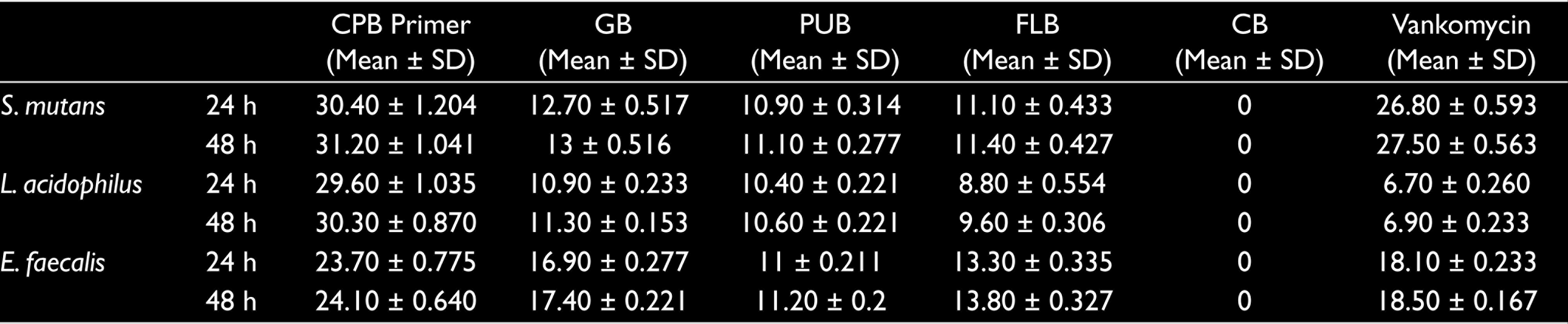
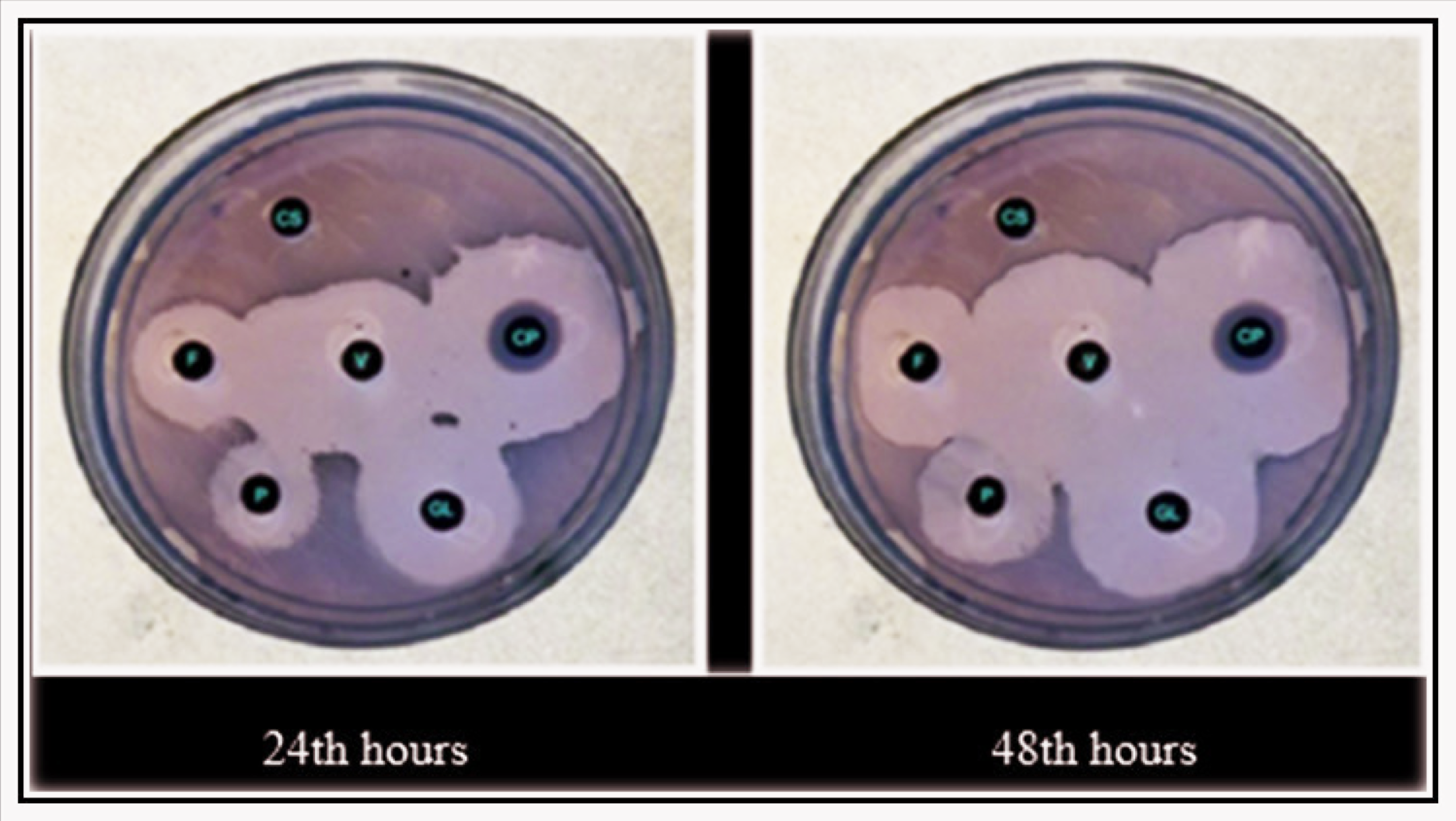
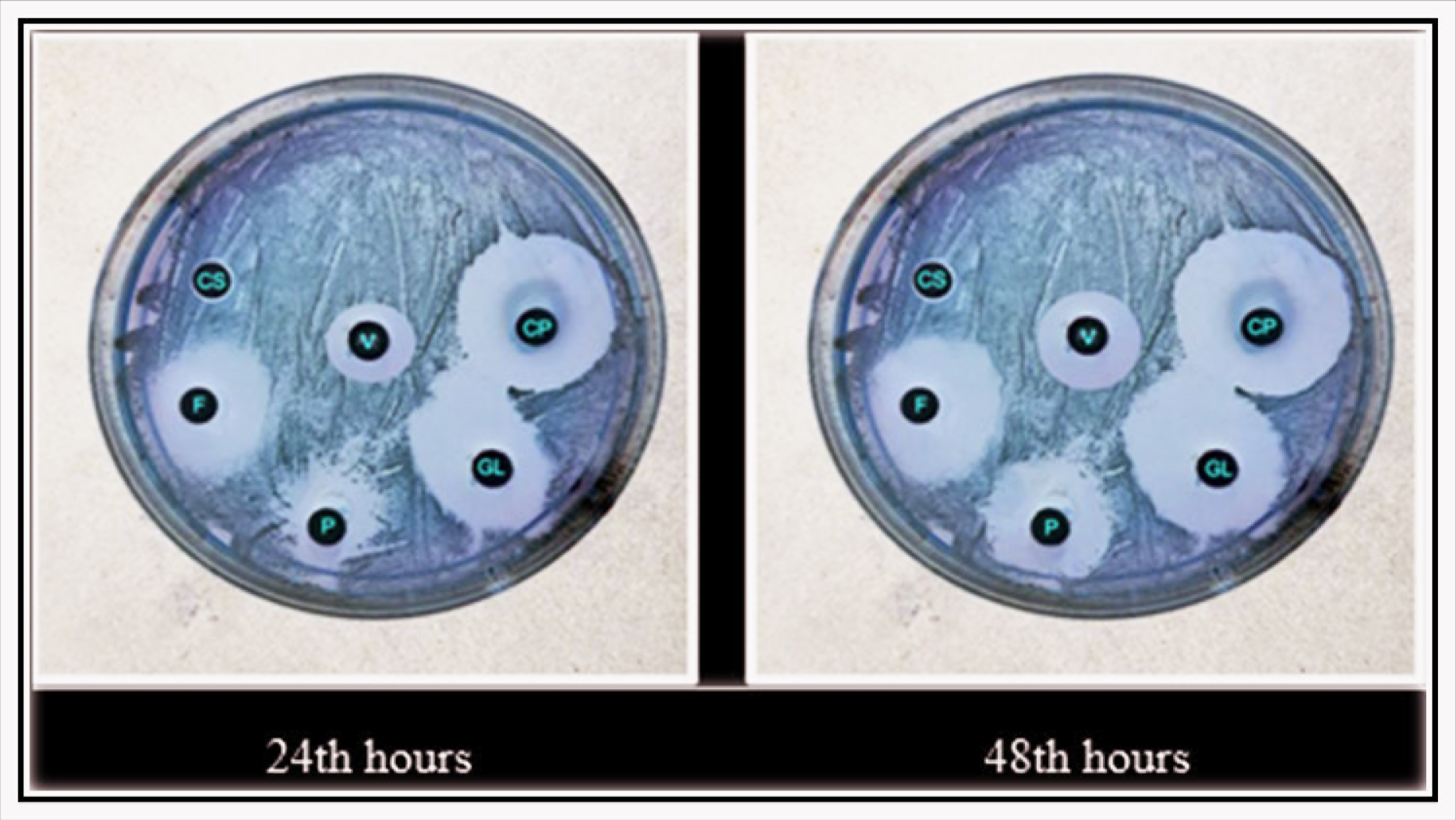
The mean in millimeter and standard deviation values of the inhibition zone diameters of the antibacterial effects of five different adhesive agents and the vancomycin antibiogram disc on all of the bacteria at 24 h and 48 h are shown in Table 2 and Figures 1 to 3.
Standard Deviation Values and Mean of the Inhibition Zone Diameters (mm) of Adhesive Agents at 24 h and 48 h (n = 10)
Zone Diameter (mm) on S. mutans Bacteria at 24th h and 48th h
Zone Diameter (mm) on L. acidophilus Bacteria at 24 h and 48 h
Zone Diameter (mm) on E. faecalis Bacteria at 24th h and 48th h
Comparison of Antibacterial Efficacy of Different Adhesive Agents in the Different Time Periods
CPB Primer, GB, PUB, FLB, and vancomycin used in the study were found to have significant antibacterial effects on all tested bacteria (P < .05). It was found that CB did not have an antibacterial effect on any bacteria.
When the antibacterial activity of the test materials on Streptococcus mutans and Enterococcus faecalis bacteria was evaluated within the 24th h and 48th h, it was found that there was an order from highest to lowest, which was CPB Primer > vancomycin > GB > FLB > PUB.
It was observed that the antibacterial activity of the test materials on Lactobacillus acidophilus in the 24th h and 48th h was ranked from highest to lowest as CPB Primer > GB > PUB > FLB >vancomycin.
Comparison of Antibacterial Efficacy of the Groups Applying the Same Adhesive Agent According to Different Time Periods
The effect of all groups used in the study on all of the bacteria at the end of the 48th h was found to be higher than the effect they showed at the 24th h. However, when the time periods were compared statistically, no significant difference was observed (P > .05).
Comparison of Antibacterial Efficacy of Adhesive Agents Applied in the Same Time Periods on Different Bacteria
In the paired comparisons, a statistically significant difference was found between the degrees of antibacterial activity of all adhesive agents on all bacteria in the same time period (P < .05).
In our study, the bacterium on which CPB Primer showed the most effect at 24 h and 48 h was S. mutans. It was observed that this was followed by L. acidophilus and E. faecalis, respectively. E. faecalis was the bacterium on which GB, PUB, and FLB showed the most effect at 24th h and 48th h, followed by S. mutans and L. acidophilus, respectively.
The vancomycin-impregnated antibiogram disc, which constituted the positive control group of the study, showed the greatest effect on S.mutans in both time periods. This was followed by E. faecalis and L. acidophilus, respectively.
Discussion
The occurrence of dental caries, which can cause pain and aesthetic and chewing dysfunction, depends on the presence of many factors. The presence of microorganisms with cariogenic properties, which is one of these factors, in the cavity walls can cause microleakage, secondary caries formation, postoperative sensitivity, and discoloration of the teeth. 13 Today, with the increasing use of composite resins, adhesive resins have gained importance. Although composite resins have many advantages, such as aesthetic appearance and ease of use, the problem of polymerization shrinkage, which is the biggest disadvantage, has not been eliminated. The use of cavity disinfectants is recommended to prevent bacterial colonization in the cavities caused by resin shrinkage. However, the need for additional time and increased cost of these disinfectants in the clinic have been tried to be solved by adding disinfectant components to adhesive agents. Thanks to the bactericidal effect of the adhesive systems, it was thought that the formation of secondary caries could be prevented, and thus the restorations would have a longer lifespan. Studies on the subject have shown that antibacterial activity can be increased by adding some materials, such as MDPB monomer, glutaraldehyde, chlorhexidine, and fluorine to dental adhesives. 14
The antibacterial activity of adhesive agents has been the subject of many studies in recent years. MDPB is an antibacterial monomer consisting of quaternary ammonium and a methacrylol group. Oba et al. 15 compared the effects of CPB Primer containing antibacterial effective MDPB monomer and two different self-etching adhesive agents without antibacterial content on S. mutans, L. acidophilus, and L. casei bacteria. In their study, where they used 2% chlorhexidine solution as a positive control group, they stated that CPB Primer had a strong antibacterial effect similar to chlorhexidine against S. mutans and L. casei. However, self-etching adhesive agents (Adper Easy Bond and Xeno V) showed higher antibacterial activity against L. acidophilus than CPB Primer. The reason for this was explained by the fact that both adhesives are agents with acidic monomers. 15
Feuerstein et al. 16 reported that CPB Primer showed bacterial inhibition on S. mutans within a period of 14 days, whereas the antibacterial activity of self-etch adhesive systems that were polymerized in vitro lasted longer. 16 In similar studies, Özer et al. 17 , Türkün et al. 18 , and Kaya et al. 19 found the antibacterial activity of CPB Primer to be statistically significant.17,18,19 Imazato et al. 20 , Poggio et al. 21 , and Öztürk et al. 22 found that CPB Primer has a strong antibacterial effect on S. mutans.20,21,22 Consistent with the studies in the literature, the MDPB-containing CPB Primer, which we used in our study, showed a significant antibacterial effect on all test bacteria, especially on S. mutans bacteria.
Glutaraldehyde, which has a wide spectrum of antibacterial action, prevents transmission by binding to the cell wall of bacteria, causes cross-linking of protein amino acids, and inhibits RNA, DNA, and protein synthesis. Gluma 2 Bond with added glutaraldehyde is an adhesive agent produced to provide disinfection in the cavity. Andre et al. 23 investigated the antibacterial activity of dental adhesives with or without antibacterial components on S. mutans using the SEM imaging technique. In their studies, they found that GB, CB Primer, and PUB, which contain antibacterial components, are quite effective. The efficacy of CPB Primer and GB adhesives was found to be statistically significantly higher than that of CB, which does not contain antibacterial components. 23 Our study is in agreement with the results of the study of Andre et al. In a similar study, Ikemura et al., 24 who examined the antibacterial activity of the FLB adhesive agent, claimed that this agent would reduce the development of caries thanks to the high fluorine it contains. 24
In line with literature information, it is preferred to use CPB Primer containing MDPB, GB containing antibacterial effective glutaraldehyde, PUB containing chlorhexidine, and FLB containing fluorine. As for the negative control group, it is preferred to use CB, which is known to have no antibacterial properties.
Bagis et al. 25 emphasized that the antibacterial activity of different adhesive agents on S. mutans at the end of 24 h and 48 h increased over time. 25 Similarly, in our study, it was observed that adhesive agents with antibacterial content were effective on S. mutans, L. acidophilus, and E. faecalis bacteria when used at the end of both 24 h and 48 h. However, there are insignificant statistical differences.
Many different methods have been tried in studies for the determination of antibacterial activity. In the selection of the appropriate method, it is necessary to know the properties of the material well and to evaluate the superiority of the method to be used over other methods. The most commonly preferred method for measuring the antimicrobial activity of fluid materials in dentistry is the simple and easy-to-apply disc diffusion test method. 26 The main advantage of this method is that different inhibition zone values of different materials placed on the same agar medium can be compared at the same time. However, the most important disadvantage is that the effect of the tested materials cannot be differentiated as bactericidal/bacteriostatic, and the duration of the antibacterial effect cannot be known exactly. 27
Conclusion
Within the limitations of the current in vitro study, it was concluded that CPB Primer exhibited the strongest antibacterial action on the three tested bacteria. Accordingly, its use as an adhesive agent in cavitated enamel and dentin caries is highly recommended.
Footnotes
Acknowledgements
This article was taken from the specialty thesis in dentistry named “Evaluation of the Antibacterial Effects of Four Different Adhesive Materials in Different Time Periods” supervised by Assistant Professor Şeyhmus Bakır.
Declaration of Conflicting Interests
The authors declared no potential conflicts of interest with respect to the research, authorship, and/or publication of this article.
Funding
The authors received no financial support for the research, authorship, and/or publication of this article.