
Abstract
Aim:
Survivin is a multifunctional protein chiefly involved in apoptosis and cell cycle regulation. Increased expression of survivin in tumors and fetal tissue determines its antiapoptotic activity. The aim of the study is to identify the immunoexpression of survivin in metastatic and nonmetastatic oral squamous cell carcinoma (OSCC) and also to evaluate and compare the expression of survivin in metastatic and nonmetastatic OSCC of buccal mucosa.
Materials and Methods:
In total, 40 histopathologically proven cases of OSCC, including 20 metastatic and 20 nonmetastatic cases, are selected. Among the 20 metastatic and nonmetastatic cases, 10 well-differentiated and 10 moderately differentiated squamous cell carcinoma cases were included and were subjected to immunohistochemical staining for survivin expression. The results were analyzed by SPSS version 11.5 using chi-square test.
Results:
The expression of survivin in metastatic and nonmetastatic tumors is 15%–70% and 15%–60%, respectively. When comparing the cases of moderately differentiated squamous cell carcinoma in metastatic and nonmetastatic tumors, 70% cases show moderate staining intensity.
Conclusion:
The survivin expression was comparatively high in metastatic OSCC. Also based on the aforementioned results, survivin expression was high in increasing grades of OSCC.
Abbreviations
BIR: Baculovirus inhibitor apoptosis protein
DAB: DI amino benzidine
DPX: Dibutyl phthalate xylene
H&E: Hematoxylin & eosin
MOSCC: Moderately differentiated squamous cell carcinoma
OSCC: Oral squamous cell carcinoma
WOSCC: Well-differentiated oral squamous cell carcinoma
Introduction
Oral squamous cell carcinoma (OSCC) is one of the most common cancer worldwide and accounts for about 90% of malignant cases in oral cavity. The mortality rate of OSCC is relatively high with a 5-year survival rate of 50%. Increased incidence of OSCC seen in males when compared to females with a ratio of 3.26:1. In India, the most common site of OSCC occurrence is seen in buccal mucosa. Beyond prevention, early detection is the most crucial determinant for successful treatment and better prognosis.1–3 The early progression and metastasis reduces the 5-year survival rate to 50%. 4 The incidence of metastasis depends on the degree of cellular differentiation, deep invasion, and site of the primary tumor. 1 Survivin is an apoptosis inhibitor protein seen in cell division. Survivin is also known as baculoviral inhibitor of apoptosis protein repeat-containing 5 or BIRC5 protein; it is a member of the inhibitor of apoptosis protein (IAP) family. 5 Survivin is a multifunctional protein involved in regulation of cytokinesis and cell cycle progression, and participation in a variety of pathways such as the p53, Wnt, hypoxia, TGF, and Notch signaling pathways.6–8 Survivin is considered to be good prognostic marker of OSCC 9 and is highly expressed in most human tumors of the oral cavity, lung, colon, breast, liver, gastrointestinal, and prostate. 8
In this study, investigation of immunohistochemical expression of survivin in metastatic and nonmetastatic tumors is done. The comparison between the two is done based on the staining intensity and different grades of OSCC.
Materials and Methods
Setting and Design
In this laboratory, an in vitro study comprised of 40 histopathological proven cases of squamous cell carcinoma. Out of 40 cases, 20 metastatic and 20 nonmetastatic cases with varying grades of differentiation were selected. The study was done in Central Research Laboratory, Vinayaka Missions Sankarachariyar Dental College, Salem, for 1 year. From the archival tissue blocks, thin sections around 3–4 µm were sectioned by using rotary microtome (Wiswas Optik, SMT-1090A). From each block one section is subjected to H&E staining and the other with immunohistochemical staining.
Sampling Criteria
The study samples include the cases taken from common tumor site—buccal mucosa, commissure, and retro molar area with patient’s age above 40 years. The histopathological samples taken from patients who underwent chemotherapy or radiotherapy were excluded. Also, other variants of squamous cell carcinoma, carcinoma other than buccal mucosa, and recurrent cases are excluded.
Study Method
Two slides were prepared for all the specimen, confirmatory diagnosis was made in one slide by routine hematoxylin and eosin stains, and another one is taken for immunohistochemistry (IHC) staining. In total, 40 histopathologically proven cases of squamous cell carcinoma were considered for final diagnosis. Sections of all the 40 cases were taken for immunohistochemical evaluation of survivin expression, the usual procedure of IHC was followed step by step. Sections were kept for 1 hour at 60 °C on the slide warmer for deparaffinizing, then in two changes of xylene for 15 min to remove wax. Later, subjected to descending graded of alcohol for rehydration. After that, the sections were kept in pressure cooker with citrate buffer for antigen retrieval. The pressure cooker was allowed to cool down to room temperature before removal of slides. The advantages of the pressure cooker antigen retrieval method were even distribution of heat over the slides, minimal reagent evaporation, excellent heat-source regulation with temperature range between 25 °C and 125 °C. During immunohistochemical staining, all the reagents should be kept at room temperature after taking out from refrigerator, humidifying chamber should be used for all the incubation procedures.
The excess buffer should be removed from sections, then sections were covered for 10 min with 3% hydrogen peroxide, after this for another 10 min, sections were treated with power block (DAKO REAL EnVision, Denmark), this will reduce cross reactions. The sections were then washed with phosphate wash buffer to prevent nonspecific binding, after removing the excess buffer, sections were kept for 1.5 hours in optimally diluted mouse monoclonal survivin antibody in 1:50 dilutions in phosphate buffered saline (PBS). For next 10 min, sections were gently washed in PBS buffer bath then, for 30 min, treated with secondary antibody tagged with poly horseradish peroxide enzyme (HRP) (DAKO REAL EnVision, Denmark). The sections were then washed with PBS (pH 7.5) and treated 5 min in freshly prepared DAB solution used for immunostaining, excess chromogen was removed by washing slides in distilled water, followed by counter staining with Harris hematoxylin for 7 min. Finally, the sections were dehydrated with ascending grades of alcohol, cleared in xylene, and mounted in DPX.
Interpretation of Staining
Positive target immunohistochemical staining was evaluated by brown stain under 10×/40× magnification and recorded by microphotograph. The evaluation was done by grading the stained sections based on the expression of the survivin in tumor cells as follows: Grade 1 = 1%–25%; Grade 2 = 25%–50%; Grade 3 = 50%–75%; Grade 4 = 75%–100%. Based on the intensity of staining again it is graded as mild, moderate, and severe.
Statistical Analysis
The results were analyzed using SPSS version 11.5 (SPSS Inc., Chicago, IL) and assessed by the chi-square test. The P value less than .05 is considered to be statistically significant. The level of significance (P < .05) was employed in all statistical comparisons. Quantitative data were recorded as mean ± standard deviation. The expressions of survivin between metastatic and nonmetastatic OSCC were analyzed.
Results
The immunostaining of survivin expression was seen in both cytoplasm and nucleus of tumor cells (Figure 1). The overall survivin expression in metastatic and nonmetastatic carcinoma is as follows (Figures 2 and 3).
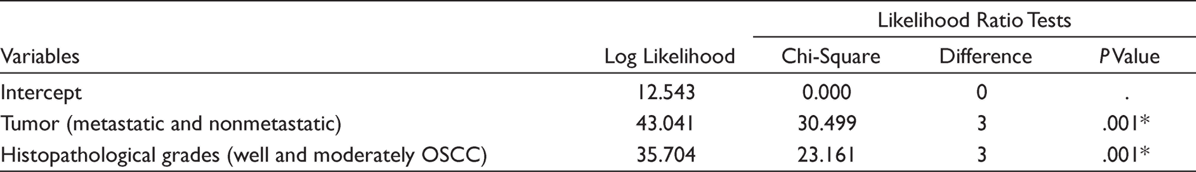
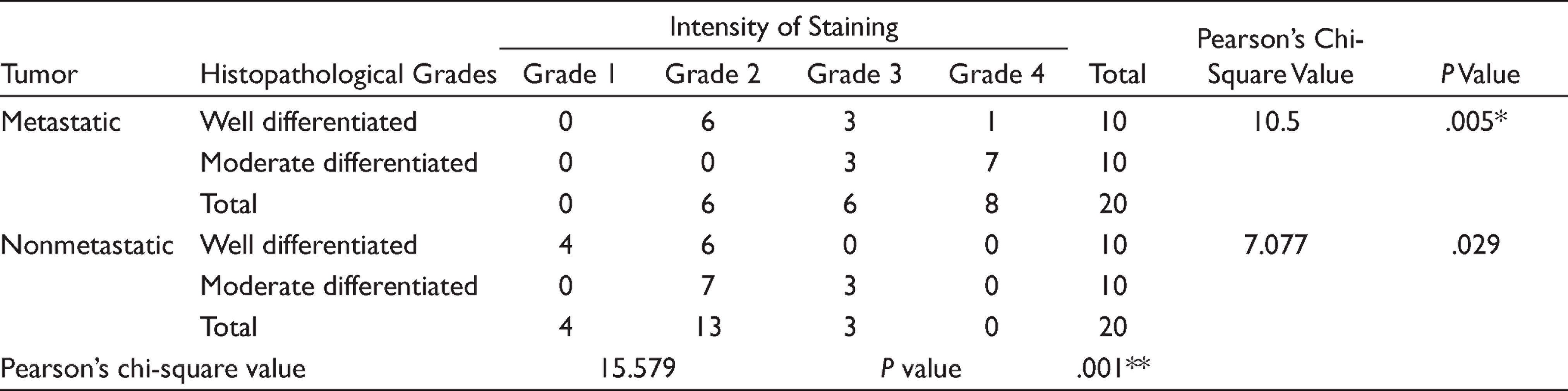
Multivariate analysis shows likelihood significant difference considering histopathological grades (WOSCC and MOSCC) and tumor (metastatic and nonmetastatic). Based on histopathological grades, there is increased staining intensity in moderately differentiated OSCC when compared to well-differentiated OSCC in metastatic carcinoma, Same thing goes for moderately and well-differentiated OSCC in nonmetastatic carcinoma (Table 1).
Overall comparison based on type of metastatic and nonmetastatic tumor shows highly significant P value (.001**), metastatic tumor shows more intensity of staining when compare to nonmetastatic tumor (Table 2).
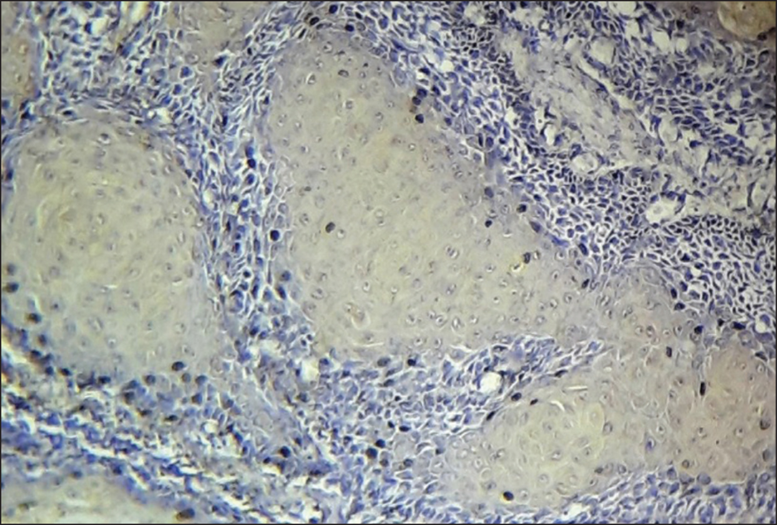
Survivin Expression Seen in Both Cytoplasm and Nucleus in Metastatic Oral Squamous Cell Carcinoma (Photomicrography 40×)
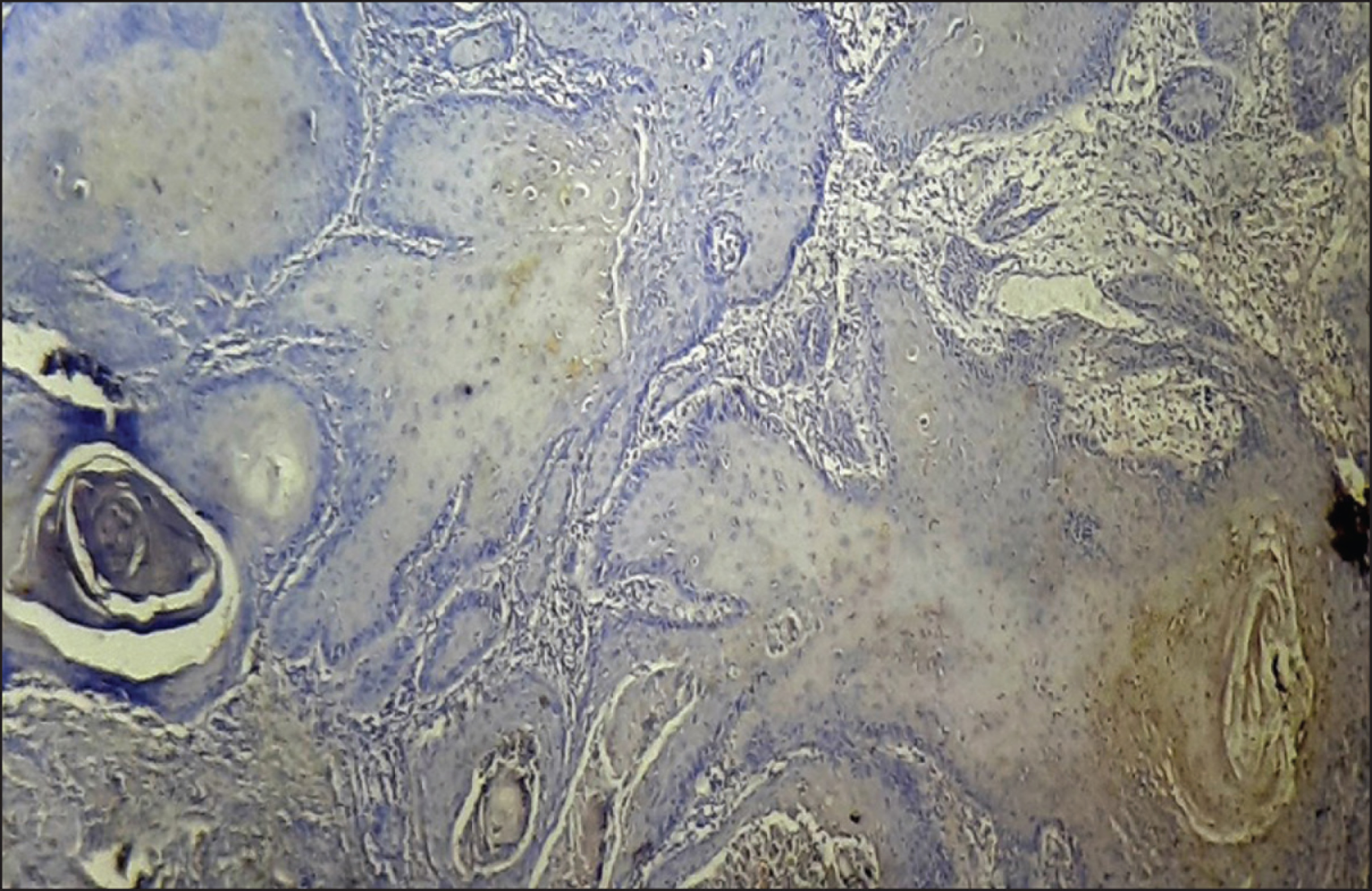
Histopathological Image Shows Grade 1 (26%–50%) Expression of Survivin in Nonmetastatic Well-Differentiated OSCC (Photomicrography 10×)
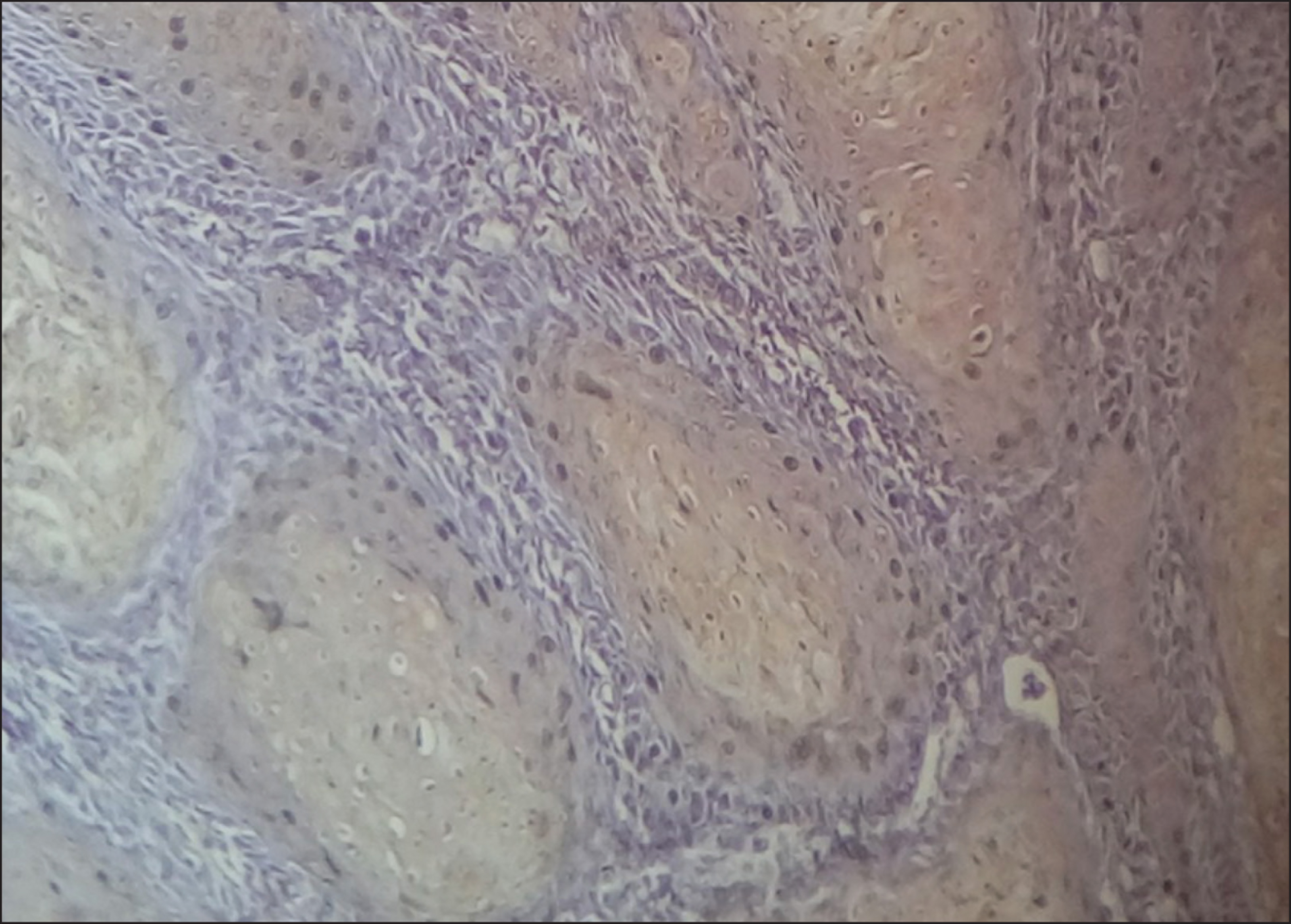
Histopathological High-Power Image Shows Grade 3 (50%–75%) Expression of Survivin in Metastatic Moderately Differentiated OSCC (Photomicrography 40×)
Multivariate Logistic Analysis of Survivin Expression Between Metastatic and Nonmetastatic OSCC
Comparison of Survivin Expression Between, Metastatic/Nonmetastatic and Histopathological Grades of OSCC Using Pearson’s Chi-Square Test
Discussion
Survivin, a unique and smallest member of inhibitor of apoptosis family, functions mainly by suppressing the activity of caspases. In embryonic tissues, survivin is highly expressed during G2-M phase of cell cycle, in tumor, depletion of survivin due to alteration in cell cycle by oncogenes causes defective cell division. 10 Based on its expression in different locations (cytoplasm and/or nucleus), the function of survivin varies. 11 In nucleus the survivin is chiefly involved in promoting cell proliferation, and in cytoplasm it participates in cell survival. In tumor cells, cytoplasmic expression is indicative of cytoprotective role, and nuclear expression represents impaired function of the cell.12,13 In various studies, authors have proposed that increase in level of nuclear survivin indicates increase in proliferative activity of tumor. Also, reduced cytoprotective survivin in tumor may be associated with favorable prognosis. Both cytoplasmic and nuclear expression of survivin serves as an indicator of active survivin in tumor cells, which relatively correlates with intracellular localization of survivin. 12 Invariably, functional activity of survivin in tumor cell and its significance in prognosis denote its dynamic role in molecular mechanism of cancer progression.11,12,14
In normal adult tissue the levels of survivin is low or nondetectable, and overexpression of survivin has been linked to its increased antiapoptotic activity in tumors.15,16 In previous studies, silencing survivin inhibits the VEGF expression, which indicates its possible role in proliferation of endothelial cells, and promotes tumor angiogenesis.17,18 High frequency of survivin was found in precancerous lesions or initial stages of cancer. 18 Based on the expression of p63 and survivin in oral cancer, Lauxen et al. suggested the possible role of survivin in malignant transformation of oral epithelium. 19 The differential expression of survivin suggests its possible role in targeting drug therapy in recent studies,5,15,20 and serves as a helpful tool for prognosis of head and neck carcinomas.
In the present study, the overall expression of survivin is positive in both metastatic and nonmetastatic OSCCs. The moderately differentiated squamous cell carcinomas of metastatic and nonmetastatic cases showed significantly increased survivin expression, which was similar to the study conducted by Deo et al. 9 This result is in contrast to the study conducted by Kulkarni et al., 21 where the survivin expression is high in poorly differentiated squamous cell carcinoma cases.
The overall survivin expression is significantly high in metastatic tumors when compared to nonmetastatic squamous cell carcinomas. Also, survivin expression was noticed to increase with increasing grades of squamous cell carcinoma, similar results seen in studies conducted by Muzio et al., 1 Jane et al., 22 and Rong et al. 23 .
The staining intensity was high in poorly differentiated cases but was not statistically significant, and the survivin expression was seen in both cytoplasm and nucleus, which was similar to the study conducted by Gayathri et al. 24 Based on the aforementioned results, balanced expression of survivin in nucleus and cytoplasm denotes the subcellular localization of survivin, which determines the overall prognostic significance of the tumor. Comparing the expression of survivin in metastatic and nonmetastatic oral cancer, increased expression in metastatic cases confirms the role of survivin in cancer progression and metastasis. This suggests that survivin can serve as a better prognostic marker in oral cancer.
Still, cytoplasmic and nuclear expression of survivin denotes active survivin in tumor, hence quantitative expression of survivin in both cytoplasm and nucleus has to be determined, to analyze the prognostic relevance of tumor. Hence standardization of immunohistochemical assays is mandatory to analyze and compare the prognostic role of survivin in cancer, which was a limitation in our study.
Conclusion
The functional significance of survivin enhances our understanding in the complex mechanism of cell division and cell survival. The cytoprotective function of survivin is chiefly by the antiapoptotic role in cancer cells. Nuclear function promotes its increases in proliferative activity. Hence, high expression of survivin in metastatic and nonmetastatic tumor helps to predict the prognostic impact in tumor progression. Further studies with the increased sample size are required to identify its significant correlation with clinicopathological features, lymph node metastasis, and potential therapeutic approach in OSCC.
Future Scope
Survivin target immunotherapy using survivin vaccines (SurVaxM) have been used against multiple myeloma and recurrent malignant gliomas. In future, probably survivin may serve as a lead target for treatment of OSCC patients.
Footnotes
Acknowledgements
The authors would like to thank Dr Mahesh J, senior lecturer, Department of Public Health Dentistry, Karpaga Vinayaga Institute of Dental Sciences, for statistical assistance.
Author Contributions
All the author contributed equally to the work.
Data Availability Statement
All the data have been attached in main manuscript.
Declaration of Conflicting Interests
The authors declared no potential conflicts of interest with respect to the research, authorship, and/or publication of this article.
Ethical Policy and Institutional Review Board Statement
Ethical clearance for the study was issued by the ethical committee of Vinayaka Missions research foundation (DU) (VMSDC/IEC/Approval No. 153) on February 2020.
Funding
The authors received no financial support for the research, authorship, and/or publication of this article.