
Abstract
Aim:
To evaluate the effect of the caffeic acid phenethyl ester (CAPE) as a root canal irrigation agent on the push-out bond strength of the AH-Plus sealer.
Materials and Methods:
A total of 75 single-rooted teeth were decoronated and were randomly divided into 5 groups of 15 roots for irrigation protocols: Group NaOCl: 5.25 percent NaOCl; Group CAPE: 0.5 percent CAPE; Group NaOCl + ethylenediaminetetraacetic acid (EDTA): 5.25 percent NaOCl-17 percent EDTA; Group NaOCl + CAPE: 5.25 percent NaOCl-0.5 percent CAPE; and Group CAPE + EDTA: 0.5 percent CAPE-17 percent EDTA (for 3 min each group). All root canals were then obturated and 1-mm-thick horizontal slices were obtained from different root thirds of the root canal (coronal, middle, and apical, respectively). The groups were challenged with push-out tests. Modes of failure were determined under a stereomicroscope.
Results:
The CAPE-EDTA-treated group presented the highest mean bond strength in the coronal region of root dentin (P < .05). The CAPE-treated group had a higher mean bond strength than the NaOCl-treated group (P < .05). The mixed mode of failure was most predominant in all groups.
Conclusion:
Under the presented in vitro conditions, CAPE alone or in combination with EDTA or NaOCl demonstrated a positive effect that increased the push-out bond strength of the AH-Plus sealer to root dentin.
Introduction
Effective irrigation agents are essential for debris removal, root canal disinfection, microleakage minimization, and in the improvement of the outcome of endodontic treatment. Mechanical endodontic debridement results in the formation of a smear layer that adheres to the root canal walls. 1 This layer reduces the bond strength of resin-based filling materials and increases bacterial microleakage. 2
Sodium hypochlorite (NaOCl) has been the most widely used agent for root canal treatment 3 ; however, it has some disadvantages such as unpleasant taste and inability to remove the smear layer. 4 Ethylenediaminetetraacetic acid (EDTA) is best known for its smear layer removal effect.5–7 However, the main drawbacks of EDTA are its reduced antibacterial and solvent activities of hypochlorite. 8 Although EDTA and NaOCl are the standard irrigation protocols, providing excellent smear layer removal and root canal dentin-cleaning, this irrigation protocol reduces the bond strength of the epoxy resin sealer to the root canal dentin. 9 However, the combination affects the covalent bonds formed by the epoxy-resin-based sealer and causes collagen destruction, thereby reducing the push-out bond strength to the root canal dentin. 10
After long clinical service, the biodegradation of collagen in dentin becomes a major problem with resin-based materials, leading to the situation that causes a failure in interfacial bond strength. 11
Protecting resin–dentin interfaces from degradation is critical for the longevity of adhesive materials. In recent years, several strategies have been tested in vitro.12–14 The host-derived matrix metalloproteinases are one of the factors leading to this degradation in dentin. 13 Caffeic acid phenethyl ester (CAPE) is an active derivative of propolis, which is a potent antioxidant, and its mechanism is based on the inhibition of metalloproteinases. 15 A study on dentin has shown that CAPE can be used as an antioxidant before the restorative procedure. 16 It also possesses several biological activities such as antioxidant, antiviral, anti-inflammatory, and carcinostatic activities. 17 The antimicrobial activity of CAPE has been reported against gram-positive and gram-negative bacteria,18–20 even in the field of dentistry.16,21,22 CAPE showed no effect as an irrigation agent on the bond strength between the filling material and the root canal walls in either in-vitro or in-vivo studies. Therefore, the aim of this in-vitro study was to evaluate the influence of different irrigation procedures on the bond strength of the endodontic sealer. The null hypothesis was that the push-out bond strength of the AH-Plus sealer to root dentin would not be influenced by the application of a CAPE irrigation agent.
Materials and Methods
Setting and Design
Preparation of Solution
A 0.5% (wt/vol) CAPE (Aldrich Chemistry, St. Louis, USA) irrigation solution was prepared by dissolving the 100 ml 0.01 percent saline-ethanol solution.
Irrigation Procedure and Obturation
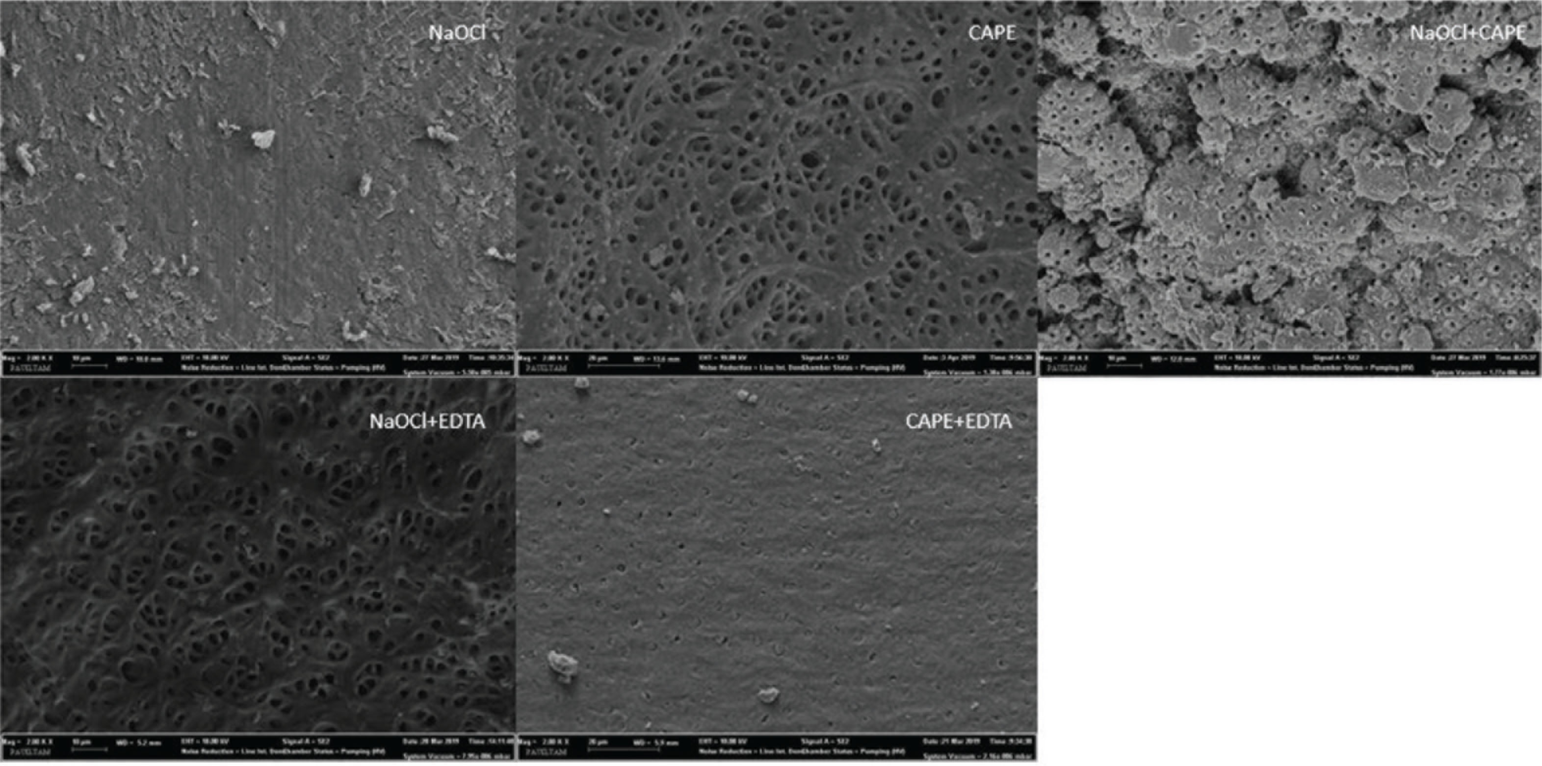
A total of 75 extracted human maxillary central incisor teeth were used for the in vitro study. Teeth with any abnormalities such as immature apices, cracks, calcification, or resorption were excluded. After the removal of the coronal part of the teeth, roots were standardized at 13 mm. The roots were randomly subdivided into 5 groups of 15 roots for irrigation protocols: NaOCl: 3 mm of 5.25 percent NaOCl for 3 min; CAPE: 3 mm of 0.5 percent CAPE for 3 min; NaOCl + EDTA: 3 mm of 5.25 percent NaOCl for 3 min, followed by 1 mm of 17 percent EDTA; NaOCl + CAPE: 3 mm of 5.25 percent NaOCl for 3 min, followed by 1 mm of 0.5 percent CAPE; CAPE + EDTA: 3 mm of 0.5 percent CAPE for 3 min, followed by 1 mm of 17 percent EDTA. The root canals were prepared with an R25 file (Munich, Germany) by using in reciprocating motion. The needle tip of the irrigating syringe was positioned at 3 mm away from working length with the passive irrigation modality. After the root canals were irrigated with irrigation protocols, the root canals dried with absorbent paper points. After root canal preparation, the one root selected at each group and splited along its long axis was examined under the scanning electron microscope (SEM). Representative images from all groups were taken at 2000× magnification to determine the presence or absence of the smear layer from the middle region of root dentin (Figure 1). The root canals were then filled with the AH-Plus (Dentsply, Konstanz, Germany) root canal sealer and gutta-percha by means of the cold lateral condensation technique using a single-cone technique. After seven days and 100 percent humidity for the complete setting of the sealer, the roots were cut into 1-mm slices, which were evaluated quantitatively using the universal test machine.
Representative SEM Images of Middle Third of the Root Canal Surface (2000×)
Push-out Test
Similar to our previous study, 23 after obturation, each root was horizontally cut into 1-mm slices from the apical, middle, and coronal thirds with a slow-speed diamond saw (Isomet 1000, Buehler, USA) underwater. The slices of 1 mm were sectioned from the apex at 3, 7, and 11 mm, respectively. The slices were attached to a universal testing machine (AGS-X, Shimadzu, Kyoto, Japan) for the push-out bond strength test, which was performed in the coronal topical direction using suitable diameter stainless steel plungers. The loading speed was 0.5 mm/min. These data were recorded in Newton and transformed into push-out strength (MPa) by dividing the force by the area of the adhesive bond with the following formula 24 : MPa = F/SL, where SL was the sealer adhesion area and was calculated using the following equation: SL = π. (R + r)g, where r is the mean radius of the apical canal (in mm), R is the mean radius of the coronal canal (in mm), g is the height (in mm), and π = 3.14.
Mode of Failure Evaluation
After performing the bond strength test, the specimens were examined under an optical microscope (Leica Microsystems, Wetzlar, Germany) at ×30 magnification. According to the previous study, 25 the failure modes were divided into three basic categories as follows: adhesive, cohesive, and mixed failure mode.
Statistical Analysis
The data of the push-out bond strength (MPa) are subjected to statistical analysis using SPSS 22 (SPSS Inc, Chicago, IL, USA), showing normal (Shapiro-wilk). The push-out test data were analyzed using the one-way analysis of variance (ANOVA) and Tukey test. These results were reported as means ± SD for each group and values for P ≤ .05 were considered statistically significant.
Results
Push-out Bond Strength
Table 1 presents the results of the mean and standard deviation of the push-out bond strength in all root thirds at treatment groups. The results were obtained as follows. For the coronal third of the root canal, the CAPE-treated group showed significantly greater bond strength than the NaOCl- and NaOCl-EDTA-treated groups (P < .05). The CAPE-EDTA-treated group had significantly higher push-out bond strength than NaOCl-, NaOCl-EDTA-, and NaOCl-CAPE-treated groups (P < .05). For the middle third of the root canal, no statistically significant difference was found for bond strength between the NaOCl-EDTA- and CAPE-EDTA-treated groups (P> .05). In the apical third of the root canal, no statistically significant differences were found between the groups (P> .05). Overall, the NaOCl-treated group resulted in the lowest bond strength compared with the other treated groups (P < .05).
Mode of Failure
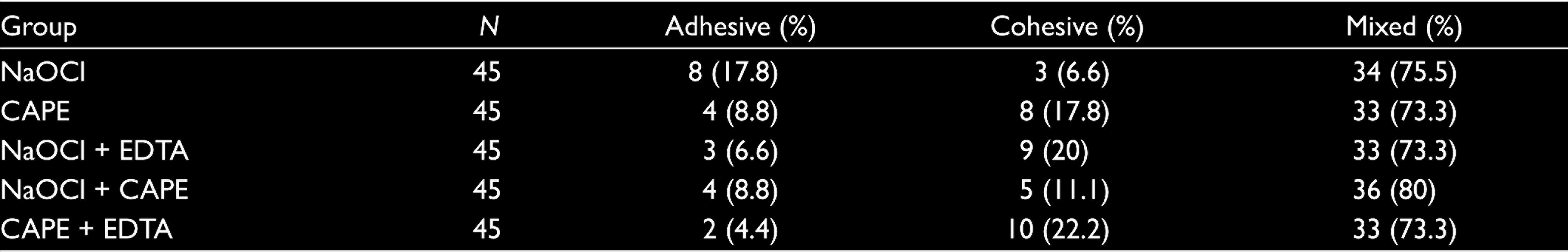
As shown in Table 2, failure mode analysis, all three types of adhesive, cohesive, and mixed failure were found for each group. Moreover, mixed failure was most common in all groups.
Comparison of Push-out Bond Strength Values for Each Group in the Different Root Thirds
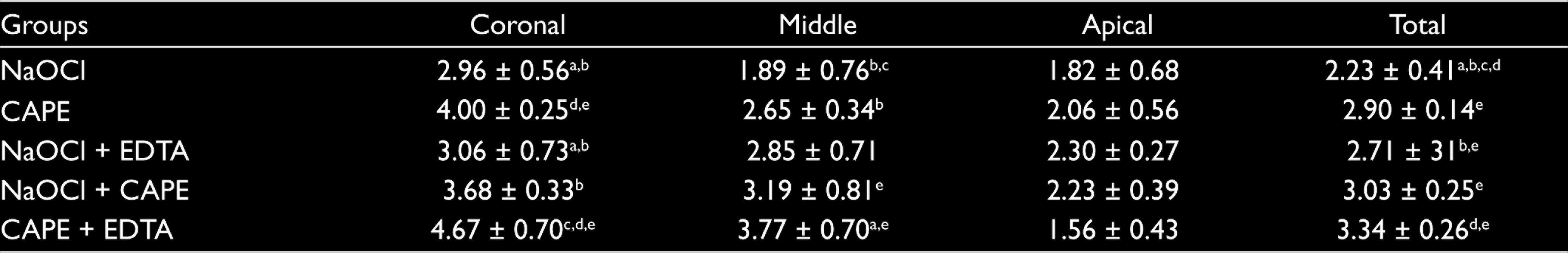
a Statistically significant difference (P < .05) vs. the CAPE group.
b Statistically significant difference (P < .05) vs. the CAPE + EDTA group.
c Statistically significant difference (P < .05) vs. the NaOCl + CAPE group.
d Statistically significant difference (P < .05) vs. the NaOCl + EDTA group.
e Statistically significant difference (P < .05) vs. the NaOCl group.
Mode of Failure, Number of Specimens and (%) for Each Group
Discussion
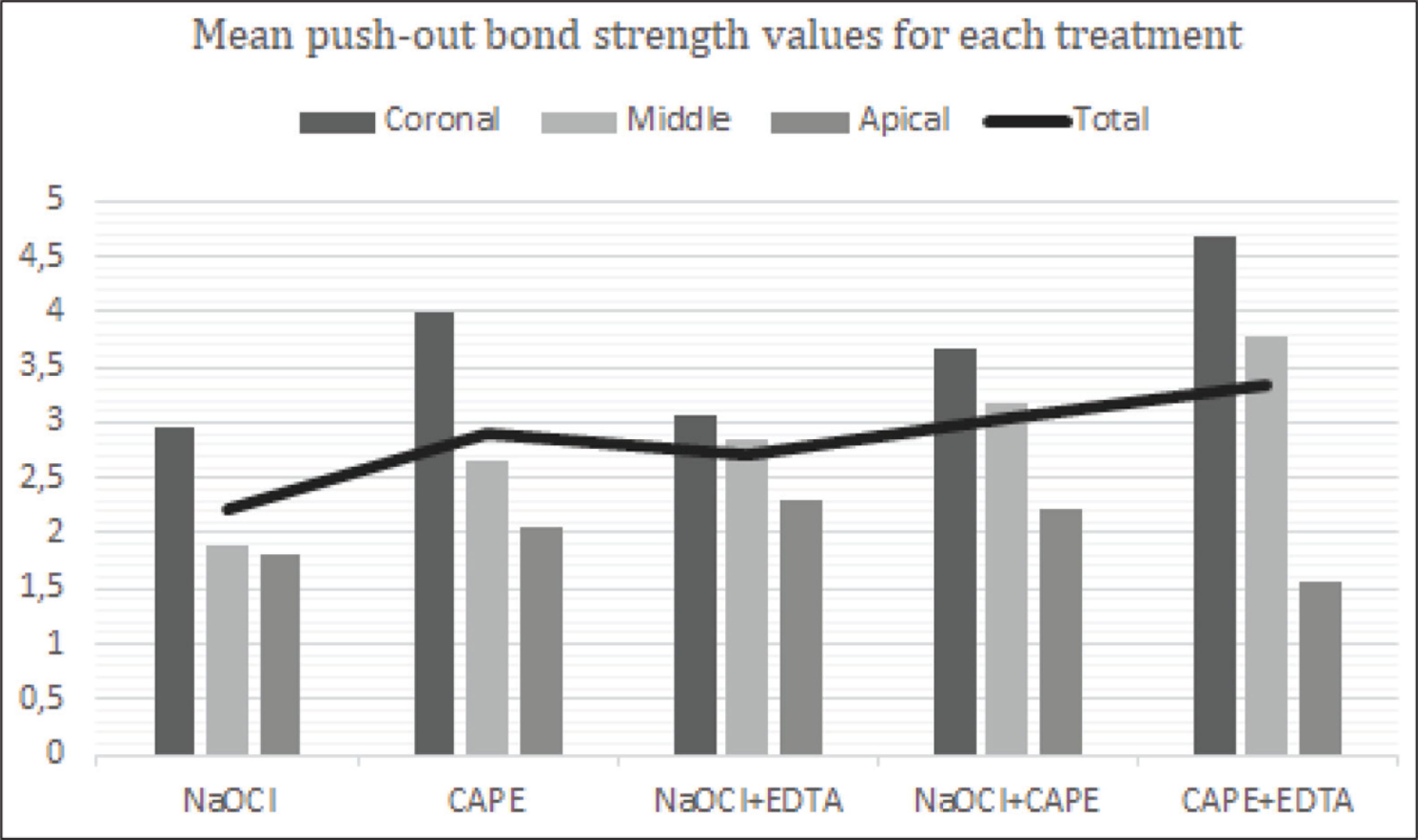
The null hypothesis was rejected as the CAPE-treated group had a higher mean bond strength than that of the NaOCl-treated group and, when the CAPE solution was also used in combination with NaOCl, bond strength values increased. The result of the mean push-out bond strength to root dentin of the AH-Plus sealer is summarized in Figure 2.
Chemical irrigation changes the dentin collagen network, thereby affecting the bond strength of the epoxy resin sealer.13,26 Moreover, the use of different irrigation solutions with the capacity to demineralize the dentin surface may cause changes in the chemical and structural composition of the dentin and hence may affect the adhesion capability of the sealer to the dentin. 27 We found that AH-Plus is a higher means push-out bond strength values, which is consistent with earlier reports.13,23,28 On the other hand, lower bond strength after NaOCl irrigation has also been reported to result from its residues that have an adverse effect on polymerization. 29 A similar situation is also observed in our study. Overall, it was found that the NaOCl-treated group recorded the lowest bond strength as compared to the other groups in the present study.
In the present study, the combination of NaOCl and EDTA was used for irrigation. This protocol had higher bond strength values than those of the NaOCl-treated group. These results could be related to the removal of the smear layer (Figure 1B).
Alternatively, in the present study, CAPE, NaOCl-CAPE, and CAPE-EDTA combinations were used for irrigation in treatment groups. Sodium ascorbate, an antioxidant, has been demonstrated to reverse the negative effects of oxidants in many in vitro experiments.30,31 Similar studies have tested epigallocatechin-3-gallate, a group of polyphenol with powerful antioxidative activity and inhibiting collagenase activity.13,32 CAPE is structurally similar to flavonoids and also has powerful antioxidative activity. 33 It has been reported that CAPE inhibits endogenous MMPs that cause hybrid-layer degradation. 16 In overall evaluation, the effect of the CAPE solution alone, or in combination, on bond strength was higher than that of the NaOCl solution as intracanal irrigation. It can be attributed to the antioxidant ability of CAPE which helps to neutralize and reverse the oxidizing effects of NaOCl. The CAPE solution can be considered a possible alternative to the NaOCl solution for intracanal irrigation or as a final rinse. Moreover, in the present study, the effect of both CAPE-and CAPE-EDTA-treated groups in push-out bond strength testing was greatest in the coronal and middle thirds. Most probably, the removal of the smear layer may also play a role (Figures 1B and E, respectively).
In all groups, the mixed failure mode is the dominant one, followed by adhesive failure and least adhesive failure, respectively, except for the NaOCl–treated group. These results were similar to other studies showing the predominance of mixed or cohesive bond failure.34,35
The Mean Push-out Bond Strength Values (in MPa) for Each Group
In order for our study to be compatible with clinical endodontic practice, the application time of the irrigation solution was chosen in a manner similar to the previous studies that generally used irrigation agents for 3–5 min.7,36 On the other hand, another study reported the use of irrigation agents at different application times: 1 percent NaOCl at 30 min and 17 percent EDTA at 5 min. 37 CAPE is a recently introduced material in the dental field, and its possible effects on the adhesion of endodontic filling materials have not been previously studied. Further research on the optimal concentration and application time for CAPE is recommended. Moreover, CAPE has additional antibacterial, anti-inflammatory, and antioxidant properties, 17 which may be useful as a root canal irrigation agent to improve the success of endodontic treatment. Future study is needed to evaluate the effect of CAPE on the long-term bonding resistance of epoxy resin-sealer canal fillers.
Conclusion
The CAPE-treated group had a higher mean bond strength than the NaOCl-treated group. The adhesion of AH-Plus to dentin was positively influenced by CAPE which has an antioxidant effect. Therefore, when using AH-Plus as a root canal sealer, CAPE can be considered as an effective alternative irrigation agent of the root canals.
Footnotes
Declaration of Conflicting Interests
The authors declared no potential conflicts of interest with respect to the research, authorship, and/or publication of this article.
Funding
This study was supported by Pamukkale University-BAP-BSP program (2018BSP002).