
Abstract
Objective:
To evaluate microleakage under the orthodontic adhesives applied following two version of erbium:yttrium aluminum garnet (Er:YAG) laser-aided enamel conditioning after thermal and thermomechanical simulators.
Materials and Methods:
A comparative analytical study based on metal braces bonded on the enamel of extracted teeth (n = 160) etched with acid, Er:YAG laser and Er:YAG laser with an X-Runner handpiece, and self-etch adhesives. An arch wire was ligatured to samples which were embedded in acrylic blocks by two with periodontal ligaments. The specimens were subdivided into two groups: those aged with thermal cycling and thermomechanical aging procedures. The samples were immersed in basic fuchsin solution (0.5%) for 24 h. Buccolingual sections were performed on the mesial and distal wings of the braces. The color penetration at the gingival and occlusal margins of the adhesive-bracket and enamel-adhesive was evaluated under a stereomicroscope. The median and mean values of microleakage in both groups were evaluated with Kruskal–Wallis and Mann–Whitney U tests (P < .05).
Results:
The highest microleakage was recorded in the gingival part of the samples aged with the thermomechanical aging procedure (P = .001). The amount of microleakage generally increased in the samples subjected to thermomechanical loading, but the only significant difference was recorded in the gingival part in each four different conditioning methods.
Conclusion:
Microleakage of the phosphoric acid-etched groups was recorded with lower values for both aging methods. Thermomechanical aging should be included to microleakage studies due to increased microleakage on gingival side for all etching groups.
Keywords
Introduction
Demineralization of the teeth enamel surfaces during fixed orthodontic treatment is a crucial aesthetic problem. Demineralization reported measurable decalcification areas around fixed treatment appliances just one month later, together with surface demineralization and the genesis of white spots due to mineral loss on the surface or underneath it. 1 The demineralization and debonding of fixed attachments has been previously related to microleakage between the enamel-adhesive and the bracket surfaces.
Microleakage is defined as the passage of mouth fluids, bacteria, ions, and molecules between the bracket, adhesive, and surface of enamel. 2 The prevention of leakage between composite-to-tooth and composite-to-bracket is important for the success of the bonding procedure. An ideal adhesive material must be well-adhered to the enamel surface and should provide good insulation. 3 A marginal gap area resulting from insufficient insulation will lead to plaque accumulation and the migration of bacteria and toxins. This microleakage may result in undesirable conditions, such as edge coloring or decay. 4
Microleakage was shown to be related to chemical and physical interactions during the curing process, such as polymerization shrinkage, which is thought to cause microcracks between the material and the enamel’s surface. Another factor resulting in microleakage is the low tensile stress, which causes physical changes in composites. 2
In the literature, different etching methods had been compared and found to affect the amount of microleakage. The acid-etching procedure was reported most effective, preventive method for microleakage.5–8 Recently, laser applications have been accepted as an alternative to conventional acid etching of the enamel. 9 The erbium:yttrium aluminum garnet (Er:YAG) laser has been reported to ablate the tissue while creating minimal thermal side effects for both soft and hard tissues. These lasers are effective on enamel and dentin because both water and hydroxyapatite absorb their output. 10 Er:YAG laser has shown more microleakage compare to acid etch. 11 On the other hand, it has been reported that laser-etched enamel is more resistant to caries formation than acid-etched specimens, although there is more microleakage.12,13 Er:YAG lasers form an enamel surface with heterogeneous micropathies when compared to an X-Runner digital laser scanning handpiece, 14 which has not been tested from the aspect of microleakage yet.
The advantage of the X-Runner handpiece is that the shape and size of the ablation area can be selected, enabling dentists to work more precisely and to avoid unnecessary laser irradiation.
Dental adhesive materials may exhibit static or dynamic fatigue failures that vary depending on the nature of loadings or residual stress. When the composite is exposed to cyclic stress for a long time, microscopic cracks develop in the structure, leading to fatigue failure. In static or dynamic fatigue failure cases, failure begins as a crack that continues until a catastrophic fracture occurs. 15
High fatigue strength means that dental materials are durable and their clinical performance can last for a long time. It is important for dental adhesives exposed to chewing forces to be constructed with their fatigue properties in mind. In addition, the environment in which the material is located is also important for determining fatigue properties, 16 which can be affected by moisture, biological agents, saliva, and pH changes. Therefore, if the adhesive materials are tested in an environment where these properties can be generated in vitro, fatigue data will be more meaningful.
According to our literature review, no former studies included thermomechanical aging to their study design. The aim of our study was to compare the microleakage quantities of an Er:YAG laser with a conventional probe and a new handpiece, X-Runner, to conventional etching methods on the samples aged with a thermal cycle alone and those aged using the thermal cycle and chewing simulation in in vitro conditions.
Materials and Methods
This presents a comparative cross-sectional analytical study conducted at the Orthodontic Department of Bezmialem Vakif University during 2015–2016. The study was performed with 160 maxillary and mandibular premolar teeth extracted for orthodontic or periodontal reasons. Written consents were taken from participants whose teeth were included in the study.
The extracted teeth crowns with free of caries and no restorations or fracture lines were included in the study. Any teeth with enamel hypoplasia, crown fracture, badly decayed, or attrition were excluded. All of the teeth were cleaned and polished with pumice and were divided in equal four etching groups (N: 40) using a simple random sampling technique: acid-etch (Group 1), self-etch (Group 2), Er:YAG laser (Group 3), and laser with X-Runner handpiece (Group 4) were four groups. Teeth were stored in distilled water.
The specimens selected for the four etching groups were also subdivided into two aging subgroups (N:20) for thermal and thermomechanical aging to simulate the thermal cycling and thermomechanical simulation.
Group 1: the enamel surface was etched with 37% phosphoric acid (Reliance Orthodontic Products, Inc., Itasca, USA) for 15 s, rinsed with water, and dried for the next 15 s. The etched enamel surface had a uniform, frosty, and dull appearance.
Group 2: 3M ESPE Adper Prompt L-Pop Self-Etch Adhesive (3M ESPE, St. Paul, USA) was rubbed onto the surface of enamel for 3 s supplied with the system.
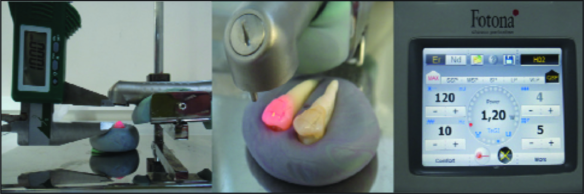
Group 3: a 2,940 nm wavelength Er:YAG (LightWalker, Fotona, Slovenia) laser was used for etching with the quantum square pulse mode at 1.2 W power for 15 s and a pulse repetition rate of 15 per second (10 Hz; Figure 1).
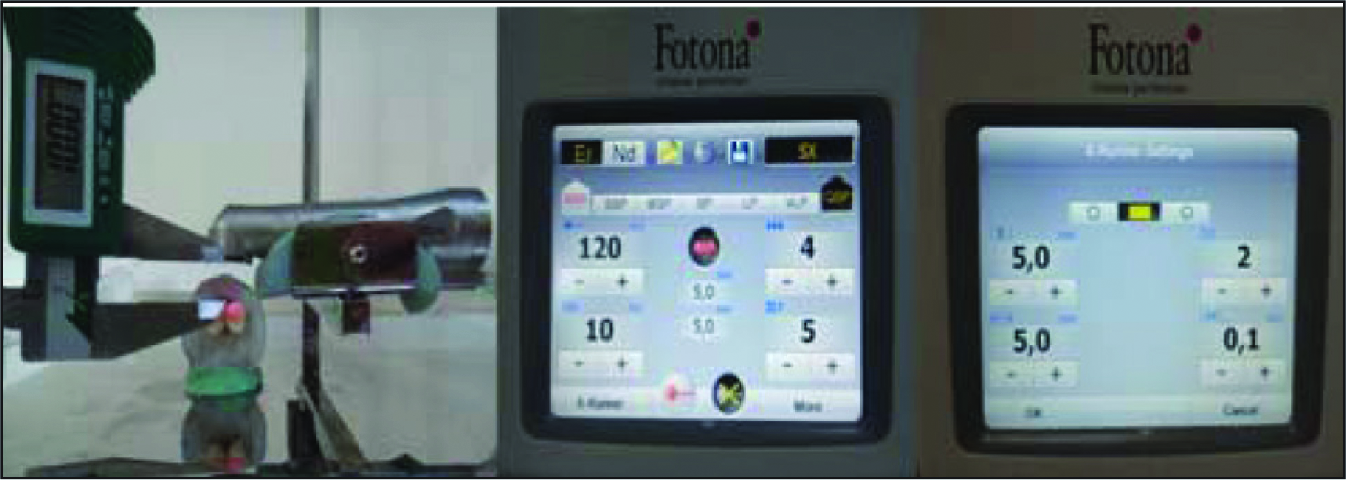
Group 4: a 2,940 nm wavelength Er:YAG laser was used for etching with an X-Runner handpiece (LightWalker, Fotona, Slovenia) at 1.2 W power and 10 Hz for 15 s in an area of 5 × 5 mm. All laser applications were performed with sufficient air and water cooling (Figure 2).
Preparation of Er:YAG Laser Group Specimens
Preparation of X-Runner Handpiece Group Specimens
After conditioning the enamel surface, bonding resin (Transbond XT Primer, 3M Unitek, Monrovia, CA, USA) was applied, and then mini Roth prescription premolar brackets (American Orthodontics, Washington, USA) were bonded with a light-curing adhesive (Transbond XT, 3M Unitek, Monrovia, CA, USA). Light was applied using a light-emitting diode (VALO, Ultradent Products Inc., South Jordan, USA) source for 3 s through the mesial and distal surfaces.
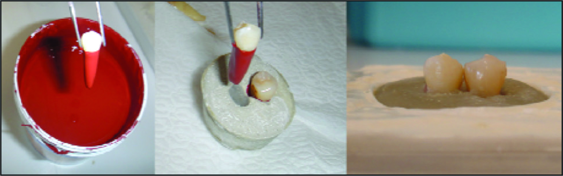
Prior to the chewing simulation, to simulate the human periodontium, the roots of the teeth were coated with wax. The teeth were then fixed in twos by 15 mm-diameter metal rings using fast-setting polyester resin (Technovit 4000, Heraeus Kulzer, Wehrheim, Germany) that simulated the human alveolar bone. To prevent overheating, teeth were submerged in water for 5 min during resin polymerization. 17
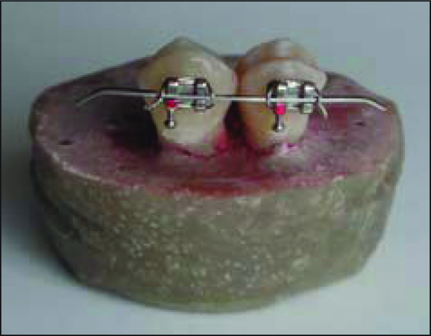
Then the teeth were easily removed from the resin molds by means of wax and the roots of the teeth were covered with a 0.1 mm-thick layer of autopolymerizing silicone (Anti-Rutsch Lack, Wenko-Wenselaar, Hilden, Germany) 2 mm away from the cemento-enamel junction (Figure 3). The brackets were bonded to the paired teeth and a 0.016 × 0.022-inch nickel titanium arch wire (Ortho Organizers Inc., Carlsbad, CA, USA) was ligated with the help of a ligature wire (Figure 4).
Preparing Specimens for Thermomechanical Loading
Teeth Ligatured With Arch Wire
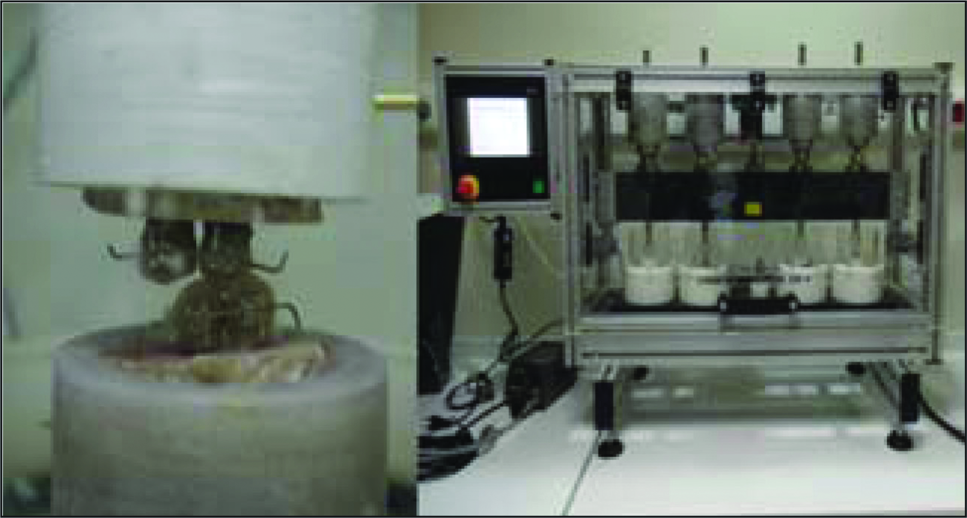
The prepared samples were subjected to a force simulating a clinical period of approximately two years under a force of 50 N with the aid of a dual axle chewing simulator (2 mm vertical, 1.5 mm horizontal). According to the literature, 240,000 cycles in the mechanical simulator are equivalent to one year in the oral environment. 18 For this reason, 500,000 cycles of chewing, equaling 2 years of clinical use, have been found suitable for the samples considering the average orthodontic treatment duration (Figure 5).
Thermomechanical Aging
Gale et al reported that the use of thermal cycling (5°C and 55°C) was conducted 10,000 times to mimic one year of natural use. 19 All specimens were subject to 20,000 thermal cycles (5 ± 1°C and 55 ± 1°C) with 10 s transfer time between baths and 30 s bathing time, considering that the average duration of orthodontic treatment is 2 years.
After aging, all specimens were removed from the implanted Technovit 4000 autopolymerizing acrylic blocks. Before the dying procedure, the apices were sealed with sticky wax and the specimens were coated with two coats of nail varnish up to 1 mm from the bracket margins. The specimens were once again placed in a soft acrylic block to allow for precise cutting.
The specimens were immersed in 0.5% basic fuchsin solution for 24 h.20,21 After this immersion, the teeth were carefully rinsed with distilled water.
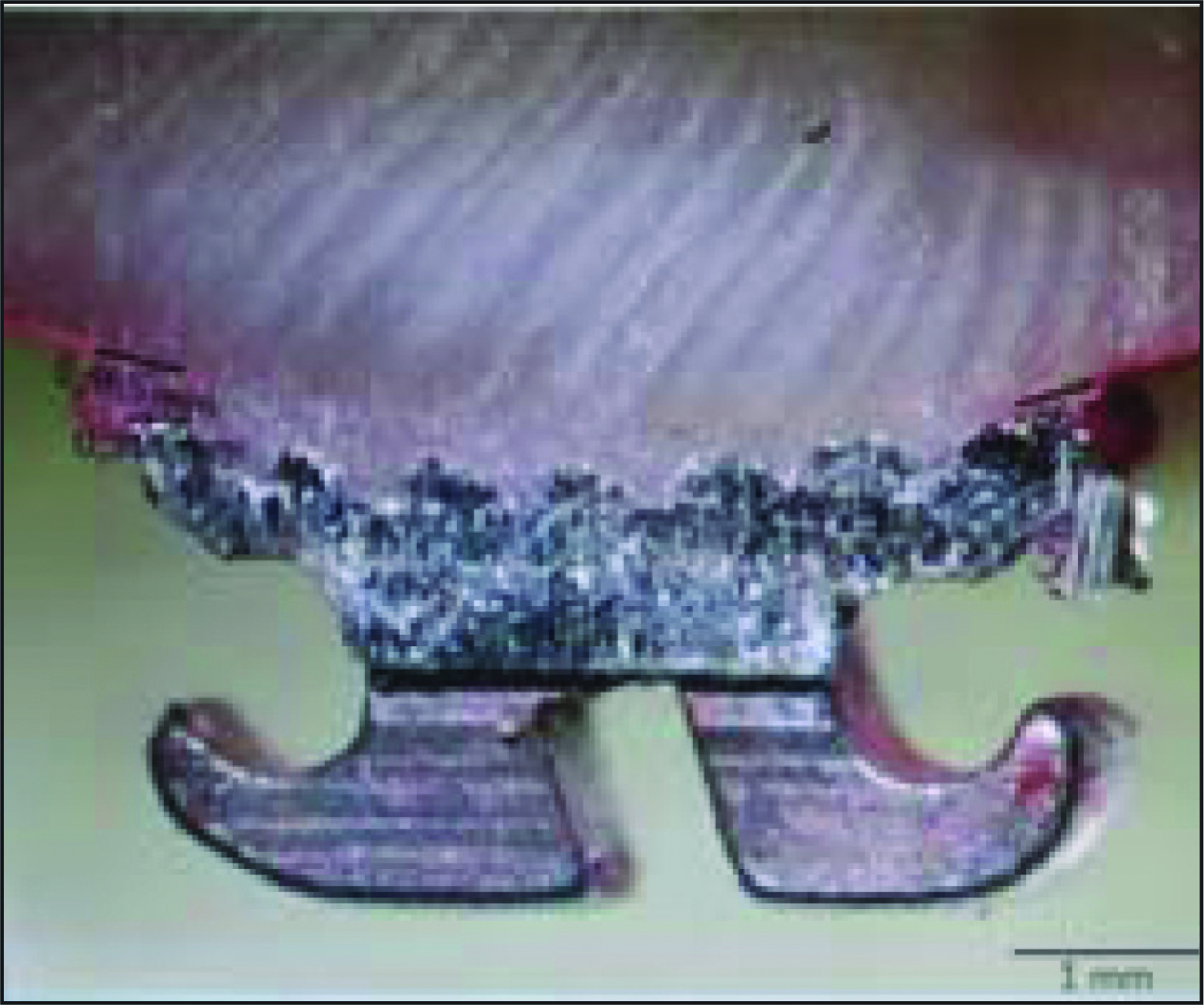
Buccolingual sections in the center of the brackets were performed using a diamond bur (Metkon 19-100 [101 × 12.7 × 0.3] Metkon Endüstriyel San. Tic. A.Ş. Bursa, Turkey) and a cutting instrument (Mecatome T180 PRESI-Métallographie, Eybens, France) along with adequate water. The penetration of color in all samples at the gingival and occlusal margins of the adhesive-bracket and enamel-adhesive was evaluated under a stereomicroscope (Olympus SZX7; Olympus optical, Tokyo, Japan) at 30× magnification. All measurements were photographed with a Phototonic PL200 (Photonic Optische Geräte GmbH & Co. KG, Vienna, Austria) and the measurements were taken using the Kameram (version: 2.8.5.0) computer software (Argenit Akilli Bilgi Tek. Ltd Şti, Istanbul, Turkey; Figure 6).
Statistical Analysis
We recorded the microleakage score of each tooth for occlusal and gingival portions. The microleakage values were evaluated statistically between the test groups using the Mann–Whitney U and Kruskal–Wallis tests with significance set at P < .05 by using SPSS software version 16.0 (SPSS Inc., Chicago, III).
Results
All groups exhibited microleakage values between the bracket-adhesive and adhesive-enamel interface.
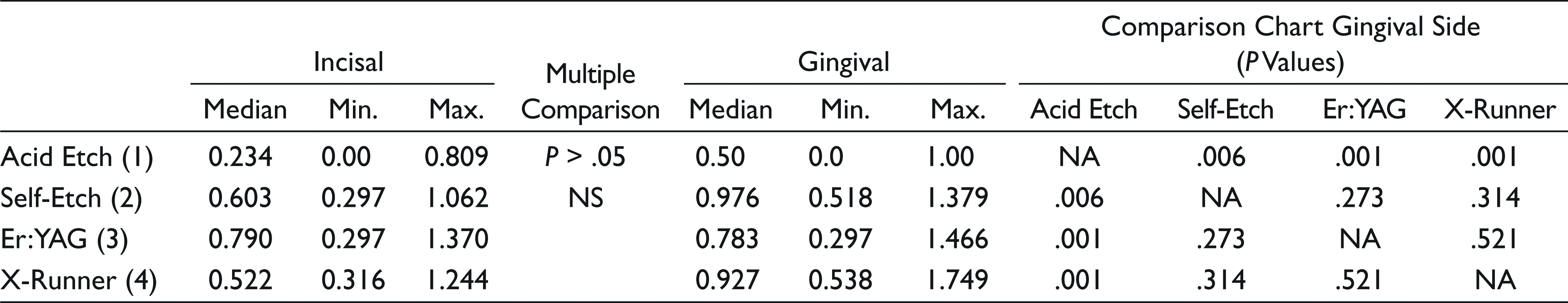
For the adhesive-enamel and adhesive-bracket interfaces of the samples aged with thermal cycling only, on the occlusal and gingival sides, significant differences were evaluated between the acid-etched group and the other etching procedures (Table 1).
Evaluations of Microleakage on Enamel-Adhesive Interface, Aged With Thermal Cycling
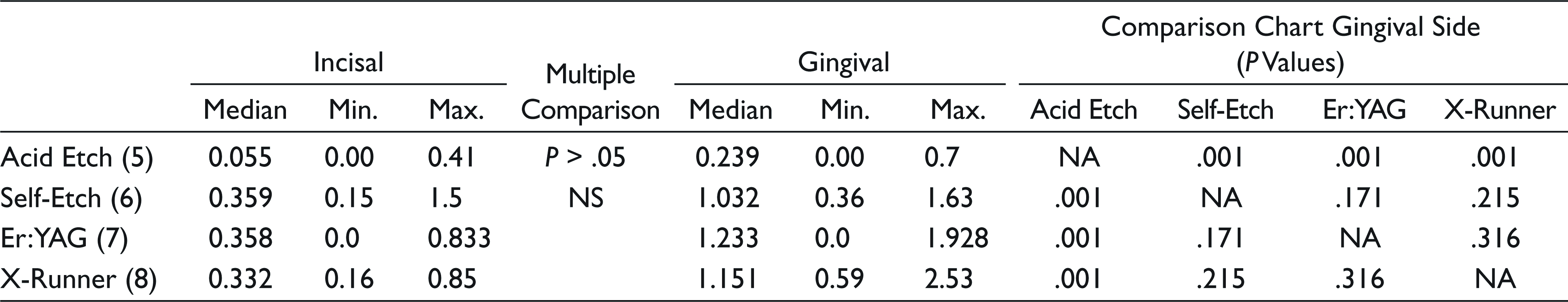
Similarly, for the enamel-adhesive and adhesive-bracket interfaces of the samples aged with chewing simulation and thermal cycling, significant differences were observed on the occlusal and gingival sides for the acid etching and the other etching procedures (Table 2).
Evaluations of Microleakage on Enamel-Adhesive Interface, Aged With Thermomechanical Cycling
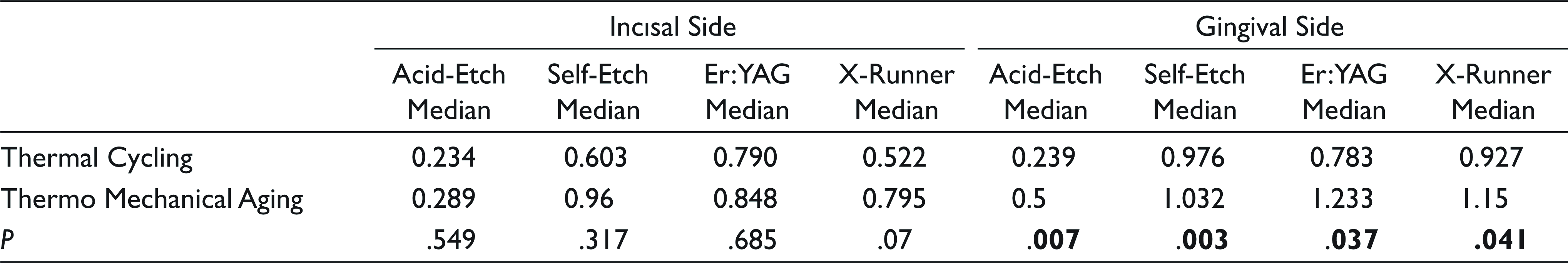
To compare the data, Mann–Whitney U test was used obtained after aging using the chewing simulator with the thermal cycle and the data obtained using the thermal cycle alone. Statistically significant differences were observed only in the enamel-adhesive interface on the gingival side in all groups (Table 3).
Comparison of the Microleakage Between Groups (Enamel-Adhesive Interface)
Discussion
Microleakage between the bracket and the adhesive plays a role in bracket loss by reducing the mechanical bond strength. However, microleakage at the enamel-adhesive interface has other negative effects on the integrity of the enamel surface since it can cause white lesions. 22 Patients receiving fixed orthodontic treatment risk having more white spot lesions than untreated patients 23 and this demineralization process can be seen in as short a period as a single month. 1
In 2014, Toodehzaeim et al reported that the highest microleakage value occurred between the enamel-adhesive interface of the gingival portion for three different etching methods, sandblasting, an Er:YAG laser, and acid etching. 24 Hamamcı et al compared acid and laser-etching methods and reported that there was more microleakage in the gingival part than in the occlusal part. These findings are in accord with studies by Arhun et al 25 and Ramoglu, 26 who evaluated the differences between the scores of incisal and gingival to the surface curvature, which may result in comparatively thicker adhesive at the gingival margin. These findings were supported by the comparison of different adhesive systems by Alkis et al; they reported that there was no difference between the adhesives in the study, but found more microleakage between the enamel and the gingival side. 27 Similarly, Yagci et al evaluated the microleakage with indirect bonding techniques and reported that there was more microleakage in the gingival part, which was explained by the increased composite thickness. 28 In our study, the highest microleakage value for all of the etching methods was recorded between the enamel-adhesive surfaces in the gingival side, similar to the results found in the literature.11,24–27,29
Several studies have reported that the enamel-adhesive bond provided by self-etch adhesives is weaker than that of phosphoric acid.30,31 In addition, different researchers have noted that the rough appearance provided by self-etch adhesives is much more superficial than for phosphoric acid. This superficial and very specific etching is based on the lack of penetration of the self-etch adhesives into the enamel surface. 32 Uysal et al, who studied metal and ceramic brackets to evaluate microleakage using acid- and self-etch adhesives, reported more microleakage between groups for self-etching adhesive enamel. 33 In our study, the microleakage was found to be higher in samples bonded with self-etch adhesives, which is in line with the related studies.11,33
Studies have shown that enamel roughening with Er:YAG lasers forms an enamel surface with characteristically irregular heterogeneous micropathies when compared to self-etching and acid etching. 14 It has been reported that laser-etched enamel is more resistant to caries formation than acid-etched specimens, although there is more microleakage. Other available studies suggest that white spot lesions and caries formation can be avoided by laser application.12,13 For this reason, it is thought that the decay resistance on the surface of enamel roughened by a laser is of great importance for orthodontists.34,35 On the other hand, in 2013, Hamamci et al studied Er:YAG laser and acid etching on the enamel surface to evaluate the microleakage under the bracket and reported a significant increase in microleakage in the laser group. 11
No studies in the orthodontic literature have subjected braces adhered by a laser-etching process to a chewing simulator to assess microleakage. Some studies have evaluated the microleakage of surfaces treated with laser etching in operative dentistry; in some of these studies, the microleakage was reported to decrease,6,36-38 while others reported increased microleakage.5-8 Excessive microleakage after laser etching can be explained by power outputs and heterogeneous surface characteristics. 14 The heterogeneous roughening of the surface structure can initiate different adhesive thicknesses in different areas of the bonding. Composite adhesives of varying thickness have been found to be effective in microleakage formation.28,39
Self-etch systems are known to cause more microleakage than laser systems, but the difference in the literature is not statistically significant.6,7,40,41 Our results are similar to the results of these studies. We recorded the highest microleakage values with laser- and self-etched enamel surfaces.
In the orthodontic literature, no studies have evaluated samples submitted to aging procedures other than thermal cycling. In the field of restorative dentistry, Poitevin et al examined the influence of dynamic forces on binding strength at filler interfaces and reported that mechanical fatigue of the adhesive may cause the resulting connection to be weakened. 42 This information is supported by the study by Rütterman et al, in which the surface energies of resin-based dental materials were reportedly weakened by the chewing simulator. 43 Ana Karina et al evaluated the effect of the chewing simulator aging process on microleakage in composite pads and reported that there was more microleakage in the samples aged with mechanical and thermal cycles than those only subjected to thermal cycling or mechanical aging. This is related to the mechanical fatigue of the weakest points of the adhesive. 44
In our study, statistically more microleakage supporting the above-mentioned studies was found but only in the gingival part between the enamel-adhesive surfaces. No significant difference was found in other measurements. It is thought that the reason for this is that the composite thickness on the other side is higher.
Limitations
In the literature, only the thermal cycle was used in orthodontic studies and no mechanical aging was used. In this study, it was reported that mechanical effects had an effect on microleakage. The study was conducted in vitro and variables such as saliva, diet, and eating habits could not be evaluated.
Conclusion
We evaluated the microleakage after thermal cycling and thermomechanical aging on specimens containing brackets adhered to the enamel surface etched by four different methods. The following results were obtained:
Microleakage values in all groups were found to be greater in the gingival part of the bracket. Microleakage values in gingival and occlusal sides measured for the phosphoric acid-etched groups were lower in both aging methods. The amount of microleakage was generally higher for the samples submitted to thermomechanical aging, but the only statistically significant difference was recorded for the gingival part for all the conditioning methods. According to the results, thermomechanical aging might be advised to be included to further study designs.
Declaration of Conflicting Interests
The authors declared no potential conflicts of interest with respect to the research, authorship, and/or publication of this article.
Funding
The authors disclosed receipt of the following financial support for the research, authorship, and/or publication of this article: All funding sources for the reported research have been covered by Ufuk OK. All consumables used in the study were paid by Ufuk OK. The acquisition, measurement, and analysis of the data in the study were carried out at the labs of Bezmialem Vakif University.