
Abstract
Introduction:
The aim of study is to determine if laser irradiated enamel may be a viable alternative to acid etching for orthodontic bonding of molar tubes.
Method:
Sixty extracted permanent molars were divided into 3 groups of 20 each. Group I was irradiated with Er:YAG laser for 15 s, group II with Er;Cr:YSGG laser for 15 s, and group III was etched with 37% phosphoric acid. Ten samples from each group were observed under scanning electron microscope and later all 60 samples were bonded with molar tubes. Molar tubes were debonded and shear bond strength was recorded. Adhesive remnant index was measured under electron microscope (2000×).
Results:
Shear bond strength with Er:YAG laser etching was significantly higher than with acid etching and Er;Cr:YSGG laser etching. Er:YAG laser etching, and Er;Cr:YSGG laser etching showed Type III etching pattern with microcracks while acid etching showed Type I etching pattern. Adhesive remnant index was significantly higher for Er:YAG laser etching as compared to conventional acid etching.
Conclusion:
Er:YAG and Er;Cr:YSGG laser etching is a viable alternative to acid etching for bonding of bondable molar tubes.
Keywords
List of Abbreviations
ARI: Adhesive Remnant Index
Er,Cr:YSGG: Erbium, chromium: yttrium-scandium-gallium-garnet laser
Er:YAG: Erbium: yttrium-aluminum-garnet laser
SBS: Shear bond strength
SEM: Scanning electron microscope
Introduction
Buonocore, in 1955, introduced a method to adhere acrylic material to enamel while Newman, in 1965, used this method to bond brackets to the teeth.1, 2
Acid etching acts by erosion of hydroxyapatite structures and thus helps in penetration by formation of resin tags. The micro-mechanical bonds increase the bond strength, but the disadvantage is the potential risk of decalcification, thus making it prone to caries.3, 4
Four-ruby laser was introduced by Maiman in 1960, following which CO2 laser and Nd:YAG laser were invented and introduced to dental practice. 3 Lasers consist of an ion and a crystal matrix. This gives thermo-optical and also mechanical properties. Erbium lasers consist of erbium ion (Er3+) and have wavelength of 2,780 to 2,940 nm. YSGG and YAG form the matrix for laser. Hydroxyapatite crystals and water absorb it and thus there is ablation of hard and soft tissues. 5 Wavelength of Er:YAG laser is 2,940 nm while that of Er,Cr:YSGG laser is 2,790 nm. Thus, Er:YAG laser has higher absorption and smaller penetration depth, while Er,Cr:YSGG laser is absorbed less and thus penetrates more. 6 Diaci and Gaspire in 2012 observed that Er:YAG laser has a penetration of 7 µm in enamel while 5 µm in dentin; while Er,Cr:YSGG laser has a deeper penetration, 21 µm in enamel, and 15 µm in dentin. 6 Erbium laser does not require priming of tooth. The fiber optic tips can be reused after autoclaving. The size of laser unit is large and also very costly. 5
Laser irradiation can (a) remove smear layer, (b) melt and recrystallize hydroxyapatite structures, and (c) cause appearance of pores in dental hard tissue. Adhesive material infiltrates these nonuniform, uneven surfaces. 7
Numerous reports have proven the laser etching technique to be effective for bonding of brackets and tubes.3, 4, 7–10 However, very few studies have compared both Er:YAG laser etching and the Er,Cr:YSGG laser etching. 11
Hence, this study was conducted to determine whether Er,Cr:YSGG and Er:YAG laser irradiation may be an alternative method for etching of an enamel surface to bond the molar tubes.
Aim
To determine whether irradiating enamel with a laser can be an alternative for etching with acid, for orthodontic bonding of molar tubes.
Objective
To study and compare surface characteristics along with the shear bond strength (SBS) after bonding of molar tubes using Er,Cr:YSGG laser irradiation, Er:YAG laser irradiation, and conventional acid etching.
Material and Method
Experimental Design
The research was reviewed and approved by institutional research committee of Bharati Dental College and Hospital, Bharati Vidyapeeth Deemed to be University, Pune, India (ECR/328/Inst/MH/2016) dated November 27, 2018. This study was conducted on 60 extracted permanent molars having an intact buccal surface. Extracted molars were preserved in artificial saliva. Extracted molars were implanted in acrylic resin blocks, such that 2 mm of buccal surface was visible over the resin.
All 60 extracted molars were arbitrarily divided to form 3 groups of 20 each.
Group 1: Er:YAG laser (2W) irradiated the enamel for 15 s from a distance of 1 mm.
Group 2: Er,Cr:YSGG laser (2W) irradiated the enamel for 15 s from a distance of 1 mm.
Group 3: 37% orthophosphoric acid was used for etching of enamel for 15 s.
Etching Procedure
Pumice and rubber cup were used to cleanse extracted molars. Rinsing was done with water and later they were air-dried.
The Er:YAG laser, used for etching the buccal surface of group 1, has wavelength of 2,940 nm at 2 W for 15 s and was highly absorbed by dental hard tissue. The Er,Cr:YSGG laser, used for etching the buccal surface of samples of group 2, has wavelength of 2,780 nm at 2 W for 15 s and was absorbed by dental hard tissue. The third group had been etched with 37% orthophosphoric acid, rinsed, and air-dried.
Scanning Electron Microscopy examination
Ten representative samples from each group were examined using Scanning Electron Microscopy (SEM; Quanta200, ThermoFischer Scientific, Waltham, MA, USA) and EDX (EDAX, Ametek Inc., Berwyn, PA, USA) in Icon Analytical Equipment Pvt. Ltd., Mumbai, Maharashtra, to assess the surface texture of enamel. Each tooth was kept on a flat surface with a black background. This platform went into the SEM machine. Each sample was examined at 2000× magnification at the center of etched surface.
Bonding Procedure
Bonding of all 30 samples of SEM analysis along with the remaining 30 samples was done. Bondable molar tubes (3M, Unitek, USA) were bonded to buccal surface using primer (Transbond XT) and composite (3M Unitek, USA) followed by light curing with blue LED light for 12 s.
Shear Bond Strength Testing
Samples were stored in artificial saliva at 37 ºC for 24 h, following which thermocycling was done of 500 cycles at 5 ºC to 55 ºC and dwell time of 30 s. For SBS, the teeth were mounted on testing machine (Universal testing machine UNITEST-10). The machine has a sharp edge which hits the tooth where the tube is attached, with a speed of 1 mm/s. This force decay with which debonding occurred was measured. Bond strength was measured by dividing the force decay by the base area of the molar tubes. Base area of molar tube is 15.81 mm2.
After debonding was done, the debonded surface of molar was observed under stereomicroscope of 10× magnification (Apex Industrial Electronics, India).
Adhesive remnant index (ARI) was calculated using the method introduced by Artun and Bergland (1984)12. ARI score has a scale from 0 to 3;
0 = no adhesive is present on the tooth;
1 = adhesive is present only on less than half of bonding site;
2 = adhesive is present only on more than half of bonding site;
3 = adhesive completely covers bonding site.
Method of Data Examination
Sample size derivation
Significance level = 5%,
Power = 80%,
Test type = Measuring sample size using 2-sided formula (sample size for clinical trial) outcome variable on ratio scale and testing null hypothesis: M1 = M2 = M3 (means of 3 intervention)
n = 2 [S2 (Z1 + Z2)2/(M1 − M2)2]
Here,
(M1) Mean test intervention = 7.650,
(M2) Mean control intervention = 5.150,
(S1) Standard deviation of M1 = 3.600,
(S2) Standard deviation of M2 = 2.500,
Pooled SD = 3.09919,
α) Set level of confidence. Usual values 0.95; 0.99 = 0.95,
β) Set level of power of test. Usual values 0.8, 0.9 = 0.8,
(Z1) Z value associated with alpha = 1.64485,
(Z2) Z value associated with beta = 0.84162,
(n1) Minimum sample size = 20.
Statistical Product and Service Solutions (SPSS) version 16 for Windows (SPSS Inc, Chicago, IL) was used to perform the statistical analysis. Descriptive quantitative data was demonstrated in mean and standard deviation respectively. Data normality was checked by using Shapiro-Wilk test. Confidence interval set at 95.00% and probability of alpha error (level of significance) set at 5.00%. Power of the experiment set at 80.00%.
Comparisons of the SBS were accomplished by analysis of variance (ANOVA) test. Multiple pair-wise intergroup comparison among three groups was done with the help of Tukey’s post hoc test. ARI score differences were assessed with chi-square test.
Results
Comparison of SBS in Er:YAG Laser Etched Group; Er,Cr:YSGG Laser Etched Group, and Acid Etched Group
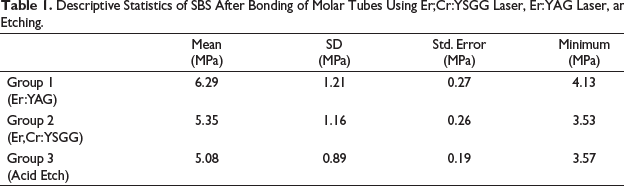
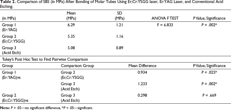
SBS associated with Er:YAG laser, Er,Cr:YSGG laser, and acid etch was 6.29 ± 1.21 MPa, 5.35 ± 1.16 MPa, and 5.08 ± 0.89 MPa, respectively. The minimum and maximum SBS with Er:YAG laser was 4.13 MPa and 8.05 MPa, respectively, while minimum SBS was 3.53 MPa and maximum SBS 6.97 MPa with Er,Cr:YSGG laser. The minimum and maximum SBS with acid etch was 3.57 MPa and 6.68 MPa, respectively as shown in Table 1. SBS with Er:YAG laser was significantly increased than Er,Cr:YSGG laser (0.025). SBS with Er:YAG laser was significantly increased than acid etching (0.002). The results have been displayed in Table 2.
Descriptive Statistics of SBS After Bonding of Molar Tubes Using Er;Cr:YSGG Laser, Er:YAG Laser, and Conventional Acid Etching.
Comparison of SBS (in MPa) After Bonding of Molar Tubes Using Er;Cr:YSGG laser, Er:YAG Laser, and Conventional Acid Etching.
Comparison of Etching Pattern in Er:YAG Laser Etched Group, Er,Cr:YSGG Laser Etched Group, and Acid Etched Group
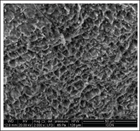
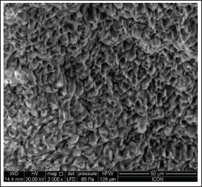
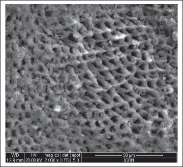
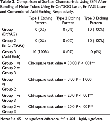
Both the laser etched groups (Figures 1 and 2) showed Type III etching pattern while Type I etching pattern (Figure 3) was shown by acid etching as shown in Table 3. The differences between Er:YAG laser etching pattern and acid etching pattern (0.001) and between Er,Cr:YSGG laser etching pattern and acid etching pattern (0.001) were highly significant, while there was no difference between Er:YAG laser etching pattern and Er,Cr:YSGG laser etching pattern, as shown in Table 3.
Type III Etching Pattern Seen With Er:YAG Laser Etching.
Type III Etching Pattern Seen With Er;Cr:YSGG Laser Etching.
Type I Etching Pattern Seen With Acid Etching.
Comparison of Surface Characteristic Using SEM After Bonding of Molar Tubes Using Er;Cr:YSGG Laser, Er:YAG Laser, and Conventional Acid Etching, Respectively.
Comparison of ARI in Er:YAG Laser Etched Group, Er,Cr:YSGG Laser Etched Group, and Acid Etched Group
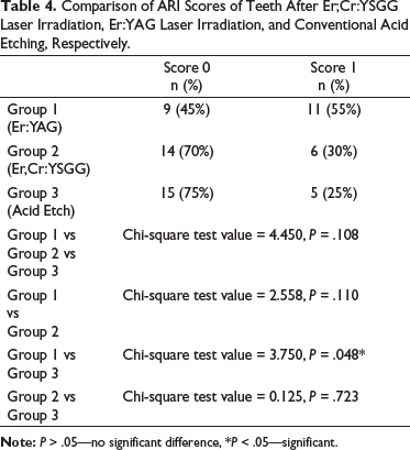
Out of 20 samples of Er:YAG laser group, 9 samples showed score 0 and 11 samples showed score 1 while in Er,Cr:YSGG laser group, 14 samples showed score 0 and 6 samples showed score 1. In the acid etched group, 15 samples out of 20 samples showed score 0 and 5 samples showed score 1. The results have been shown in Table 4. A statistically significant difference between Er:YAG laser etched group and acid etched group (0.048) was observed, as shown in Table 4.
Comparison of ARI Scores of Teeth After Er;Cr:YSGG Laser Irradiation, Er:YAG Laser Irradiation, and Conventional Acid Etching, Respectively.
Discussion
Reason for Selecting Molars
Banding of molars (a) requires the making of interproximal space for fitting of bands, (b) is a time-consuming procedure, (c) causes discomfort to the patient, (d) when the treatment comes to an end, the spaces created due to banding have to be closed again, (e) banding generally causes trauma to the tissues, (f) causes increased plaque accumulation, and (g) causes gingival inflammation.13, 14 Also, Zachrisson (1977) concluded that the failure rate is more for banding of molar tubes than bonding of molar tubes. 15 Thus, to prevent these issues, bonding of the tube can be done. Various studies related to SBS of laser etching and acid etching have been done on premolars, while very few studies have been done on the molars.3, 4, 7–10, 16–19 Hence, molars were selected to determine if laser etching is a viable alternate method to bond the molar tubes.
Scanning Electron Microscopy Analysis
SEM gives high-resolution images which assess materials for surface roughness, fractures, texture, impurity, foreign material, or erosion.
In this study, at 2000×, acid etched enamel had Type I etching pattern, which is hollowed prism centers with relatively intact peripheral region. It resembled a honeycomb-like pattern which aids penetration of resin. Phosphoric acid dissolved the hydroxyapaptite crystals which led to eroded plane and formation of tags and thus helped in mechanical locking of the resin material.
Both laser etched groups showed Type III etching pattern, which is hollowed prism centers adjoining to areas which had loss of prism peripheries along with microcracks. Laser irradiation causes many stress concentration sites, which not only gives rise to an uneven and inhomogeneous surface and aids in locking resins but also causes microcracks. Thus, surface destruction was more eminent with Type III etching pattern and microcracks. But, in this study, the SBS was increased when Type III etching pattern was seen and it may be because of increased depth of etched enamel due to laser.
Berk et al 7 has compared only Er,Cr:YSGG laser and acid etching and observed Type III pattern and microcracks by laser irradiation with 2W Er,Cr:YSGG laser and type I pattern on acid etching, while we have compared two types of lasers, Er:YAG laser and Er,Cr:YSGG laser with acid etching and observed type III etching pattern with both, Er:YAG laser and Er,Cr:YSGG laser and type I etching pattern with acid etching.
Shear Bond Strength
SBS measured using universal testing machine was higher for Er:YAG laser etched group, followed by Er,Cr:YSGG laser group and acid etched group.
Berk et al 7 showed that SBS with Er,Cr:YSGG laser at 1.5W (7.13 ± 1.67 MPa) and 2W (7.17 ± 1.69 MPa) is almost equal to that obtained with acid etch technique (7.65 ± 1.38 MPa), while we observed that SBS with Er:YAG laser at 2W (6.29 ± 1.21 MPa) is significantly greater than that achieved with Er,Cr:YSGG laser at 2W (5.35 ± 1.16 MPa) which is greater than that obtained with the acid etch technique (5.08 ± 0.89 MPa).
Adhesive Remnant Index
ARI was measured to further investigate bond strength. This study observed greater ARI for Er:YAG laser, followed by Er,Cr:YSGG laser and then acid etch group. Some authors believe that if more adhesive is present on the tooth then more time is required to clean it when brackets are debonded, thus making it undesirable, while others believe that bond failure at adhesive-enamel interface is less desirable because it may lead to fragmentation and formation of microcracks on enamel, particularly in ceramic brackets.20, 21 Debonding at bracket-resin interface indicates etching which gives good surface wetting. 10
Berk et al 7 observed that ARI scores obtained with Er,Cr:YSGG laser and with acid etching showed that composite remaining on the enamel surface after debonding was either less than half or more than half; however, we observed that ARI scores achieved with Er:YAG laser were 0 (45%) and 1 (55%), with Er,Cr:YSGG laser were 0 (70%) and 1 (30%), and with acid etch were 0 (75%) and 1 (25%), indicating that for Er:YAG laser etching, more composite was present on the enamel, while for acid etch technique, more composite was present on the bracket.
Since in this study ARI score was better for the laser etched group, it can be concluded that good surface wetting occurred with the laser etched group.
Conclusion
This study was conducted to compare Er:YAG laser etching, Er,Cr:YSGG laser etching, and conventional acid etching for bonding of molar tubes.
Within the limits of this study, the following conclusions were drawn:
Mean SBS of bonded molar tubes obtained after Er:YAG laser etching was significantly higher than that observed with acid etching (P = .002) and Er,Cr:YSGG laser etching (P = .025).
Enamel surface prepared by Er:YAG laser etching and Er,Cr:YSGG laser etching showed Type III etching pattern with microcracks while acid etching showed Type I etching pattern.
ARI was significantly higher for Er:YAG laser etching as compared to conventional acid etching (P = .048).
Thus, Er:YAG laser etching and Er,Cr:YSGG laser etching can be a good alternative to acid etching for bonding of molar tubes.
Footnotes
Figures 1 to 6 along with their citations have been deleted. The section ‘Limitations of Current Study’ has been deleted.
Please see
Author Contributions
AHR, AS, VIB, and AP conceived the research idea. Literature review was done by AHR. AHR collected the samples. AS and AP helped in conducting scientific experiment. AHR and AP analyzed the samples. AHR prepared the initial draft of the manuscript. All authors reviewed and edited the final manuscript prior to submission.
Declaration of Conflicting Interests
The authors declared the following potential conflicts of interest with respect to the research, authorship, and/or publication of this article:
All authors of the manuscript have read and agreed to its content and are accountable for all aspects of the accuracy and integrity of the manuscript. That the article is original, has not already been published in a journal, and is not currently under consideration by another journal.
Ethical Approval
The research was reviewed and approved by institutional research committee of Bharati Dental College and Hospital, Bharati Vidyapeeth Deemed to be University, Pune, India (ECR/328/Inst/MH/2016) dated November 27, 2018.
Funding
The authors received no financial support for the research, authorship, and/or publication of this article.
Informed Consent
The participant has consented to the submission of the article to the journal.