
Abstract
Aims and objectives:
The aim of this study was to determine the effects of chitosan, Ankaferd Blood Stopper® (ABS, Ankaferd Health Products Ltd, Turkey), and ferric sulfate (FS) on the shear-bond strength of the self-etch adhesive to primary tooth dentin.
Materials and methods:
The occlusal surfaces of 80 extracted human primary teeth (stored in a 0.5% chloramine-T solution at 4oC) were ground flat, exposing the dentin. The teeth were divided into 4 groups: chitosan, ABS, FS, and control. Hemostatic agents were applied to the teeth, and then they were rinsed with distilled water and air dried. In the control group, the teeth were only rinsed with distilled water and slightly air dried. A self-etch adhesive was applied, and the composite cylinder was created on all the samples. Shear-bond strength was tested with a universal testing machine. Failure mode analysis was performed using scanning electron microscopy.
Results:
The study revealed no statistically significant difference between the hemostatic agent and control groups in terms of bond strength (P >.05). The highest bond strength was observed in the chitosan group. In this group, a statistically significant difference between chitosan and ABS was observed (P <.05). The adhesive failure was the predominant failure mode in all the groups.
Conclusions:
These findings suggest that chitosan, ABS, and FS have no adverse effects on the bonding of the self-etch adhesive to primary teeth dentin.
Introduction
Human primary teeth require pulp treatment more often than permanent teeth because they have a low amount of enamel and dentin thickness, and a relatively large pulp chamber. Also, in primary teeth, the pulp horn is closer to the surface compared with that in the permanent teeth. Pulpotomy is a common technique used for carious expo-sures of asymptomatic vital primary teeth. The purpose of this technique is to amputate the inflamed coronal pulp and preserve the radicular pulp’s vitality. 1 To date many pulpotomy materials have been used, including formocresol (FC), ferric sulfate (FS), glutaraldehyde, calcium hydroxide, zinc oxide eugenol, mineral trioksit aggregate, and bioceramics.
Several problems have been identified in the use of different pulpotomy agents, such as postoperative systemic transport, the possibility of toxicity, harmful effects on the succedaneous tooth enamel, solubility in tissue fluids, internal resorption, and autoantibody formation due to reversible fixation. 2 These complications have led clini-cians to search for alternative pulpotomy agents. In recent years, new hemostatic agents such as Ankaferd Blood Stopper® (ABS) and chitosan have begun to be used in pulpotomy treatments.3,4
FS is commonly used in pulpotomy treatment and induces hemostasis by agglutinating the blood proteins with ferric and sulfate ions. This agglutinated complex (FS protein) mechanically seals the capillary orifices and prevents clot formation. However, it has been reported to have cytotoxic effects and cause tissue necrosis. 5
ABS is a hemostatic agent consisting of 5 different herbal ingredients. 6 It has various effects on the endothelium, angiogenesis, cellular proliferation, and blood vessels. 7 This agent is effective in both patients with normal hemostatic parameters and those with primary/secondary hemostasis deficiency. 6 Although it is safely used in the medical field, studies about its dental use have been limited. ABS is indicated as an alternative pulpotomy agent because it is biocompatible and yields successful results. 8 However, it presents slightly lower clinical and radiographic success when compared with FS and FC. 4
Chitosan is a biodegradable, biocompatible, and non-toxic derivative of chitin. This hemostatic polymer in the form of a granular sea-product powder is produced to control mild, moderate, and severe bleeding. 9 It has been proven to control bleeding in the absence of clotting factors or thrombocytes; thus, it is useful for patients receiving coagulopathic or anticoagulant therapy. 10 In addition, this product exhibits antiviral and antiphage activities, and inhibits bacterial infections. 11 It can be used in wound dressing and bone tissue engineering,3,12,13 and it recently started to be used in dentistry for inhibition of cariogenic bacteria, enamel remineralization, tissue regeneration, and pulpotomy treatments.14,15
Adhesive restorations are preferred frequently after primary tooth pulpotomy due to the more conservative cavity preparation need and its superior aesthetics. The bond strength between adhesive and dentin is an important factor for the success of a restoration. It has been shown that adhesive’s bond strength to primary teeth is less than that to permanent teeth due to their inherent morphological and physiological differences. 16 In addition, various agents used during dental treatments can affect bond strength.
Although there are several studies on ABS and FS, no study has been conducted on the effect of chitosan on bond strength in adhesive restorations. Therefore, the aim of this study was to compare the effects of chitosan, ABS, and FS on the shear-bond strength of the self-etch adhesive to the dentin of primary teeth.
Materials and Methods
This study was approved by the Clinical Research Ethics Committee of Erciyes University (approval number 2014/421). A total of 80 noncarious extracted primary teeth were used in the present study. The number of specimens to be studied was estimated on the basis of a power analysis with the G*Power package program that assumed a power of 90% and a significance level of .05. The sample size was determined as n = 20.
After the removal of soft and hard tissue residues, the teeth were stored in a 0.5% chloramine-T solution at 4°C. The teeth were then embedded into autopolymerizing acrylic resin blocks up to the cementoenamel junction. The smooth dentin surface was exposed by eroding all the occlusal enamel and some superficial dentin (grooves at a depth of 1 mm formed with diamond burs on the occlusal surface intermittently, and these grooves were removed by diamond disks. This way the removed dentin layer was measured), using a diamond-coated disk under water cooling, in the mesiodistal direction parallel to the cemento-enamel border.
The process of bonding is crucially dependent on the angle of application, the amount of time etched, and ulti-mately on the smear layer. A smear layer can be produced on a dentine surface using low- or high-speed burs/disks. 17 In our study, we erode enamel and dentin using a diamond-coated disk, thereby forming the smear layer. To obtain the homogeneous dentin surface, the exposed dentin surface was ground flat with 600-grit sandpaper for 10 seconds, and then rinsed with distilled water to remove debris.
Experimental Groups
The teeth were divided into the following 4 experimental groups:
Chitosan: Chitosan (Celox; Medtrade Products Ltd, Crewe, United Kingdom) was moistened with distilled water and applied on the dentin surface for 20 seconds using an applicator; the surface was rinsed with distilled water for 15 seconds and gently air dried for 10 seconds (Figure 1a). ABS: ABS (Ankaferd Health Products Ltd, Istanbul, Turkey) was applied on the dentin surface using an applicator for 20 seconds, and then the surface was rinsed with distilled water for 15 seconds and gently air dried for 10 seconds (Figure 1a). FS: FS (AstringedentTM Ultradent Products Inc., Salt Lake City, UT, USA) was applied on the dentin surface using an applicator for 20 seconds, following which the surface was rinsed with distilled water for 15 seconds and gently air dried for 10 seconds (Figure 1a). Control: The dentin surface was rinsed with distilled water for 15 seconds and gently air dried for 10 seconds.
A 1-step self-etch bonding system (3M ESPE; 3M Company, St. Paul MN, USA) was applied on the dentin surface and cured according to the manufacturer’s instructions.
Schematic illustration of the specimen preparation and experimental design used to measure shear bond strength. (a) Hemostatic agents were applied to dentin, which was then rinsed with distilled water and gently air dried. The control group was only rinsed with distilled water and gently air dried. (b) Self-etch adhesive was applied, and composite cylinders were created for all samples. (c) Shear-bond strength was measured using a universal testing machine.
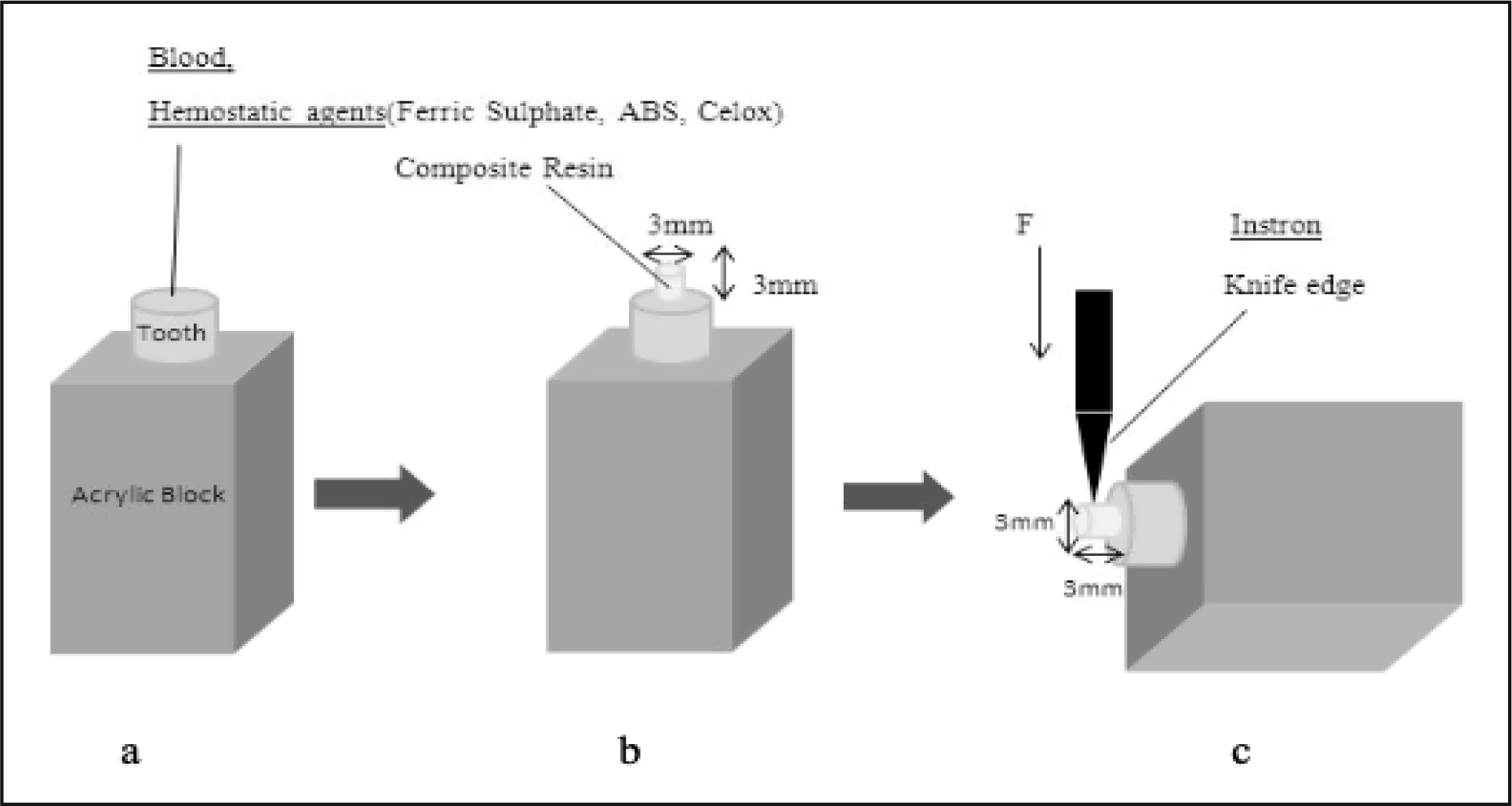
A micro-hybrid composite resin (3M ESPE Filtek P60) was applied on the dentin surfaces of each sample, with an internal diameter of 3 mm and height of 3 mm (by using a plastic tube), using an incremental technique (first 2 mm, and then 1 mm). Each layer was cured for 40 seconds using a light-emitting diode (3M ESPE Elipar S10) (Figure 1b). All the samples were stored in distilled water at 37°C for 48 hours.
After 48 hours, the samples were thermocycled for 500 cycles between 5°C and 55°C, with a dwell time of 30 seconds each and transfer time between the 2 baths of 5 to 10 seconds.
After thermocycling, the shear-bond strength was measured using a universal testing device (Instron; AGS-1000 kg/W; Shidmadzu Corp; Chiroda-Ku, Tokyo, Japan) applying a force of 1 mm/min until the composite cylinder was dislodged from the tooth (Figure 1c). The maximum loads at bond failure were recorded in newtons (N), and the bond strengths were calculated in megapascals (MPa).
All fractured sample surfaces were coated with gold palladium and examined using scanning electron microscopy (SEM; LEO 440; Oxford, England) under 2500× magni-fication. Surface images were obtained to identify the mode of bond failure. Failure modes were classified as adhesive fracture (failure at the dentin-resin interface), cohesive fracture (failure within the resin composite or dentin), or mixed fracture (a combination of adhesive and cohesive fracture).
Statistical Analysis
All statistical analyses were completed using SPSS 15.0 (SPSS Inc., Chicago, IL, USA) software. One-way analysis of variance and Tukey’s posthoc multiple comparison tests were used in the statistical evaluation of the data. A P value <.05 was considered to indicate a statistically significant difference.
Results
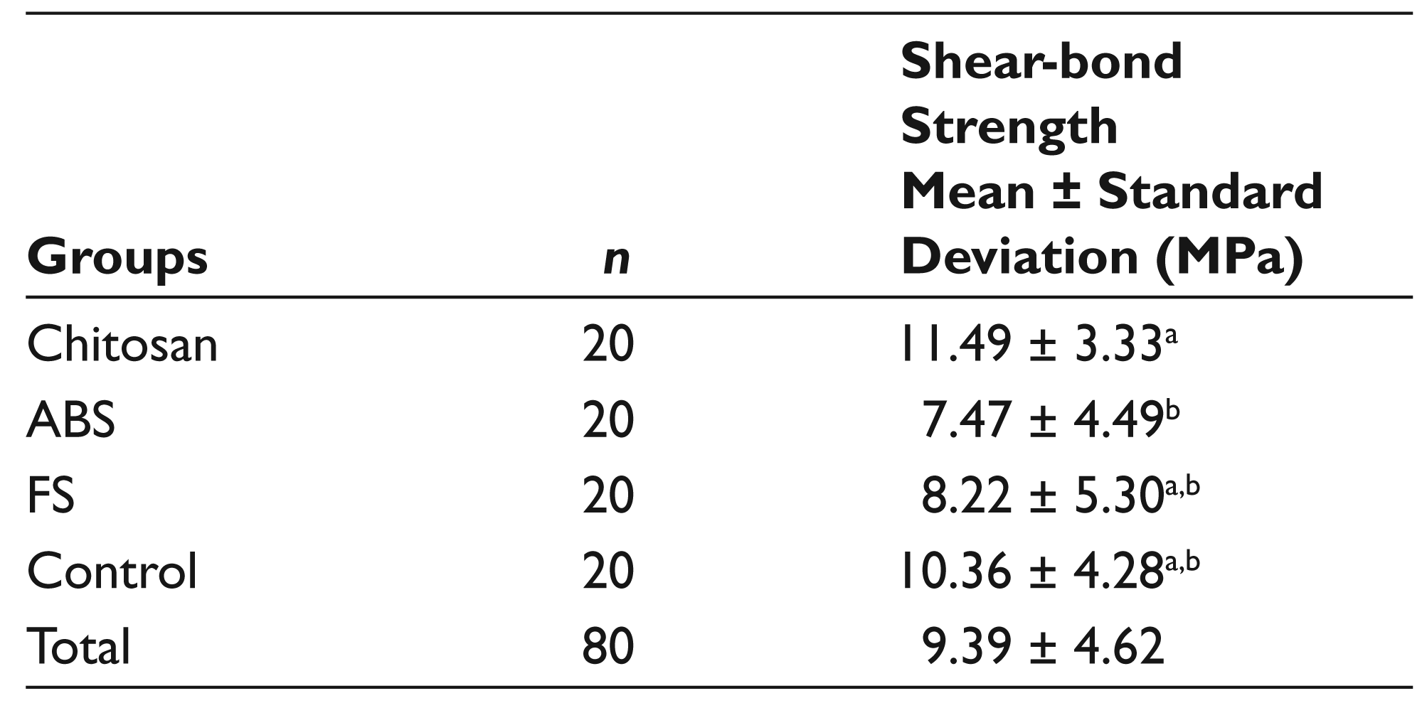
The mean and standard deviation values of the shear-bond strength of the study groups are presented in Table 1. In the intergroup comparison between the mean shear-bond-strength values, chitosan, and ABS samples showed a statistically significant difference (P < .05).
Among the hemostatic agents, the highest bond-strength values were observed in chitosan, and the lowest bond-strength values were observed in ABS (Table 1). There was no statistically significant difference between the control and hemostatic agent groups.
Mean and Standard Deviation Values of Shear-bond Strength of All Groups
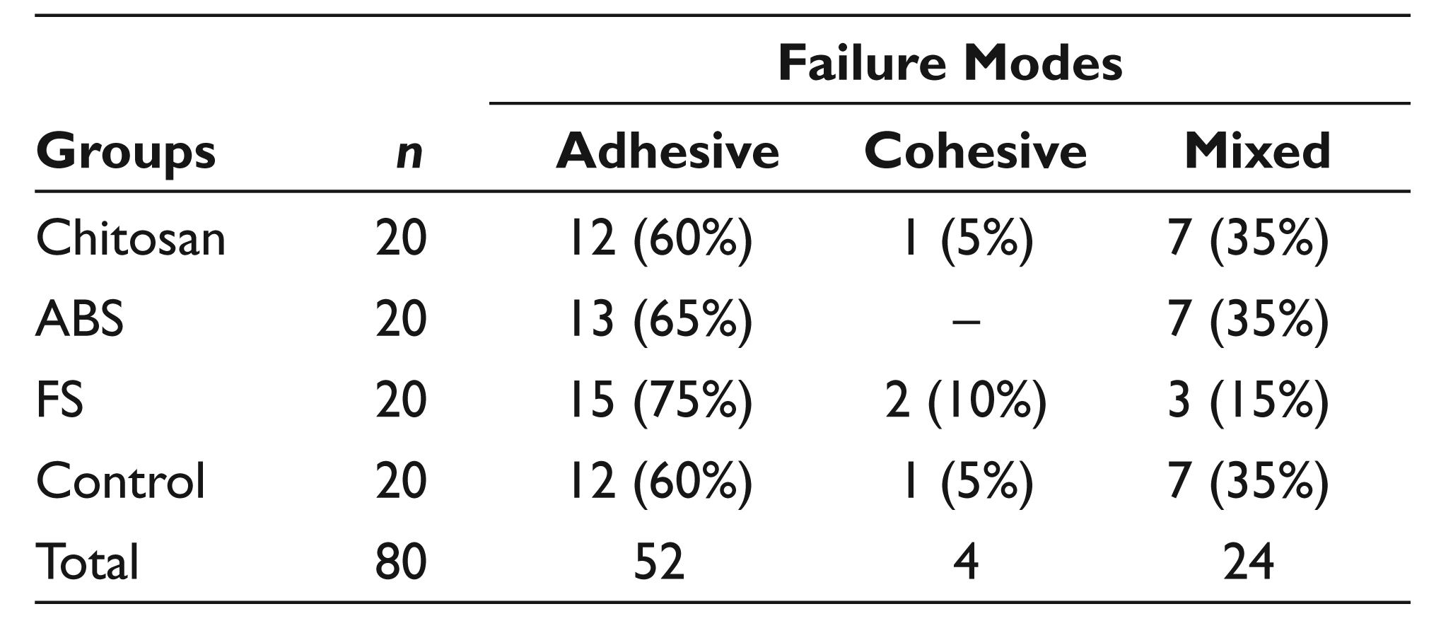
The failure modes of the groups are presented in Table 2. Adhesive failure was a predominant failure mode in all groups. Cohesive failure modes were rarely seen.
Fracture Modes of the Groups
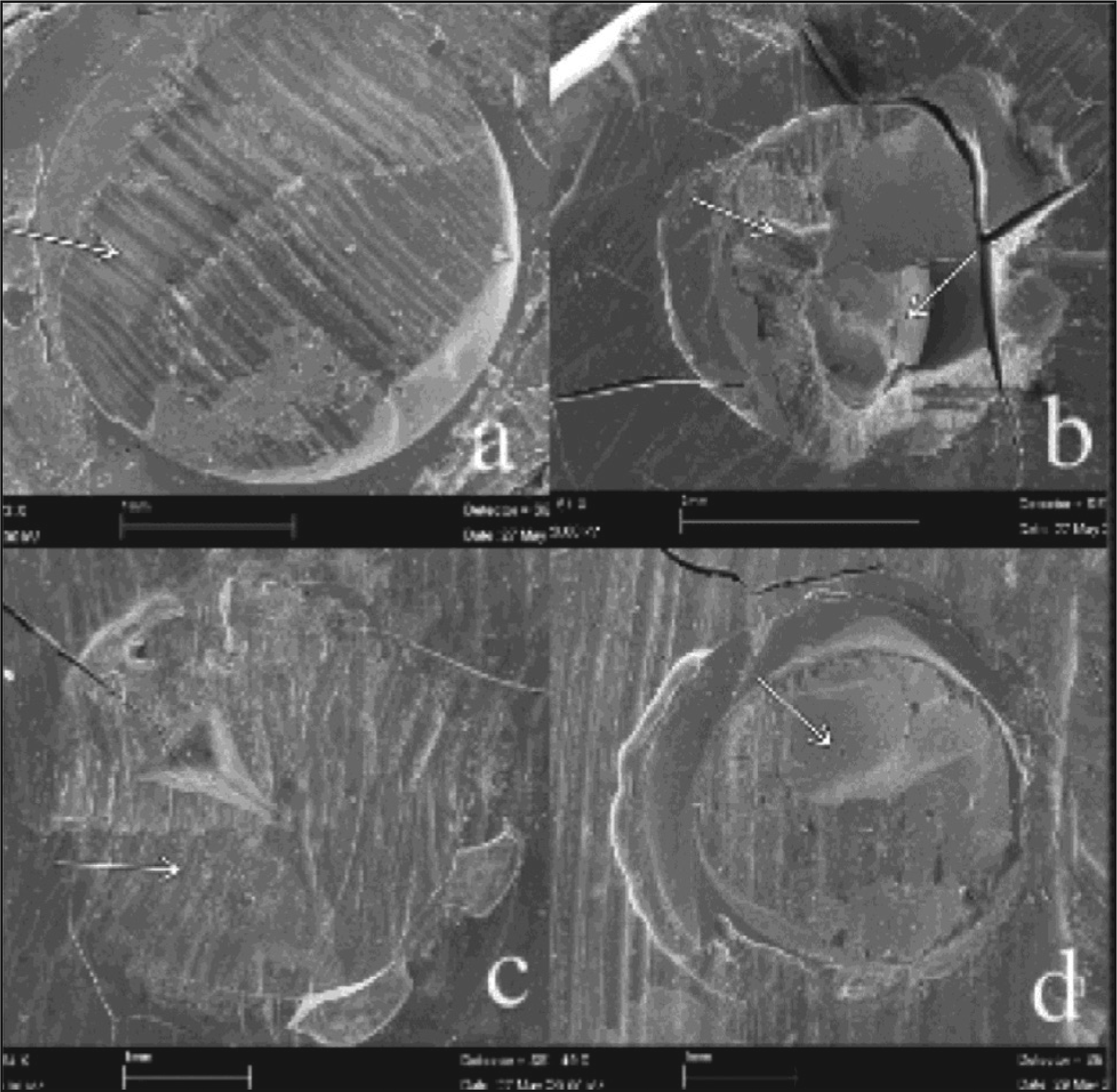
Morphological aspects of the fractured surface of samples at 2500× magnification are shown in Figure 2. The SEM analysis of debonded surfaces showed that adhesive failure occurred most frequently in all the groups. Adhesive fracture SEM images in the chitosan and FS groups are shown with arrows in Figures 2a and 2c. A mixed fracture SEM image in the ABS group is shown in Figure 2b. A cohesive fracture SEM image in the control group is shown in Figure 2d.
Morphological aspects of the fractured surface of samples at 2500× magnification. (a) An image of the chitosan group that showed adhesive failure. (b) An image of the ABS group that showed mixed failure. (c) An image of the FS group that showed adhesive failure. (d) An image of the control group that showed cohesive failure. ABS, Ankaferd Blood Stopper; FS, ferric sulfate.
Discussion
Pulpotomy is the most frequently performed endodontic treatment in children. The treatment success depends on the clinical properties of the pulpotomy agents. Durable bond strength between the restoration and tooth interface is essential for providing long-term success. Blood and saliva contaminations are important risk factors for bond failure during the adhesive restoration process. 18 Therefore, pro-viding bleeding control by using a hemostatic agent is important to increase the bond strength. Different hemostatic agents such as FS, ABS, and chitosan have been used for this purpose. Although there are several studies in the literature about the effects of ABS and FS on shear-bond strength,3,4 no study has been performed that considers chitosan’s effect on the bond strength in adhesive restorations. The present study was undertaken to evaluate the hemostatic agents (chitosan, ABS, and FS) effects on the bond strength of self-etch adhesive systems to primary tooth dentin.
It has been shown that FS, which is classified as a “protective” agent in primary teeth pulpotomies, can signi-ficantly lower the bond strength of self-etch adhesives. 19 Although in the current study the FS group showed a lower bond strength, this result was not statistically significant. This finding is in accordance with the study by Kimmes et al. 20
ABS has effective hemostatic, anti-inflammatory, and antibacterial properties. Due to these favorable charac-teristics, it has begun to be used in pulpotomy treatments in pediatric patients. 8 Trakyali and Oztoprak 21 indicated that the ABS group showed the second highest bond-strength values in their study. Samples contaminated with blood showed lower bond-strength values. The blood and ABS groups exhibited significantly lower values than the control group. This result is in line with our study, which showed that the ABS group exhibited the lowest bond-strength values. Although there was no statistically significant difference between the ABS, FS, and control groups in terms of bond strength, the difference between the ABS and chitosan groups was statistically significant.
Chitosan has positive effects on bleeding control and wound healing. In order to benefit from its positive features, chitosan has been used in dental and maxillofacial surgery and in conservative and endodontic treatments. This hemostatic agent can be safely used in patients receiving coagulopathic or anticoagulant therapy, because its hemostatic feature is independent from the body clotting mechanism. 22 The clinical efficacy and local hemostatic effects of ABS and chitosan were reported to be satisfactory because of great success related to their use in intraoperative and postoperative bleeding control. In addition, the tissue factor activities of chitosan-applied tissues were significantly higher than those of ABS-applied tissues. 23 Although there have been many studies in medicine related to chitosan, the use of this agent in dentistry has increased in recent years due to its anti-bacterial, antifungal, and antimicrobial properties.22,24-26 It has begun to be used in enamel remineralization, pulp regeneration, dentin tubule penetration increase, and vital pulpotomy treatments.3,14,15 Balata et al 3 indicated that the formulation of propolis into biodegradable chitosan chips is an effective alternative agent for the treatment of vital pulpotomy. In addition, Elsaka 27 prepared an experimental adhesive by adding different percentages of the chitosan solution to a single-bond adhesive to evaluate its anti-bacterial activity. The microtensile bond-strength values of the experimental adhesives to dentin were evaluated, and it was stated that chitosan-containing adhesive is antibacterial and does not adversely affect the adhesive properties.
There has been no study about the bond-strength effect of chitosan when used as a hemostatic agent. This is the first study to be undertaken in this regard, and it compares the shear-bond strength of chitosan with different hemo-static agents such as ABS and FS. The chitosan group showed the highest bond-strength values. This result could be attributed to the granular and hydrophilic structure of the hemostatic agent.
SEM evaluation of the failure type after shear-bond-strength testing showed a predominance of adhesive failure, primarily of the adhesive systems, on dentin sur-faces. This finding is not consistent with a previous study, indicating that cohesive failure is the dominant failure in primary teeth with FS and glutaraldehyde contamination. 28 However, it is consistent with a study reporting that adhesive failure is more common on the ABS-applied dentin surface. 29
This in vitro study was performed on extracted teeth, and properties of them from alive teeth might differ. This was the limitation of the study.
Conclusion
According to the results of this study, chitosan, ABS, and FS had no negative effect on the bonding of the self-etch adhesive to primary teeth dentin. Furthermore, when compared with other hemostatic agents, the highest bond-strength values were observed in chitosan.
This in vitro study on the effects of the use of hemostatic agents on adhesive restorations as a pulpotomy agent in primary teeth would be a guide for restorations with better prognosis, especially in pediatric dentistry. However, there is a need for further in vitro and in vivo studies to better elucidate the effect of chitosan in adhesive dentistry.
Footnotes
Declaration of Conflicting Interests
The authors declared no potential conflicts of interest with respect to the research, authorship, and/or publication of this article.
Funding
This study was supported by the Erciyes University Research Fund, Turkey (project number TDH-2015-5467).