
Abstract
Purpose:
The study aim was to evaluate gap healing after medial open-wedge high tibial osteotomy (MOWHTO) using novel injectable beta-tricalcium phosphate (β-TCP) as gap filler. We also aimed to evaluate radiographic and clinical outcome of MOWHTO using injectable β-TCP.
Methods:
Consecutive 28 patients underwent MOWHTO using anatomical locking plate fixation, and β-TCP was injected as gap filler. Serial radiographs and computed tomography were taken at postoperative 3 and 12 months, and gap healing was assessed. Lower extremity alignment was measured on radiographs, and clinical outcome was evaluated by determining International Knee Documentation Committee, Western Ontario and McMaster Universities Arthritis Index, and visual analogue scales for pain scores.
Results:
Progress of bone union was found on plain radiographs, and the mean ratio (β-TCP/host bone) of computed tomography attenuation values significantly changed from postoperative 3 months to 12 months, which indicates maturation of β-TCP. The average mechanical femoro-tibial angle changed from 4.1° varus (preoperative) to 4.8° valgus (3 months) and maintained until 12 months (4.3° valgus). All clinical outcome scores were significantly improved and no significant complication occurred.
Conclusion:
Using injectable β-TCP as gap filler for MOWHTO resulted in satisfactory gap healing without complication. Radiographic and clinical results were satisfactory. The injectable β-TCP can be a safe and effective option for gap filling after MOWHTO.
Introduction
High tibial osteotomy is an established procedure for the treatment of patients with medial compartmental osteoarthritis and varus alignment of the knee, particularly in young and/or active individuals. 1 Two osteotomies, lateral closed wedge and medial open wedge, are possible surgical options. Compared to lateral closed wedge osteotomy, medial open-wedge high tibial osteotomy (MOWHTO) has advantages including relatively easier performing, preservation of bone stock, correction of the deformity close to its origin, avoidance of fibular osteotomy, and predictable and adjustable correction. 2 However, previous studies concerned about risk of nonunion, early collapse, or loss of correction after MOWHTO and suggested that some type of osteotomy gap filler is needed. 3,4 Using bone substitutes as gap filler for MOWHTO has been tried to avoid some drawbacks of using autogenous or allogenic bone graft. 5,6 However, synthetic bone substitute does not provide primary stability, so it must be used in a mechanically stable environment. 7 The introduction of locking plate enabled rigid fixation after MOWHTO 8 and provided a chance to use bone substitute as gap filler. Beta-tricalcium phosphate (β-TCP) is a well-known bone substitute with high biocompatibility and osteoconductivity and rigid wedge or granule type of β-TCP has been used as gap filler for MOWHTO. 7,9 –11 Using injectable β-TCP can be a simple and effective method and showed accelerated bone healing in some animal model studies 12 –14 ; however, its efficacy as gap filler for patients has not been reported yet to the best of our knowledge. The main purpose of this study was to evaluate whether using novel injectable β-TCP, which consisted of 100% β-TCP and thermosensitive hydrogel to provide injectability, is safe and effective as gap filler for MOWHTO. We also aimed to evaluate radiographic and clinical outcome of MOWHTO using injectable β-TCP. Our hypothesis was that MOWHTO using injectable β-TCP as gap filler would result in satisfactory bone union and radiographic or clinical outcome after the surgery.
Materials and methods
This prospective cohort series was performed according to the declaration of Helsinki for medical research involving human subjects and was organized and approved by the institutional review board of our hospital (protocol number: BD2013-014M). Consecutive patients who were scheduled to undergo MOWHTO were assessed for eligibility. Patients who had grade 3 or 4 Kellgren & Lawrence osteoarthritic changes on lateral or patellofemoral compartment of knee joint, who had valgus or less than 2° of varus limb alignment, who had more than 10° of flexion contracture or less than 90° of knee range of motion, or who refused to participate were excluded from the study. We also excluded patients with cigarette smoking, diagnosed as diabetes, body mass index (BMI) higher than 35 kg/m2, and exogenous steroid use.
Preoperative evaluation
Full-length double-limb standing anteroposterior radiography, including the femoral head and ankle, was taken according to the standardized technique described by McGrory et al. 15 Mechanical femoro-tibial angle (mFTA) was measured, and amount of osteotomy to achieve target correction angle, 5° valgus of mFTA, was decided.
Surgical procedures
All surgical procedures were carried out by a single surgeon. The patient was placed in the supine position with spinal or epidural anesthesia and a thigh tourniquet was inflated during the surgery. Knee arthroscopy was performed first during which the menisci, ligaments, and articular cartilage were inspected, and concomitant arthroscopic procedures including partial meniscectomy or microfracture were carried out if necessary. Biplanar MOWHTO was performed under fluoroscopic control according to the method that previously reported. 16 Osteotomy was performed using osteotomes, and a calibrated distractor was used to open the osteotomy site in order to achieve the target mFTA, 5° valgus of mFTA, that preoperatively planned. Fixation of osteotomy was performed using an anatomical locking plate (OhtoFix; Ohtomedical Co. Ltd, Goyang, Korea). After plate fixation, 5 cc of β-TCP (EXCELOS inject; CG Bio, Seongnam, Korea) (Ca3(PO4)2) was injected into the osteotomy gap.
Characteristics of injectable β-TCP
The injectable gel-type β-TCP used in this study was prefilled in a syringe and consisted of 100% β-TCP granules with 75% porosity and 100–300 µm pore size mixed with biodegradable hydrogel. The hydrogel is highly concentrated polyoxyethylene–polyoxypropylene block copolymer (Poloxamer 407), which ensures the injectability of β-TCP and makes the sol–gel transition at 25°C (Figure 1). 17
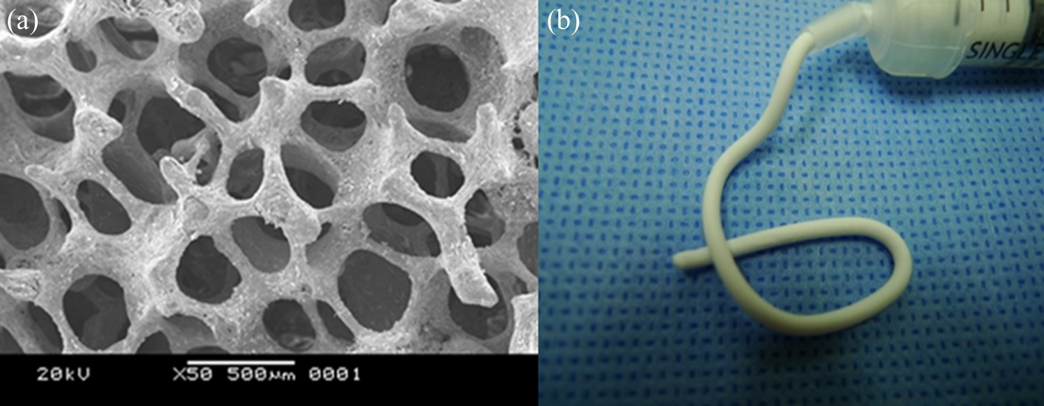
Scanning electron microscope image showing β-TCP granule with 100–300 μm pore size (a). β-TCP is prefilled in a syringe and ready for injection. The pictures are reproduced by courtesy of the manufacturer (b). β-TCP: beta-tricalcium phosphate.
Postoperative managements
Patients were encouraged to start passive range of knee motion and active quadriceps strengthening exercises the day after surgery with hinged knee brace protection. Partial weight bearing with crutches and brace was maintained for 4 weeks and after which full weight bearing was allowed as tolerated. First-generation cephalosporin was given within 1 h prior to the operation and continued until 2 days after surgery.
Postoperative radiographic evaluation
Postoperative mFTA was determined using full-length double-limb standing anteroposterior radiographs taken at 3 and 12 months after surgery. The degree of bone union and maturation of β-TCP was evaluated from knee standing anteroposterior radiographs using the modified van Hemert’s grade. 18 This rating represents grade 1 as a vascular phase with clear distinction between the β-TCP and bone; grade 2 as a calcification phase with blurred distinction; grade 3 as an osteoblastic phase with slightly visible distinction; grade 4 as a consolidation phase with no lucent signs despite recognizable osteotomy; and grade 5 as full reformation with no sign of osteotomy. We regarded grades 4 and 5 as bone union, which show no visible line between osteotomy and host bone. Computed tomography (CT) scans at 3 and 12 months after surgery were taken in order to evaluate serial change of β-TCP resorption and bony union progression. According to the method reported by Tanaka et al., 10 a CT image of osteotomy level was chosen and divided into areas of injected β-TCP and host bone. The CT attenuation values (in Hounsfield units (HU)) of each area were analyzed. Two investigators (BHK and WK) who were blinded to the study design independently evaluated all plain radiographic and CT measurements using picture archiving and communication system (PACS, Marosis, Infinity, Seoul, Republic of Korea), and interobserver reliability for each parameter was calculated.
Clinical evaluation
All patients were evaluated clinically before surgery and 12 months after surgery by using the International Knee Documentation Committee (IKDC) clinical evaluation form and the Western Ontario and McMaster Universities Arthritis Index (WOMAC). In addition, the visual analogue scale (VAS) for pain was recorded.
Statistical analysis
All continuous variables were tested for normality using the Kolmogorov–Smirnov test. We found interobserver reliability using intraclass correlation coefficient values ranging from 0.63 to 0.79 (Table 1). The difference of mFTA between 3 months and 12 months after surgery was determined using paired t-test. Change of the modified van Hemert’s grade and the CT attenuation values measured from postoperative 3 and 12 months and CT scans were compared using paired t-test. Also, the changes between preoperative and postoperative clinical scores including IKDC, WOMAC, and VAS for pain were compared using paired t-test. A p value < 0.05 was considered statistically significant. The statistical software MedCalc® (version 11.6; MedCalc Software, Mariakerke, Belgium) and R (version 2.12; Comprehensive R Archive Network, Boston, Massachusetts, USA) were used for all statistical analysis.
Interobserver reliabilities for radiographic measurements.
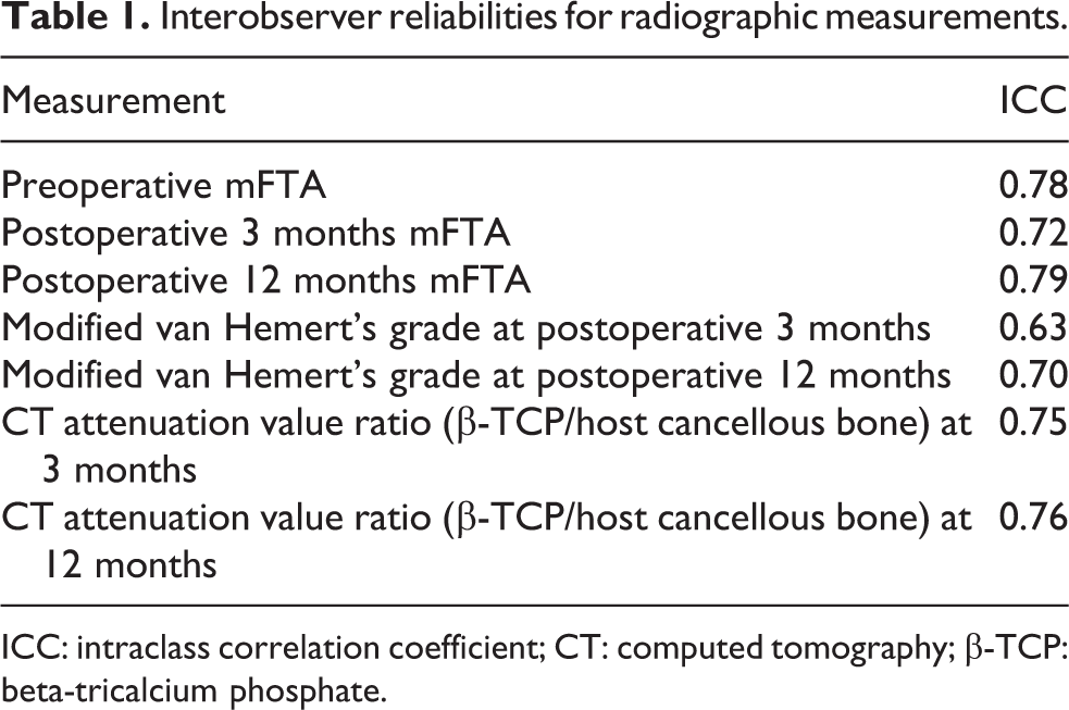
ICC: intraclass correlation coefficient; CT: computed tomography; β-TCP: beta-tricalcium phosphate.
Results
A total of 34 patients were assessed for eligibility and 6 patients who did not meet the inclusion criteria were excluded. Total of 28 patients were initially enrolled in this study; however, 3 patients (10.7%) did not take either postoperative 3 or 12 month-CT scan. Therefore, 25 patients (21 women and 4 men) were included in the final analysis. The average age of the patients was 58 years old (range 49–67 years old) and average BMI was 25.8 (range 21.9–31.1). During the follow-up period, there was no perioperative complication including wound problem, infection, or delayed union.
Bone union and maturation of β-TCP evaluated by the modified van Hemert’s grade showed progression from postoperative 3 months to 12 months in all cases (Table 2; Figure 2). The modified van Hemert’s grade at postoperative 3 months (2.3 ± 0.9, range 1–4) significantly changed at postoperative 12 months (4.4 ± 0.7, range 3–5; p < 0.001). The mean ratio (β-TCP/host cancellous bone) of CT attenuation values significantly changed from postoperative 3 months to 12 months (p = 0.012; Table 3; Figure 3). Also maturation of β-TCP and bone union was observed from the coronal view of CT scans (Figure 4). Preoperative mFTA (average 4.1° varus) was significantly different from postoperative 3 months (average 4.8° valgus) and 12 months mFTA (average 4.3° valgus; p < 0.001), while no difference was found between postoperative 3 months and 12 months (p = 0.075). Clinical outcome scores including the IKDC, WOMAC, and VAS pain scores significantly improved after surgery (p < 0.001, p = 0.002, and p = 0.002, respectively; Figure 5).
Progression of bone union and maturation of β-TCP between postoperative 3 and 12 months evaluated using the modified van Hemert’s grade.

β-TCP: beta-tricalcium phosphate.

Anteroposterior radiograph of 53-year-old female taken at postoperative 3 months (a) and 12 months (b). Bone union and maturation of β-TCP was evaluated using the modified van Hemert’s grade. Blurred distinction between osteotomy line and β-TCP was seen (black arrow) and defined as grade 2 (a). Full reformation of β-TCP was seen and defined as grade 5 (b). β-TCP: beta-tricalcium phosphate.
Change of the CT attenuation values (HU) and ratio (β-TCP/host cancellous bone) of CT attenuation values between postoperative 3 and 12 months.a
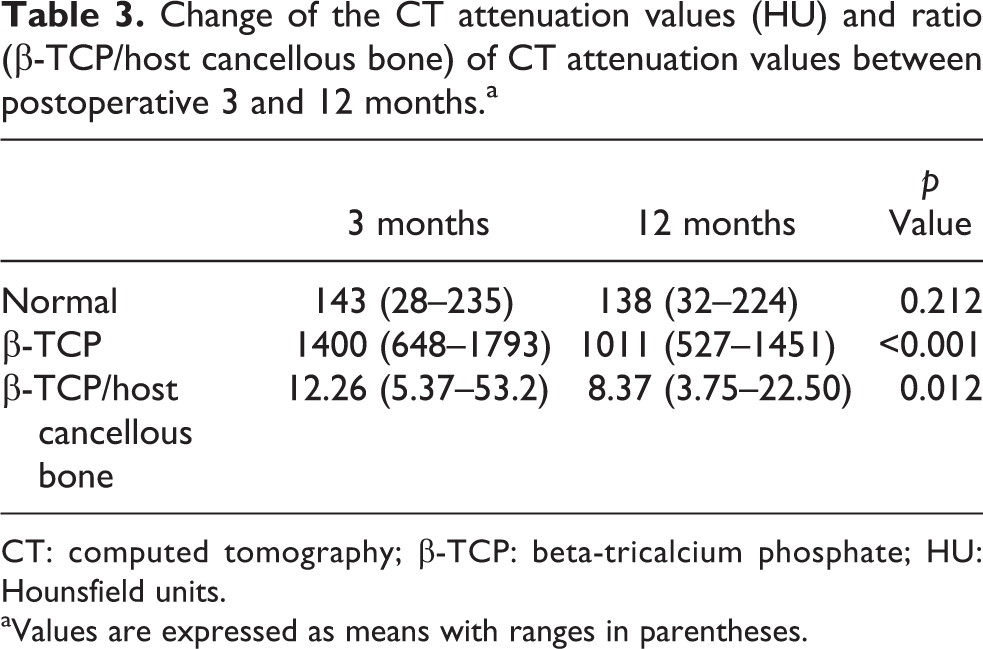
CT: computed tomography; β-TCP: beta-tricalcium phosphate; HU: Hounsfield units.
aValues are expressed as means with ranges in parentheses.

CT images of 53-year-old woman showing the center of the osteotomy plane at postoperative 3 months (a) and 12 months (b). The CT attenuation values (in HU) of the host cancellous bone area (a′) and the injected β-TCP area (b′) changed from 89 and 1327 at 3 months to 74 and 666 at 12 months. The ratio (β-TCP/host cancellous bone) of CT attenuation values was 0.067 (3 months) and 0.111 (12 months). CT: computed tomography; β-TCP: beta-tricalcium phosphate; HU: Hounsfield units.
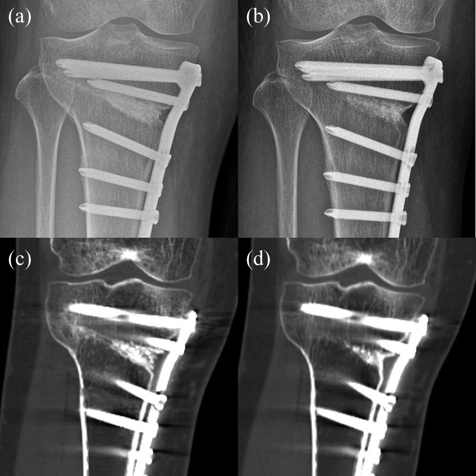
Images of 56-year-old woman showing the modified van Hemert’s grade 3 of bone union and maturation of β-TCP at postoperative 3 months plain radiography (a) and grade 4 bone union with maturation of β-TCP at postoperative 12 months (b). Progression of bone union and resorption of β-TCP is also visible on corresponding CT coronal images taken at postoperative 3 months (c) and 12 months (d). CT: computed tomography; β-TCP: beta-tricalcium phosphate.

Changes of alignment and clinical outcome scores. mFTA) (a), IKDC (b), WOMAC (c), and VAS for pain scores (d). Paired t-tests were done. mFTA: mechanical femoro-tibial angle; IKDC: International Knee Documentation Committee; WOMAC: Western Ontario and McMaster Universities Arthritis Index; VAS: visual analogue scale.
Discussion
The main aim of this study was to evaluate the safety and efficacy for gap healing of β-TCP injection after MOWHTO. Our initial hypothesis was confirmed that MOWHTO using the injectable β-TCP resulted in gap healing without complication. Also, radiographic and clinical outcomes of MOWHTO using injectable β-TCP as gap filler were satisfactory. Serial radiographs showed satisfactory bone union and maturation of β-TCP and serial CT scans showed decreased ratio (β-TCP/host cancellous bone) of CT attenuation values that suggests resorption of β-TCP. Our findings concur with previous reports that showed maturation of graft with bone union after MOWHTO using β-TCP. 7,9,10,18,19
The need for any spacer or filler to fill the osteotomy gap is an issue still on debate. Various studies reported successful clinical and radiographic outcome after MOWHTO without any spacer. 20,21 In contrast, problems including early loss of correction, delayed bone union, and failure of fixative also have been reported. 4,22 In general, autogenous bone graft is considered as the most certain filling material; however, increased operative time or donor site morbidity is not a minor problem. Allogenic bone grafting also is not free from problems like infection or disease transmission. Bone substitutes are used as alternatives in expectation of enhancing initial mechanical stability and shortening bone healing time without additional donor site issue. The β-TCP or hydroxyapatite (HA) is representative among various bone substitutes, and successful results using β-TCP or HA to fill the osteotomy gap in MOWHTO have been reported. 5,6,23 –25 Both β-TCP and HA are known to be biocompatible and have osteoconductive potential; however, several studies reported that β-TCP has relatively superior resorption rate and osteoconductivity compared to HA. 9,11,26,27 The reason for this has been suggested that β-TCP has micropores which provide microenvironment for osteoblastic cell formation. 26,28 The absorbability of β-TCP varies according to the porosity that bone formation and resorption were superior with β-TCP of 75% porosity than 60% porosity. 10,19 In our study, we used β-TCP with 75% porosity, and bone formation and resorption at postoperative 12 months were satisfactory.
Solid bone substitute wedge is generally used for spacer, since it is easy to handle and might add initial mechanical stability. However, wedge filler covers only part of the osteotomy gap since it is pre-shaped. Also, using solid bone substitute may prevent hematoma leakage which is a mechanism proposed to enhance bone healing and may delay bone ingrowth due to dense structure. 7 Meanwhile, using granule-type bone substitute has theoretical advantage that it may cover a large area of cancellous bone in the open-wedge gap and provide a loose matrix for bone ingrowth. On the other hand, concern remains that granule-type bone substitutes can only provide weak mechanical support. Since the advent of anatomical locking plate enabled rigid fixation and decreased the concern of early collapse after MOWHTO, 29 we supposed that using injectable β-TCP granule combined with locking plate would have benefits. In addition, the injectable β-TCP we used in this study was relatively easy to handle and possible to adjust the shape during the surgery. One thing should be noted that injectable filler may extensively cover the osteotomy surface which may cause limited hematoma formation and delayed bone union. Fortunately, uneventful bony union was achieved in this case series and we assume that higher porosity of the injectable β-TCP granule might have minimized this problem. Concerns about infection using synthetic bone substitutes exist and a systematic review reported variable ranges (0–2.8%) of postoperative infection that required revision after MOWHTO with the use of synthetic grafts. 30 It is possible that the rate of minor infection or wound problem may be higher. Fortunately, neither superficial nor deep infection occurred in our series, and we suppose that we could avoid this problem since nonsmoker, nondiabetic, and relatively low BMI patients were included.
This study has several limitations. First, this study is a case series without any control group, so we could not compare whether using β-TCP has any advantage over other gap filler or osteotomy without filling material. Our finding only suggests that β-TCP is safe with satisfactory bone union as gap filler after MOWHTO. Second, three patients who failed to take either postoperative 3 or 12 months CT were excluded from the analysis. Although CT data were not available, those three patients were not lost from the follow-up and bony union was achieved uneventfully after postoperative 12 months. Third, longer term effect of injected β-TCP on maintaining the correction angle and functional outcome is uncertain, even though early clinical outcome and bone union rate were satisfactory. Finally, although different sizes of osteotomy gap were needed for each case, we injected same amount of β-TCP. There is possibility that we might have injected excessive β-TCP and vice versa. However, deciding whether adequate amount of β-TCP depend on the size of osteotomy gap was beyond the scope of our study.
Conclusion
We found gap healing without any complication and satisfactory clinical outcome after using injectable β-TCP as gap filler for MOWHTO. We suppose that injectable β-TCP has theoretical merits including superior bone formation, earlier resorption, and operating convenience. Therefore, it can be an effective option as gap filler for MOWHTO when combined with rigid locking plate fixation.
Footnotes
Declaration of conflicting interests
The author(s) declared no potential conflicts of interest with respect to the research, authorship, and/or publication of this article.
Funding
The author(s) received no financial support for the research, authorship, and/or publication of this article.