
Abstract
Face masks are an effective protection tool to prevent bacterial and viral transmission. However, commercial face masks contain filters made of materials that are not capable of inactivating either SARS-CoV-2. In this regard, we report the development of an antiviral coating of polyurethane and Copper nanoparticles on a face mask filter fabricated with a spray technology that is capable of inactivating more than 99% of SARS-CoV-2 particles in 30 min of contact.
Introduction
Severe acute respiratory syndrome coronavirus 2 (SARS-CoV-2), disease termed as (COVID-19), has involved the whole world population. The potential transmission pathways, via droplet and aerosol, prompted to finding measures to prevent and contain the spread of the disease. Therefore, it is imperative to detect devices that are required to block respiratory secretion and interrupt the chain of transmission. Despite advances in pharmacological treatment and vaccine development, wearing facial masks reducing transmission of the virus by healthcare workers and the public alike remains a recommended prophylactic measure to halt SARS-CoV-2 transmission. Commercial face masks contain filters that are usually made of materials unable to inactivate either viruses like SARS-CoV-2 or multidrug-resistant bacteria. Therefore, the development of antimicrobial surface coating on facial masks or filters would represent a significant leap forward against SARS-CoV-2. In the continuous emergence of fast-spreading SARS-CoV-2 variants, it is crucial to implement measures to decrease person-to-person transmission in ordinary populations and healthcare personnel through personal safety devices. Therefore, safer and more effective personal protective equipment (PPE) will be necessary and the development of the antiviral coating for the surface of objects that are frequently used by the public could be a practical route to prevent the spread of the viral particles and inactivation of the transmission of the viruses. 1 Van Doremalen et al. 2 assessed the viability of SARS-CoV-2 in varied environmental conditions and Copper was found to be efficacious in inactivating the virus in a shorter time. In particular, Grass et al. 3 proposed that the antiviral and antimicrobial abilities of Cu are due to four processes: cell damage, the cellular membrane loses its integrity and its contents, ROS formation, and that the genetic material undergoes degradation. Ingle et al. 4 showed that metallic nanoparticles (NPs) had a broad-spectrum antimicrobial activity due to their small size and high surface to volume ratio. Further, many NP types, such as silver, 5 copper iodide, 6 and titanium dioxide, 7 interacting with the bacterial cell membrane can inactivate bacterial cells and norovirus surrogates causing release of intracellular substances and cell death, 8 crippling the viral capsid possibly by denaturing its proteins. Few reports are available on antiviral activity of copper nanoparticles, which confirms that copper nanoparticles also possess promising antiviral activity. Fujimori et al. 9 claimed that these nanoparticles can be useful for protection against viral attacks and may have novel applications for the development of filters, face masks, protective clothing, and kitchen cloths. Face mask has been considered an effective protective tool capable to blocking the passage of viral and bacterial particles. However, the addition of an antiviral substances to surface of face mask could increase significantly the protection of this tools.
This paper aimed to create a single use filter for face masks with antiviral properties based on polymer and copper NPs applied by spray technology to increase the effectiveness of PPE against SARS-CoV-2.
Methods
Copper nanoparticles (CuNP) synthesis
Three classes of CuNPs differing in diameter sizes were tested and used. Two of these classes, diameters 25 and 60–80 nm, were purchased as being commercially accessible (Io-li-tec, Germany) and generated by laser ablation in liquid. Because the generation of NPs by laser ablation is uncapable to obtain NPs with diameters lower than 25 nm, the third class of CuNPs with diameters of approximately 4–6 nm were synthesized in our laboratory by using CuCl2·2H2O (Sinopharm Chemical Reagent Co., Ltd) and L-ascorbic acid (Sinopharm Chemical Reagent Co., Ltd) as previously described. 10
All the three classes of CuNPs dispersed in water were subsequently characterized by UV-Vis absorption spectra making use of a Jasco V-550 spectrophotometer. In turn, Raman spectroscopy (DXR3xi, Thermo Fisher) was used to further characterize surface-enhanced Raman scattering (SERS) properties of CuNPs.
Coating based on CuNP and polyurethane by spray technology
A medical-grade poly(ether)urethane was dissolved in a mix of Tetrahydrofuran and dioxane a 1:1 (v/v) at a concentration of 0.5% (w/v) (Sigma-Aldrich; St Louis, MO, USA). We added 1% of CuNP after an ultrasonic dispersion of nanoparticles performed at 70°C for 10 min. The solution was sprayed on a rotating mandrel where a filter for face mask has been placed, through a spray gun mounted onto a sliding carriage.
The coating was fabricated using specific distance between the rotating mandrel and the spray guns, mandrel rotating speed, carriage speed, flow rate, and pressure as previously described. 11
At the end of the process, the mandrel with the deposited material was dried at 40°C for 3 h, cut in a circular shape with a diameter of 1.5 cm, placed in a 24 well, and were exposed to UV light in a laminar flow cabinet integrated with a UV light source for 2 h.
SEM analysis
The morphology of the filter no treated, treated with polyurethane (PU) and treated with polyurethane and CuNP (PU-CuNP) was observed using a scanning electron microscope (FlexSEM 1000, Hitachi, Tokyo, Japan). The accelerating voltage was maintained at 5 kV with magnification of 500, 1000, 2000, 3000×. The filter samples were prepared to be conductive by gold coating with a sputter coating unit.
Antiviral activity of coatings containing copper
Cells and viruses
African green monkey kidney cells (Vero-E6) were grown as described previously. 12
A clinical isolate of SARS-CoV-2, kindly gifted from the San Raffaele Hospital (Milan, Italy) and propagated in Vero-E6 cells, was used for all experiments. All SARS-CoV-2 experiments were performed at a Biosafety Level 3 laboratory.
The propagation of SARS-CoV-2 was performed in Vero-E6 cells as described by Storti et al. 12
Virus titration by limited dilution
Viral titer was calculated from the CPE induced through limited dilution of the viral stock. Briefly, 104 Vero-E6 cells were seeded in a 96-well plate and incubated overnight (ON) at standard conditions. The day after, cells were infected in eightfold replicas with 50 µl SARS-CoV-2 at 10 several concentrations obtained by serial 1:10 dilution of the stock (from D0 to D10) and incubated 1 h at 37°C and 5% CO2.
The Spearman-Karber method has been used to calculate the viral titer using and expressed by Median Tissue Culture Infectious Dose tissue/ml (TCID50/ml). The titer of virus stock was 108 TCID50/ml.
Cytotoxicity assay
The cytotoxic effect of filter coated with polyurethane and CuNP on Vero E6 cells was evaluated using Alamar Blue (Sigma-Aldrich, Milan, Italy). To avoid contamination by bacteria, mold, or other microbes possibly present on metal surfaces, disks were previously soaked in 70% Ethanol at room temperature for 30 min and air dried under a BSL2 cabinet hood. Briefly, pre-cut disks of 14 mm diameter of filter PU and PU-CuNP, were soaked in 200 μl DMEM with no serum for 1 h at room temperature. Two disks per material were examined pooled and added undiluted to a 96-well plate seeded with 5 × 10⁴ Vero-E6/well the day before and cultured in DMEM added with 10% of FBS. Tests were performed using eight replicas for each material. 10 µl/well Alamar Blue were added after 48 h incubation, and plates were incubated for two additional hours. Fluorescence was measured at 590 nm as previously described. 13
Antiviral activity assay
A limiting dilution method was carried out with Vero-E6. Briefly, filter disks of 14 mm diameter treated with polyurethane and polyurethane with CuNPs were placed in a 24-well plate. For each experiment four disks were used: two were soaked with the titered virus supernatant, and two with fresh medium. Titered viral supernatant of SARS-COV-2 was diluted in medium with no serum to reach 6 × 10⁴TCID50/ml; 200 μl of this viral suspension or DMEM alone were added to each disk. After incubation at room temperature for 0, 5, 10, 30, and 60 min, viral supernatants and medium were collected, pooled, and used as such or diluted 1:10 up to 1:1000 (dilutions 50–500 μl in DMEM with no serum). Fifty microliter of each sample were added in triplicate to a 96 well plate seeded with 104 Vero-E6 cells/well. Plates were prepared the day before and cells were cultivated in 150 μl DMEM 12.5% serum/well. Positive (e.g. viral supernatant) and negative (medium alone) controls were similarly diluted and added to the plate in triplicate as such or 10-fold diluted. Plates were incubated at 37°C in a 5% humid atmosphere for 4 h after which the medium was replaced with fresh DMEM 10% serum. Plates were incubated for additional 3–4 days until CPE was visible. The presence and extent of CPE were determined by examining the cells with an inverted microscope and the well was scored as virus positive when 75% of cells were lysed. The reduction of infectious viral load was inferred using the Reed and Muench formula. The antiviral activity was assayed in three independent experiments and using two batches of samples.
Results
Fabrication of coating with CuNP and polyurethane
We obtained a coating with 1.5 mg/cm2 of CuNP using the spray deposition technique described above. The stability of NP dispersion is the key factor for their application. In this study, we thus used CuNP with a diameter of 25 nm for coating by spray technique because the NP of 60–80 and 4–6 nm showed practical problems with poor dispersion of nanoparticles into the polymer matrix during preparation solution and spray process.
SEM analysis
Figure 1 shows the SEM images from the filter before (no treatment) and after coating with polyurethane alone (PU filter) and with polyurethane-CuNP (PU-CuNP filter) using spray technology. The morphology of the coated filter mask was clearly different from the smooth surface of the untreated filter and showed the CuNPs were uniformly distributed and attached to the rough surface all over the fibers of the filter.
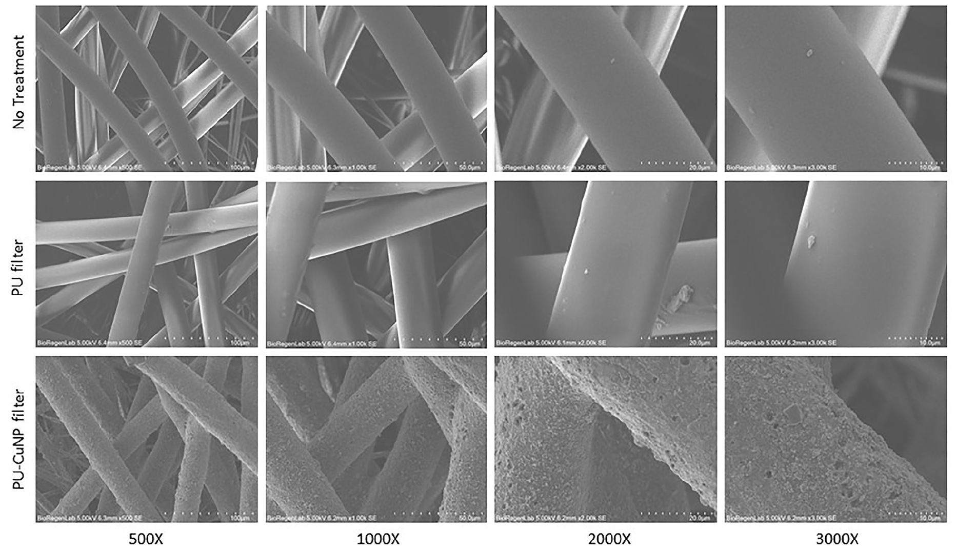
SEM images of filter no treated, treated with PU, and treated with PU and CuNP at 500, 1000, 2000, and 3000× magnifications.
Antiviral activity against SARS-CoV-2
Analysis of antiviral activity was preceded by several experiments aimed to determine the cytotoxicity of the filters coated with polyurethane and CuNP. Our data showed that filters do not release harmful substances into the culture medium and have no detrimental effects on the cells.
Antiviral tests were set in such a way to test in parallel the same viral preparation exposed at various times to the filters and then incubated with the cells until the CPE was overtly visible by microscope reading or replaced after 4 h with fresh medium. Since replacement of the medium did not alter significantly viral infectivity we used 3000 TCID50 as viral input dose, an amount that permitted precise evaluation of the antiviral activity of testing material.
The antiviral activity was assayed by the limiting dilution method in three independent experiments and using two batches of filters PU and filter PU-CuNP. Table 1 shows the residual SARS-CoV-2 infectious dose following contact with filter coated with PU and CuNP (PU-CuNP filter) expressed as reduction of TCID50 and log TCID50.
Residual SARS-CoV-2 infectious dose following contact with filter coated with PU and CuNP.

CuNP: copper nanoparticle; PU: polyurethane; TCID50: Medium Tissue Culture Infectious Dose 50%/ml.
Filter Cu-NP showed a strong virucidal activity that increased proportionally with the time of contact with the materials. As shown in Table 1, the reduction was noticeable since the first time examined, 5 min, increased three times with only five further minutes of incubation and abated 99% viral infectivity in 30 min. At 1-h contact, the virus was inactivated to a point that CPE was observed in only 1/18 wells. A similar result was obtained with the positive control virus diluted 1:1000 (CPE observed in 3/18 wells). In all, this indicated that Copper abates viral infectivity over three logs in 1 h of contact. Figure 2 shows the time-dependent reduction of viral titer with PU and with PU-CuNP filters. The reduction of SARS-CoV-2 titer after incubation with CuNP was very high in the first 5 min and then slowly decreased as shown by the reduction of the slope (Figure 2).

Reduction of viral titer after incubation with filter treated with CuNP and PU. The reduction of the SARS-CoV-2 titer was shown after incubation with a filter treated with CuNP and PU at different time points. After only 5 min of incubation, the effect produced by CuNP is relevant and decreases during the time.
Discussion
An antiviral coating on personal protective equipment such as a face mask could ensure better protection. In this regard, we developed an antiviral coating based on CuNP and PU on a filter for a surgical mask with spray technology.
This coating showed a good virucidal effect against SARS-CoV-2 on a nice and consistent time-dependent scale, eliminating over 99% viral infectivity in 30 min. Such coating could therefore provide antiviral protection on surfaces and materials in healthcare settings. These findings agree with what already reported on CoV-229E in 2015 14 where the inactivation rate was directly proportional to percentage of copper and more recently Hewawaduge et al. 15 demonstrated a SARS-CoV-2 inactivation induced by copper sulfide (CuS) incorporated three-layer mask design in a short period of exposure time. In addition, Jung et al. 16 presented the development of a copper-coated polypropylene filter face mask with antiviral capability against SARS-CoV-2 and Behzadinasab et al. 17 fabricated and tested a coating consists of cuprous oxide (Cu2O) particles bound with polyurethane that rapidly inactivates SARS-CoV-2.
Therefore, several antiviral face mask materials against SARS-CoV-2 have been recently proposed but all made with expensive materials such as graphene, 18 copper, 19 or silver. 20 Moreover, these antiviral composites are manufactured with complex and costly processes, which make them inapplicable and unsustainable during a global pandemic. More recently, Zuniga and Cortes 21 reviewed several research studies and showed that Copper is a helpful and low-cost material to reduce the transmission of several infectious diseases and limit nosocomial infectious transmission. CuNPs could be an inexpensive material that has demonstrated rapid and high microbicidal efficacy against various pathogens and spray-technology could be a rapid, continuous, cost-effective, reproducible, and scalable process.
Here, we proposed a practical and feasible large-scale preparation method to develop antiviral coating based on CuNP and PU on a filter for surgical masks using a spray technology.
However, further studies are warranted to guarantee the safety and correct use of this technology for a mass production and commercialization of this antiviral coating for facemask filter. In this regard, it is important to evaluate NP safety to achieve biocompatibility and maintain the desired activity and to assess the coating biostability for a longer period. Combining metal NPs (the copper ones in the first instance) and polymer matrices could help to tune metal release properties, and at the same time to reduce the risk of NP release into the environment. Kumar et al. 19 demonstrated that, in the absence of polymer structure, the amount of CuNP release from the face mask surface is higher than in the presence of polymer in which the CuNP remained quite stable.
Conclusion
In this study, we developed, by spray technology, a coating based on CuNP and PU with strong virucidal activity against SARS- CoV-2 that could be used to produce antiviral masks effectively preventing SARS-CoV-2 spread and halting the unrelenting chain of transmission still observed nowadays.
From these findings and considering that pandemics occur from time to time and respiratory viruses actively circulate during most time of the year, an antiviral coating could be helpful in the development of personal protection equipment with improved shielding properties and improving the safety of common touch surface. Finally, this strong virucidal coating can be used in a wide field of applications.
Footnotes
Guarantor
PL.
Author contributions
MP, IF, PL, and GS researched literature and conceived the study. PL, TAK, PQ, AC, GP, and MDA were involved in protocol development and data analysis. IF wrote the first draft of the manuscript. All authors reviewed and edited the manuscript and approved the final version of the manuscript.
Declaration of conflicting interests
The author(s) declared no potential conflicts of interest with respect to the research, authorship, and/or publication of this article.
Funding
The author(s) received no financial support for the research, authorship, and/or publication of this article.