
Abstract
Aim:
Incomplete polymerization of a resin material used for dental restoration affects the properties of the restoration. We evaluated the structural and mechanical properties of three different colored compomers cured with three different LED units to observe the characteristic changes occurring in different matches.
Methods:
Polytetrafluoroethylene molds (5 mm in diameter and 2 mm in thickness) were used to prepare disk-shaped sample specimens. Nine sample groups (each of five specimens) were prepared, three each of different compomer resin colors (gold, berry, and silver). Samples were cured using three different LED units (Optima, VALO, and Demi Ultra), according to the manufacturers’ instructions. Microstructural properties of samples were characterized by determining the degree of curing using Fourier-transform infrared spectroscopy and by analyzing sample morphology under a scanning electron microscope. The Vickers hardness, compressive strength, and elastic modulus of the samples were measured to investigate their mechanical properties.
Results:
The degrees of curing decreased in the order of silver > berry > gold for all curing units. Conversely, gold compomers exhibited poorer mechanical values than the berry and silver counterparts. The Optima 10 unit yielded slightly higher degrees of curing than the other devices, followed by Demi Ultra and VALO light-curing units, respectively.
Conclusion:
The resin color affected the structural and mechanical properties of the compomers, possibly as a result of the complex interactions and relationships between the irradiation light and resin material, such as light absorbance and reflectance; thus, depending on the color as well as the curing protocol.
Keywords
Introduction
Compomers are light-cured restorative materials with the ability of fluoride release, and were developed by combining the advantages of glass ionomers and composites. 1 They may be used extensively in restorations of anterior and posterior primary teeth, owing to their tooth-colored appearance. However, by adding a small amount of glittery particles to conventional compomer resin, compomers of various colors have also been produced to overcome numerous challenging shrinkage stress factors, and have the advantage that they are attractive to children. 2 Additionally, colored compomers have been used as fissure sealants in children’s dentistry, owing to their easy application, improved retention, and colorful appearance.3, 4
Compomers, like composite resins, include photoinitiators, such as camphorquinone, which makes it essential to use a suitable light-curing unit with a light absorption peak at 470 nm. 5 Photo-polymerization is based on the idea that all monomers in resins are converted into a polymeric matrix during the light-curing process. Although the use of curing devices with high light intensities results in a greater degree of monomer conversion, numerous residual carbon double bonds remain in the final product of all dimethacrylate-based materials. This incomplete polymerization of resin materials causes leaching of monomers, which affects the biocompatibility of the material, damages the pulp, and decreases the mechanical properties of a restoration.6–8 Thus, the process of polymerization has been extensively studied for many years.9–11 The general strategy for increasing success is mainly based on the synthesis of various resins and initiator systems using different chemical structures, as well as on improving the curing ability of polymerization devices.
There are various sources for polymerization of light-curing dental resin restorative materials. The most common light-curing units are quartz-tungsten-halogen units with 400–800 mW/cm2 output light intensity. Despite the advantage of their low cost, there are some disadvantages, such as limited curing depth, long curing time, and decreasing output light intensity over time. To eliminate these negative characteristics, LED light-curing units were introduced, owing to their high light output intensities. LED light-curing units are small wireless devices with the strongest photo-polymerization effects. There are several systems of LED light-curing unit with different advantages, such as fast-curing action, uniform depth of cure, and low temperature.
In addition to the wavelength and intensity of light used, the degree of polymerization may be affected by many factors, such as the type and amount of monomers, fillers, and initiators or catalysts, as well as the shade and translucency of the resin material. As previously reported, darker shades of composite resins reduced the degree of curing, which could cause a reduction in the hardness of the material, 12 while the color of compomer resin materials, which is related to the content of pigments and fillers, may also affect the transmission of incident light, and thus the resulting degree of polymerization.
According to the manufacturers, formulations of different colored compomers are similar to conventional compomers, except for the added pigments. However, only a few studies have been published so far on the physical and mechanical properties of colored compomers,13–15 showing that the properties of different colored compomers after polymerization with different light-curing units change because of wavelength distribution and glittering effects.16, 17
Therefore, in this study, we aimed to evaluate the structural and mechanical properties of three different colored compomers cured with three different LED light-curing units by analyzing the degree of curing, surface microhardness, compressive strength, and elastic modulus of the samples using various test methods.
Methods
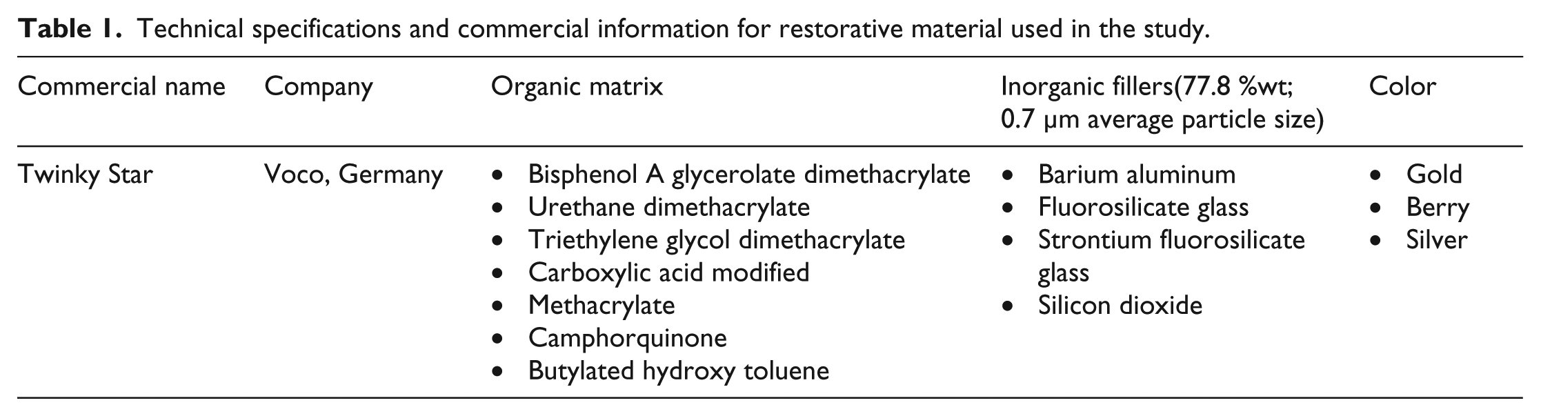
A commercial resin compomer (Twinky Star, VOCO, Cuxhaven, Germany) with three different colors; gold, berry and silver (tooth-colored) were used in this study. Some technical specifications and commercial and compositional information about the restorative material are given in Table 1.
Technical specifications and commercial information for restorative material used in the study.
Specimen preparation
As schematized in Figure 1, nine sample groups were prepared, three each of three resin colors (gold, berry, and silver). Each group consisted of five specimens. This classification enabled us to study the structural and mechanical properties of the compomers, depending on both resin color and curing conditions. Firstly, disk-shaped specimens were prepared using polytetrafluoroethylene molds, which were 5 mm in diameter and 2 mm in depth, which is the most frequently suggested depth for incremental buildup and curing of a dental resin using light-curing units.16, 18 Subsequent to covering the unpolymerized resins with Mylar strips, they were polymerized using three different LED light-curing units. The curing protocols were applied based on the procedures suggested in their manufacturers’ guides, which are henceforth denoted as 1, 2, and 3, respectively.
Optima (Optima 10, BA International, Kingsthorpe, Northampton, UK) was applied for 20 s in full power mode (1200 mW/cm2).
VALO (Ultradent, South Jordan, USA) was applied twice for 3 s each time in Xtra power mode (3200 mW/cm2).
Demi Ultra (Kerr Corporation, CA, USA) was applied for 10 s (1100–1330 mW/cm2).
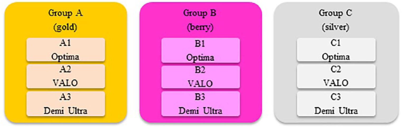
Sample groups; nine sample groups were prepared, using three resin colors and three LED light-curing units.
Microstructural characterization
Microstructural characterization of the samples was carried out using Fourier-transform infrared spectroscopy (FTIR) and scanning electron microscopy (SEM). 19 The FTIR spectra of samples were recorded in transmission mode using a Bruker Alpha infrared spectrometer with an attenuated total reflectance device and a germanium crystal within a wavenumber range of 400–4000 cm−1 with a resolution of 2 cm−1 from 32 scans. The spectra were analyzed using OPUS and Origin v. 8.5 software to calculate the degree of curing and quantify the cross-linking reactions as a function of the curing conditions. In this study, characteristic absorption peak areas were used to calculate the degree of curing, as described in equation (1) 20 :
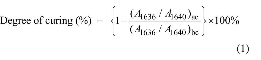
According to this equation, the characteristic absorption peaks were at 1636 cm−1 (unsaturated aliphatic C=C double bonds originated from methacrylate groups) and 1604 cm−1 (aromatic C=C double bonds). 21 The term A represents the intensity of a particular absorption peak, “ac” denotes “after curing” and “bc” denotes “before curing”.
The morphological features of the samples were characterized by SEM, which was done by imaging the samples in a field emission scanning electron microscope (FEI Quanta FEG 450, USA), and after proper sample preparation, in which the samples were sputter-coated with gold.
Mechanical properties
The mechanical properties of the samples were characterized by surface microhardness measurements and compression tests.
The Vickers hardness number was measured (HMV M-1, Shimadzu Corp., Kyoto, Japan) from the top and bottom surfaces of a total of 45 samples (five specimens in each group), which were polished using 400, 800, 1000, 1500, 2000, and 2500 grit silicon carbide papers. Then, five indentations, with a constant load of 100 g for 10 s, were inflicted on each surface, one in the center and one in each quadrant (>100 µm from each other). The average of the results was reported as the Vickers hardness number.
A universal tension–compression test machine (BWB-20, KokBir, Turkey) was used in compression mode to test the mechanical properties. In the compression tests, the cylindrical specimens, which were 5 mm in diameter and 2 mm thick, were used, based on the ISO/DIN 4049:2009 standard with cross-head or compression speed of 0.2 mm/minute. Three specimens were tested in the compression tests; mean values with standard deviations are reported.
Statistical analysis
Prior to statistical analysis, the normality and equal variance of the data were tested. Because the datasets were found to be normally distributed, one-way analysis of variance (ANOVA) and Tukey’s multiple comparisons tests were performed to analyze data using SPSS 20 software. Statistical significance was considered at p < 0.05.
Results
Microstructural characterization by FTIR and SEM analysis
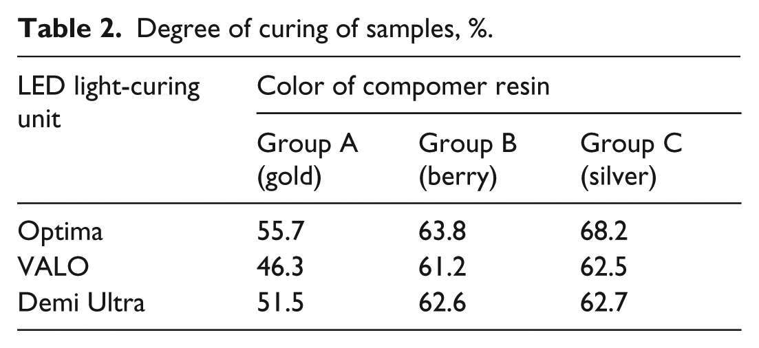
The FTIR spectra of the uncured and cured compomer specimens were obtained for a narrow range of wavenumbers (1570–1800 cm−1); the given part of the spectrum in this range was fitted with multi-peaks using the Gaussian non-linear peak deconvolution function of OriginPro 8.0 software, and the area under each corresponding peak was then calculated. According to the degrees of curing of the samples (Table 2), the Optima light-curing unit yielded slightly higher degrees of curing than the other devices, followed by Demi Ultra and VALO. Additionally, the degrees of curing of the compomers with different colors decreased in the order of silver > berry > gold for all LED light-curing units.
Degree of curing of samples, %.
As an example, the FTIR spectra of the uncured compomer specimens and those cured using the Demi Ultra LED light-curing unit are given in Figure 2(a) and 2(b), respectively. The scanning electron micrographs of the fractured cross-sections of the A3 (gold), B3 (berry), and C3 (silver) samples cured using the Demi Ultra LED light-curing unit are shown in Figure 3. The imaging magnification was ×20,000 for sample A3, while it was ×10,000 for samples B3 and C3. It can be clearly seen in these images that the filler particles were homogenously dispersed in the resin matrix. However, it may be inferred that the interfacial adhesion or interaction between the filler and resin matrix was greater in the C3 sample than in the other two specimens. The average size of the large filler particles embedded in the resin phase, as seen in the micrographs of B3 and C3 were about 4–7 µm. However, much smaller, irregularly shaped, particles, appeared. Figure 4 compares the cracking cross-sections of the A3 and B3 samples by illustrating the micrographs taken at a magnification of ×1000. As seen in the micrograph of B3, the top surfaces of the specimens were quite smooth, and no roughness or extra surface cracks can be seen. The A3 sample exhibited a layered structure, whereas the fractured cross-section of B3 was more uniform.
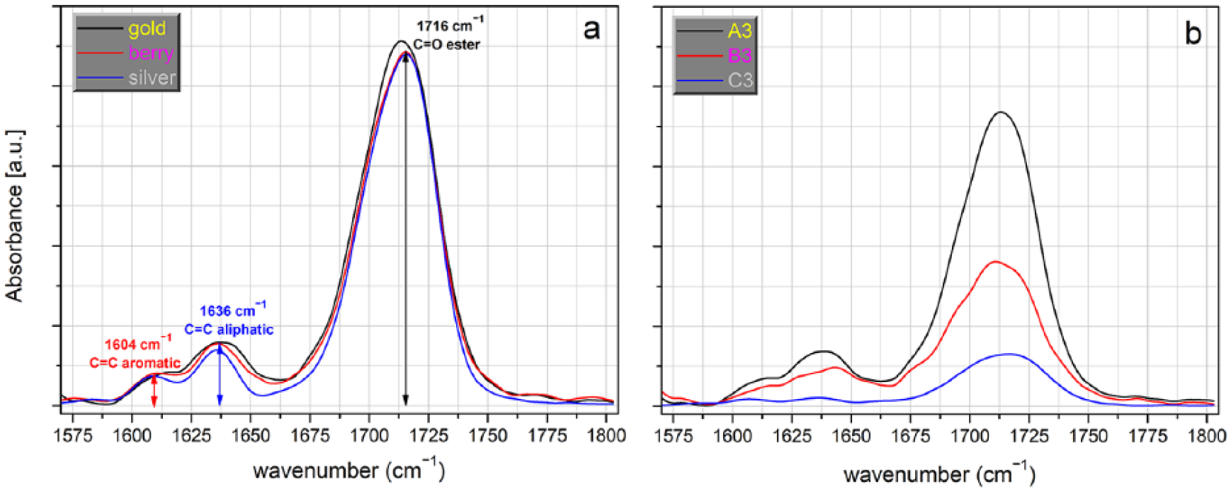
FTIR spectrum of (a) monomers and (b) samples cured with the Demi Ultra light-curing unit at 2 mm (1570–1800 cm−1).
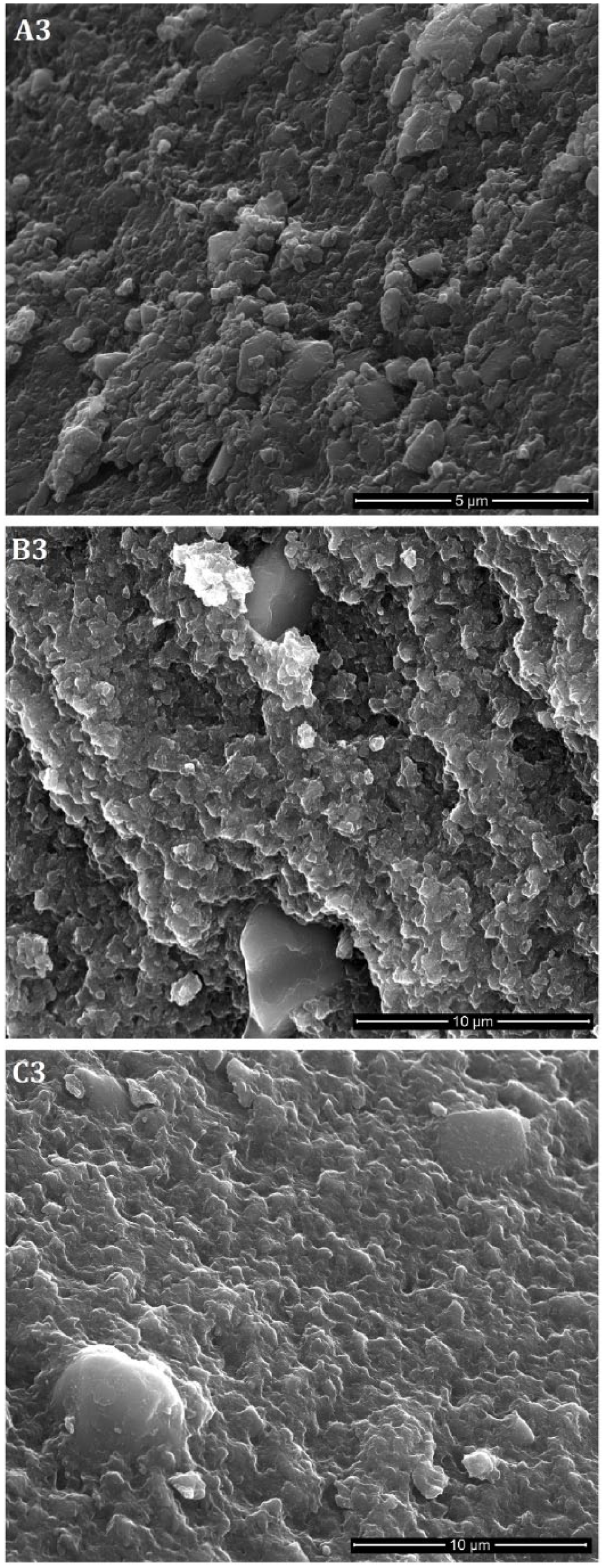
Representative scanning electron micrographs of fractured cross-sections of A3 (gold), B3 (berry), and C3 (silver) samples cured with Demi Ultra light-curing unit. The imaging magnitude was ×20.000 for the A3 sample and ×10.000 for the B3 and C3 samples. Homogenously dispersed filler particles are seen in the resin matrix for all samples; their average sizes measured about 4–7 µm in the B3 and C3 samples.
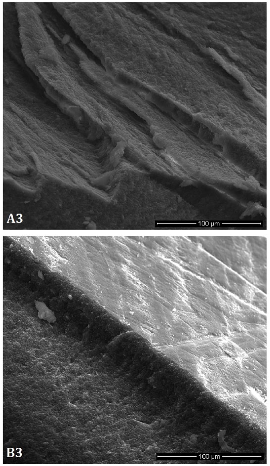
Scanning electron micrographs of fractured cross-sections of A3 and B3 samples taken at a magnitude of µ1000. Sample A3 exhibits a layered structure, whereas the fractured cross-section of sample B3 is more uniform.
Mechanical properties
The average Vickers hardness numbers of samples, with mean and standard deviation, are shown in Table 3. The top measurements of the gold- and berry-colored specimens showed no difference when they were cured with different light-curing units (p = 0.123 for Group A; p = 0.856 for Group B) but the Vickers hardness numbers of the silver-colored specimens cured with the VALO light-curing unit were found to be significantly smaller than the counterparts cured with the Optima and Demi Ultra units (p < 0.001). However, there was no difference in the bottom measurements of all colored compomer specimens cured with different light-curing units, except the gold-colored specimens cured with the VALO unit (p = 0.005 for Group A (gold); p = 0.862 for Group B (berry); p = 0.424 for Group C (silver)).
Vickers hardness numbers of samples.
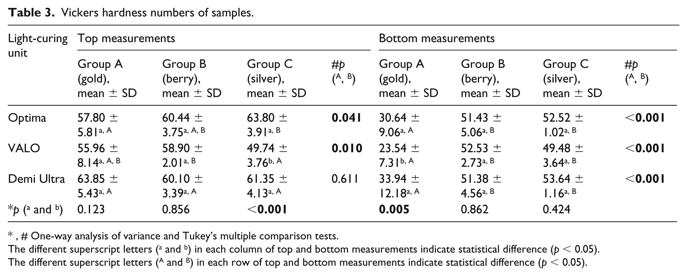
, # One-way analysis of variance and Tukey’s multiple comparison tests.
The different superscript letters (a and b) in each column of top and bottom measurements indicate statistical difference (p < 0.05).
The different superscript letters (A and B) in each row of top and bottom measurements indicate statistical difference (p < 0.05).
According to the top measurements, colored compomer specimens showed different Vickers hardness numbers when they were cured using the Optima and VALO units (p = 0.041 for Optima; p = 0.010 for VALO), meanwhile the specimens cured using the Demi Ultra unit had no significant difference in their measurements (p = 0.611).
In addition, all colored compomer specimens cured with different light-curing units showed significant differences at the bottom measurements (p < 0.001 for Optima, VALO, and Demi Ultra). Specifically, the Vickers hardness numbers were smaller for the gold specimens at the bottom surfaces. We also observed that the gold specimens with a thickness of 4 mm still remained in a soft-solid form even after irradiation, for all three curing units.
Typical stress–strain curves of samples were recorded during compression testing; the stress–strain curves for the sample series of C1 (silver-colored compomer cured using Optima unit) are given as an example in Figure 5. The characteristic mechanical properties of the samples are listed in Table 4. It was found that the compressive strengths of the samples varied with the color of the compomer. The compressive strength values decreased in the order of Group C (silver) > Group B (berry) > Group A (gold); curing with the Demi Ultra unit resulted in higher compressive strengths than the other light-curing units for the silver- and berry-colored compomers.
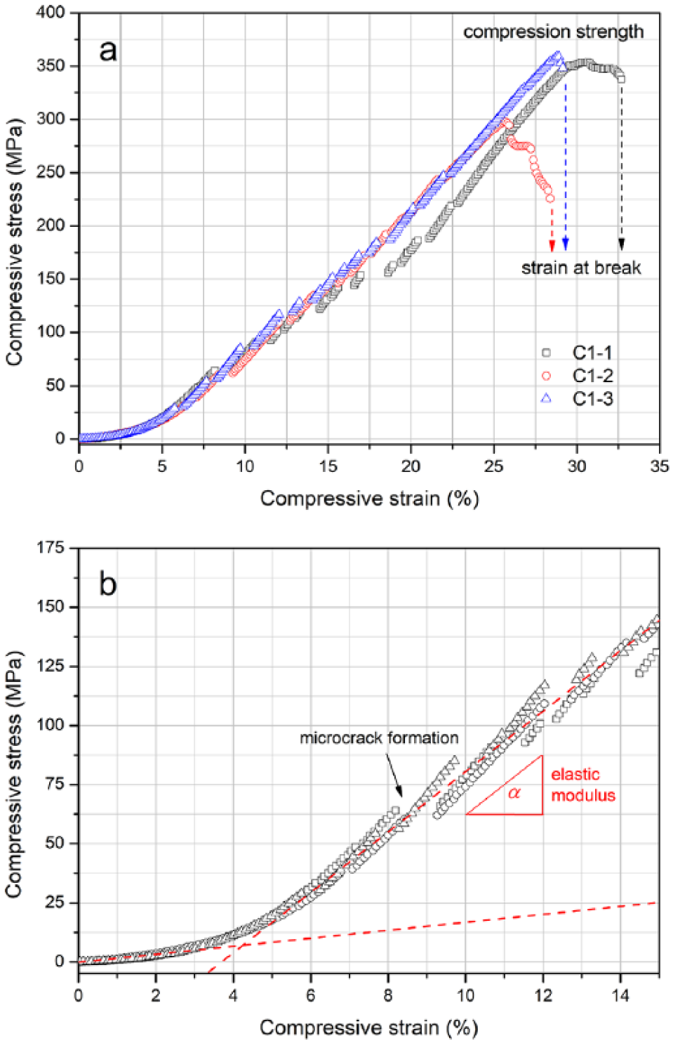
Representative stress–strain curves of C1 specimens: (a)“compression strength” – the maximum stress value just before failure (or breaking) – and “strain at break” under compressive loads; (b) definition of elastic modulus, which is the slope of the stress–strain curve.
Mechanical properties of samples.
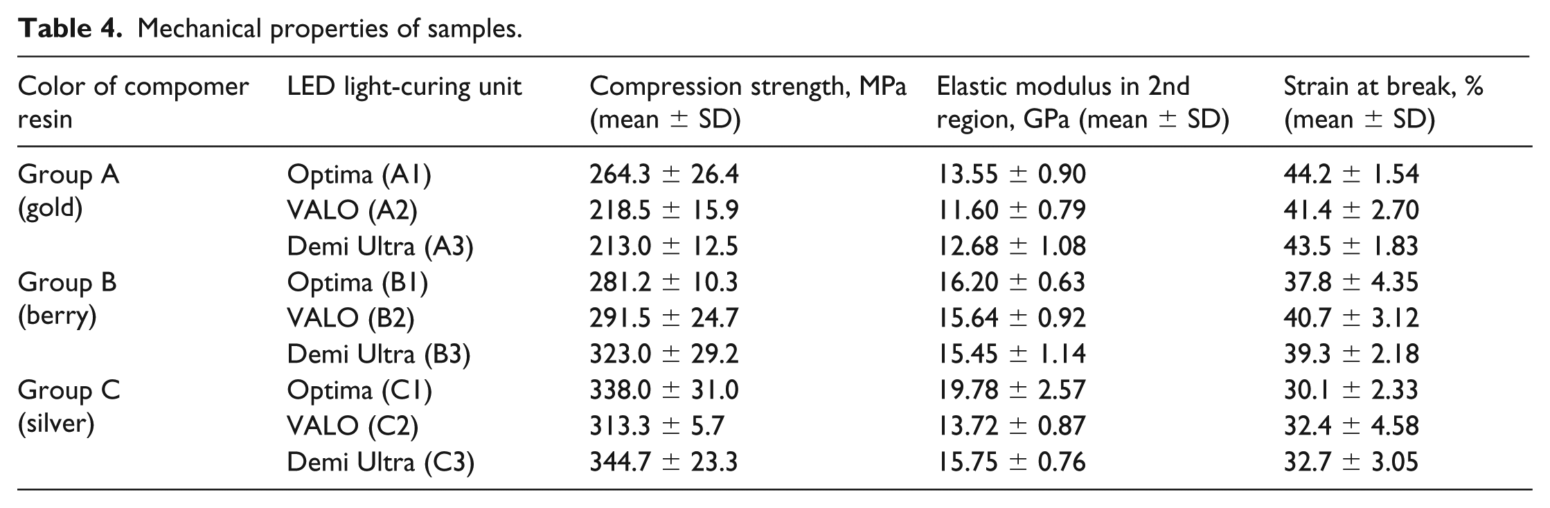
The moduli of elasticity of Group A (gold) were in the range 11.60–13.55 GPa, while the other groups exhibited higher moduli of elasticity, depending on curing device (Table 4). The moduli of elasticity of the gold- and silver-colored specimens that were cured with the VALO unit were smaller than those of the compomers cured with the Optima and Demi Ultra light-curing units. Conversely, it was found that the strain at break values of the Group A samples were greater than those of the other groups.
Discussion
The physical and mechanical characteristics of three different colored compomers were investigated in this study, as colored compomers are newly popular materials in pediatric dentistry, with the advantages of ability to release fluoride, esthetics, ease of use with light-curing and the attractiveness of different colors for children, encouraging them to cooperate during dental treatments.
Compomers are light-cured materials containing camphorquinone, which has a light absorption peak near 470 nm, as a photoinitiator. 17 Therefore, in this study, we used three different LED light-curing units with a wavelength range of 395–480 nm. The photo-polymerization process is an important stage, which affects the physical properties of the material. Moreover, the size, type, and content of the fillers may affect the transmission of light and degree of polymerization. 22 It has already been revealed that, after the polymerization process of composite resins, some dimethacrylate monomers still remain in the final product with a degree of curing in the range 55–75%. 21 Although polymerization processes in light-cured composite and compomer resins have been studied using FTIR, 23 the effects of included color particles on the polymerization process are not well known. Thus, in this study, we investigated the effects of three different LED light-curing units on the polymerization processes of compomers with three different colors, because the color of compomer, which is produced by the content of pigments and fillers within the material, could affect the transmission of incident light and the resultant degree of polymerization. FTIR spectroscopy, which can be used to determine the depth of cure, was used to evaluate the degree of curing of methacrylate monomers to polymers by comparing the peaks of C=C bands in colored compomer resins. It was found that the degrees of curing of the specimens varied with compomer color and light-curing device. According to the degrees of curing of our samples, Optima, with a 20 s curing time, had the best values, whereas VALO, with a 6 s curing time, had the worst. Because of the similarity of the wavelengths of the LED curing units used in this study, our results showed that light exposure time had a greater effect on the curing of dental resins than resin color. This was in accordance with a previous study, which suggested that the efficient curing time for polymerization of resin restorations is at least 8 s. 24 However, the degrees of curing of differently colored compomers changed in the order of silver > berry > gold, indicating that the gold color had the smallest values. An in-vitro study performed with 2-mm-thick colored compomer resin disks showed greater degrees of conversion values for blue and green colors after 20 s polymerization, whereas silver samples had the smallest degrees. 25 Contradictory results were found in our study, showing the greatest polymerization degree in the silver-colored samples, which were followed by the berry- and gold-colored samples, respectively. The reason that the greatest degrees of curing were in our silver-colored compomer samples may be related to their smaller glitter contents because of a tooth-colored appearance. Conversely, the darker (berry) color showed greater curing depths than the lighter (gold) one. Thus, this finding was attributed to the fact that light transmission may be affected by glittering because of the inclusion of irregularly shaped and sized components. 16 It has been suggested that, in addition to the type and amount of monomers, fillers, and initiator or catalyst, as well as the shade and translucency of the material, the wavelength and intensity of the light used also influence the degree of the polymerization process.
According to the surface microhardness measurements, all groups of compomers cured with the VALO unit yielded slightly lower Vickers hardness numbers than the samples cured with the Optima and Demi Ultra units. This could also be related to the shorter irradiation time of this device (6 s) in comparison with the others (10 s and 20 s). Furthermore, the Vickers hardness numbers that were measured from the top and bottom surfaces of the gold specimens were lower than those of their berry and silver counterparts. It should be noted that Vickers hardness number mainly depend on both the filler and matrix phases, rather than the color of the material. However, compomer color might indirectly influence Vickers hardness number, regarding the relationship between resin color and degree of curing. It is a well-known fact that size, geometry, amount of particles, particle size distribution, and quality of particle dispersion in the resin phase can affect specimen surface topography, and therefore the Vickers hardness number. 26 Since all differently colored compomers include the same amount of filler, 77.8 wt% as declared by the manufacturer, it could be inferred that variations in the Vickers hardness numbers of the specimens mainly originated from the different degrees of curing. It may be deduced that a more cross-linking results in more rigidity or stiffness in the resin phase, providing a higher Vickers hardness number. Compomers cured with the VALO unit yielded slightly smaller Vickers hardness numbers than the specimens cured with the Optima and Demi Ultra units for all groups of samples. This could be related to the shorter irradiation time of this device (6 s), in comparison with the others (10 s and 20 s). When we consider the light absorbed, reflected, and attenuated by resin samples, it may be concluded that the curing time that was applied using the Optima unit (20 s) might be recommended as a more valuable factor to reach sufficient degrees of curing than the other curing times. Moreover, the best values of monomeric conversion are obtained when the particle size is half of the wavelength. 27 In our study, the wavelengths of the curing units were 420–480 nm for Optima, 395–480 nm for VALO, and 450–480 nm for Demi Ultra. This would result in the VALO LED curing unit’s low degree of curing.
However, in a previous study of colored compomers, the degree of curing obtained for the blue color was found to be greater than for the gold and silver counterparts. 17 Contrary to that study’s results, in our study, the smallest degree of curing and Vickers hardness number were obtained for the gold specimens, followed by the silver and berry colors. Other studies suggested that differently colored compomers might have different proportions of glittering components, causing different hardness values.17, 28 This suggestion may also explain the effect of including pigments on the depth of curing, resulting in a change in the hardness values of the material; thus, the silver-colored compomer samples had the highest Vickers hardness number in our study.
To enlighten this controversy, we suggest considering our further mechanical analysis that were performed to obtain compression strength, modulus of elasticity and strain at break (%) values, which indicated that the lowest degree of curing and Vickers hardness number of our gold samples were in accordance with the lowest compressive strength and modulus of elasticity of the same specimens.
As seen in Figure 5, all stress–strain curves showed two distinctive regions. At the early stage of testing, the resin phase probably responds to compression loads; thus, the stress–strain curve goes up to a strain of 4% with a low slope. The shape or slope of the curve in this region depends on the strength of the matrix phase, which might be directly related to the degree of cross-linking or curing. In the second region, the stress–strain curve has a much higher slope, as compression loads are transferred to the filler particles. In fact, the mechanical property of a resin restorative depends mainly on the filler phase, as such a material includes a relatively high amount of filler. 5 Therefore, the elastic moduli of the specimens were determined by the second regions. Above a particular stress (or strain) value, small discontinuity points appeared on the stress–strain curve, as marked in Figure 5(b). These points correspond to micro-crack formations in the bulk structure, formed by increasing the compression load. Finally, the whole structure cracked, with a brittle failure. The long-term mechanical properties of a resin restorative material are strongly related to the formation of micro-cracks in the resin phase, as well as the growth of such cracks under cyclic loads.
Conversely, it was found that the strain at break values of the Group A (gold compomer) samples were higher than those of the other groups. This result is consistent with the relationship between degrees of curing and curing protocol, which indicates that a lower degree of curing results in higher strain at break.
Conclusions
This study investigated the microstructural and mechanical properties of colored compomer resin restoratives cured using different LED light-curing units. It was found that the Group A (gold) samples exhibited poorer degree of curing, surface hardness, and mechanical properties than Group B (berry) and Group C (silver). The silver-colored specimens yielded higher degrees of curing and significantly better mechanical performance. Based on the test results, we suggested that resin color might be an important compositional parameter for cross-linking reactions and interfacial adhesion between the resin phase and filler particles, and thus, result in effects on the mechanical properties of dental restoratives, as well as the curing conditions. The effects of resin color on the physical properties of a dental restorative are possibly related to transmission of incident light throughout the material.
Footnotes
Contributions of authors
Hypothesis and experimental design: MB, BY, SO
Performed the experiments: MB, BY, AD
Wrote and proofread the manuscript: MB, AD, ZD
Performed statistical evaluation: AD, ZD
Declaration of conflicting interests
The authors declare no potential conflicts of interest with respect to the research, authorship, and/or publication of this article.
Funding
The authors received no financial support for the research, authorship, and/or publication of this article.