
Abstract
This study illustrates the optimization of low-volume dispensing on a liquid handling system (LHS) to overcome the precipitation of compounds in the mammalian cytotoxicity assay with low dimethyl sulfoxide (DMSO) tolerance. All compounds at AstraZeneca Bangalore are tested in the mammalian cytotoxicity assay. In order to maintain the DMSO levels, serially diluted plates were prepared in DMSO/water. It was observed that some of the compounds precipitated. The IC50 data for such compounds were therefore erratic. To circumvent the problem of compound precipitation, the LHS was optimized to dispense low volumes (<1 µL). The plates were serially diluted using neat DMSO. Since the dilution was done using neat DMSO, there were no issues with precipitation. The serially diluted sample (0.5 µL) from the plate was stamped onto the assay plate to give the desired DMSO concentration. No significant differences in IC50 data were observed for 1 µL dispenses made from DMSO/water and 0.5 µL dispenses from neat DMSO for the samples with no precipitation issues. These data therefore gave us the confidence to switch over to 0.5 µL dispenses for the cytotoxicity assay to address the precipitation issue. However, precipitation of samples in the assay buffer is beyond the scope of this discussion.
Keywords
Introduction
Tuberculosis and malaria are two major global public health threats and cause substantial morbidity, mortality, negative socioeconomic impact, and human suffering. 1 Despite the significant increase in financial support and recent progress in addressing these two diseases, important obstacles and unmet priorities remain. Malaria is still in the top five causes of morbidity and mortality, especially among children of the age group 0–5 years. 2
Compound Management (CM) at AstraZeneca Bangalore, India, is a central and enabling function. The team supports the neglected diseases area, that is, tuberculosis and malaria, at various stages in the drug discovery cascade by dispensing compounds in the formats requested by the project teams. In the last decade or so, high-throughput screens with precision have become essential and have a major role to play in screening large compound libraries at the early stage of drug discovery programs.3,4 At AstraZeneca, we have tried to build high throughput into every possible aspect of the screens. 5
Samples are dispensed for in vitro and in vivo testing against Mycobacterium tuberculosis and Plasmodium falciparum. The in vitro cytotoxicity of compounds is measured against A549 human lung carcinoma cells to determine their safety. This assay helps to identify compounds with potential cytotoxicity issues. Structure–activity relationship (SAR) efforts are put in place to identify and modify the regions contributing to the toxicity of the flagged compounds.
The compound plate preparation process involved solubilization of solid compounds in DMSO and transfer of the required stock volume into the intermediate plate. This was followed by serial dilution of the intermediate plate and copying it onto the assay plate. The dilution and the assay plate preparation were done using the Biomek FX liquid handling system.
All compounds in the lead identification and lead optimization stage were tested in A549 cytotoxicity assay. 6 The assay has a DMSO tolerance of ≤1%. In order to achieve the desired level of DMSO in the assay, the DMSO stock was diluted with 50% water. However, some of the compounds were precipitated on addition of water. The IC50 data on compounds with solubility issues therefore may not be reliable. The focus of this study is on optimization of the Biomek FX LHS for low-volume dispensing in order to generate meaningful and consistent data for compounds with solubility issues. This would facilitate faster progression of projects.
Materials and Methods
Compound Plate Preparation
The compounds for the validation study were solubilized in DMSO at a stock concentration of 10 mM. The DMSO stocks prepared are stored under controlled conditions at 10% relative humidity and 20 °C temperature. The compound processing and distribution is tracked through a locally developed web-based dispensary application.
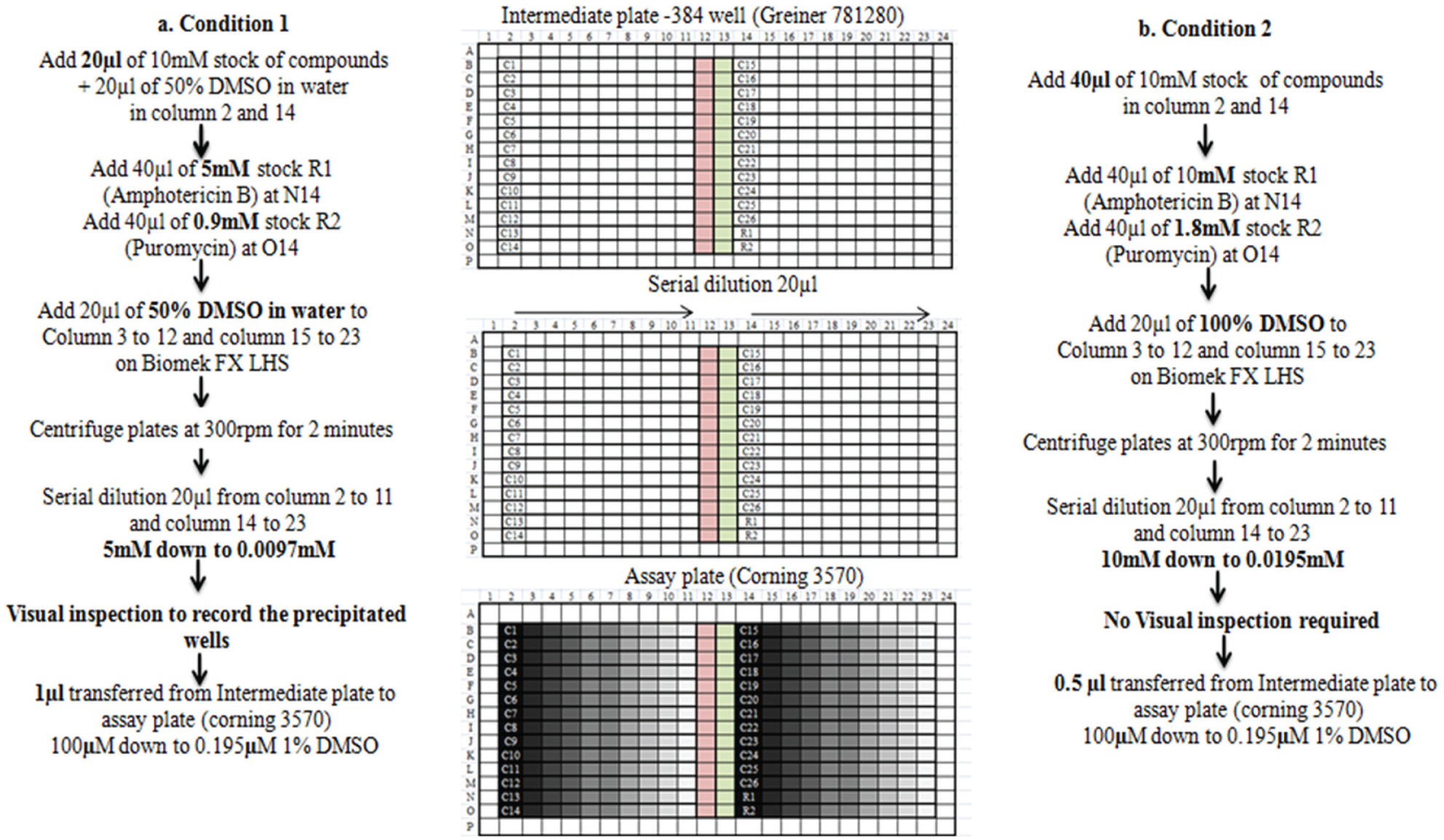
Each compound plate prepared contained 26 compounds, 14 compounds on the left side of the plate (C1–C14) in column 2 and 12 compounds on the right side of the plate (C15–C26) in column 14. The reference compounds were added at N14 (R1) and O14 (R2) wells in the plate. The assay controls, culture control, and media control were taken in columns 12 and 13, respectively. The periphery wells of the plate were left empty ( Fig. 1 ). All compound plate preparations are done in the designated clean area of the laboratory.
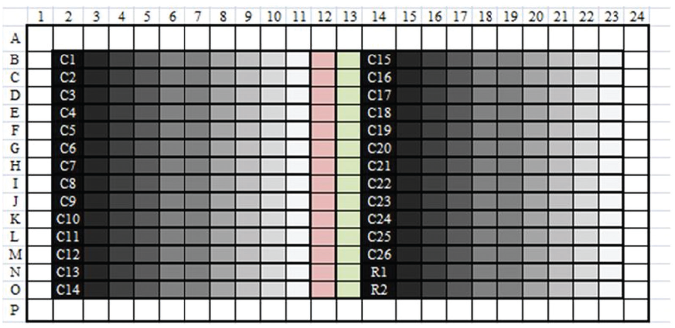
Plate format (384-well) for mammalian cytotoxicity assay. Test compounds C1–C26 in columns 2 and 14. R1, reference compound amphotericin B; R2, reference compound puromycin. Each compound serially diluted twofold from column 2 to 11 and from column 14 to 23. Column 12, culture control; column 13, media control and periphery wells empty.
DMSO stocks (20 µL of 10 mM) of the compounds were aliquoted manually into an intermediate 384-well plate (Greiner 781280) at columns 2 and 14. Amphotericin B (40 µL, 5 mM in DMSO) (1397-89-3; A4888-500MG; Sigma-Aldrich, St. Louis, MO) was added as reference 1 (R1) at N14 and 40 µL of puromycin (0.9 mM in water) (P8833 Sigma) was added as reference 2 (R2) at O14. The compounds and references were added using a single-channel calibrated manual pipette. Autoclaved Milli-Q water (20 µL) was added using a 16-channel calibrated manual pipette to the wells containing 10 mM DMSO stock of the compounds to give a final concentration of 5 mM in the intermediate plate. At this point, few compounds tend to get precipitated. For the wells from columns 3 to 12 and from columns 15 to 23, 20 µL of 50% DMSO in water was added as diluent using the transfer step on the Biomek FX LHS from Beckman Coulter (Brea, CA) with a 384-multichannel pipetting head using disposable Axygen tips (cat. no. FX-384-L-R) ( Fig. 2 ). The intermediate plate was centrifuged using Hereaus MULTIFUGE 4 KR at 300 rpm (rotations per minute) for 2 min before serial dilution. This was followed by two-fold, 10-point serial dilution (20 µL) from columns 2 to 11 and from columns 14 to 23 on the LHS. 7 The 10-point concentration ranged from 5 down to 0.0097 mM in the serially diluted intermediate plate. The wells of the compounds that precipitated were recorded after a visual inspection of the intermediate plate. The serially diluted intermediate plate was centrifuged using Hereaus MULTIFUGE 4 KR at 300 rpm for 2 min. One microliter from each well of the serially diluted intermediate plate was transferred into the assay plate (Corning 3570; Corning, Lowell, MA) using the copy plate method on the LHS. The assay plate with compounds was tested against the mammalian A549 cell line for determination of cytotoxicity.
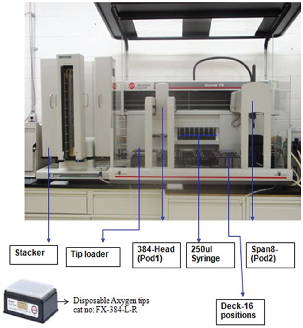
Biomek FX from Beckman Coulter with a 384-multichannel pipetting head and Span-8.
Mammalian Cytotoxicity Assay
Briefly, A549 cells (American Type Culture Collection) were grown in RPMI (Roswell Park Memorial Institute) medium (GIBCO BRL, Carlsbad, CA) containing 10% heat-inactivated fetal bovine serum (GIBCO BRL) and 1 mM
Optimization of Low Volume on Biomek FX LHS
To overcome the precipitation issues, the plate preparation was to be done with 100% DMSO (neat). To meet the cytotoxicity assay requirements at 1% DMSO tolerance, instead of 1 µL of compound in 50% DMSO/water, 0.5 µL of compound in neat DMSO was to be dispensed.
Hence, the optimization of the LHS to dispense low volumes was carried out.9,10
Briefly, the process of optimization involves the following:
Step 1: A standard curve is created with a calibrated hand pipette.
Step 2: The Biomek FX is used to pipette the desired volume using the selected technique with the default scaling factor and offset values of 1 and 0, respectively.
Step 3: The actual volume dispensed is calculated by comparison of the absorbance value from the plate dispensed with the Biomek FX to the standard curve.
Step 4: The calculated values of slope and the y-intercept replace the default values for the scaling factor and offset, respectively, in the technique.
Step 5: Another dispense is performed with the Biomek FX using the same technique with the new scaling factor and offset to determine if the accuracy requirement of the assay has been met.
Tartrazine (Sigma 1934-21-0) dye solubilized in water at 0.4 mg/mL and eosin Y disodium salt (Sigma cat. no. E6003) solubilized in DMSO at 0.5 mg/mL were used for the volume calibration study.
A tartrazine standard curve was generated using a calibrated manual pipette. Each volume, 0.5, 0.8, 1, 2, and 5 µL, was dispensed 48 times in a 384-well plate (Corning 3702). The final volume for each well was made up to 50 µL, dispensing water by multidrop (multidrop combi 5840300; Thermo Scientific, Waltham, MA). The plate was centrifuged using Hereaus MULTIFUGE 4 KR at 300 rpm for 2 min. Absorbance was measured at 405 nM using Tecan SAFIRE II. A standard curve was generated ( Fig. 3 ).
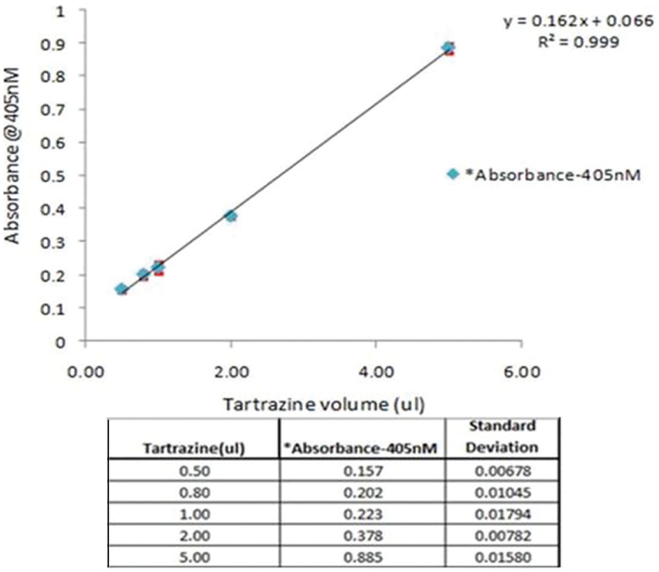
Tartrazine standard curve (regression equation). X axis: Tartrazine solution volumes dispensed using single-channel calibrated pipette. Y axis: Absorbance value. (*Each absorbance value is an average of 48 wells.)
The different volumes 0.5, 1, and 2 µL of tartrazine were dispensed in a 384-well plate using disposable Axygen tips with a 384-multichannel pipetting head on the Biomek FX LHS as in step 2. The volumes in the plates were made up to 50 µL by addition of water by multidrop. After centrifuging the plates, absorbance was measured. Actual volumes dispensed were determined by comparison of absorbance from the plate dispensed with the Biomek FX to the standard curve, as in step 3. Steps 4 and 5 were performed. Volumes (0.5 µL) were dispensed in triplicate plates on the LHS. The accuracy and precision for the 0.5 µL volume dispensed in triplicate plates ( Table 1 ) on the LHS was calculated using the regression equation from the standard curve ( Fig. 3 ).
Actual Volume Dispensed by Biomek FX Determined by Comparison to the Tartrazine Standard Curve.
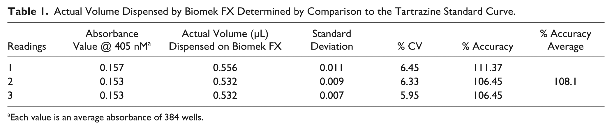
Each value is an average absorbance of 384 wells.
Similarly, volume dispense was performed with eosin dye on Biomek FX LHS (data not shown). Two concentrations (0.5 µL) of 0.5 and 0.25 mg/mL of eosin dye solution prepared in DMSO were dispensed using the LHS ( Table 2 ). The precision for these dispenses was in the acceptable range, indicating the LHS is capable of dispensing 0.5 µL volumes. To corroborate this further, 0.5 µL of the references (amphotericin B and puromycin) used in the cytotoxicity assay was dispensed at two concentrations to give 50% and 100% inhibition, respectively, with neat DMSO serving as a 0% inhibition control in the assay ( Table 3 ).
Eosin Dispenses on Biomek FX.
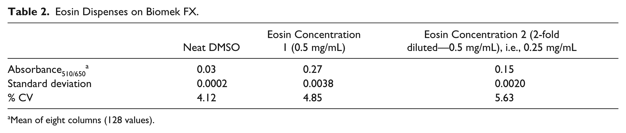
Mean of eight columns (128 values).
Dispensing Precision at 0.5 µL Volume for Reference Samples Using Biomek FX LHS.
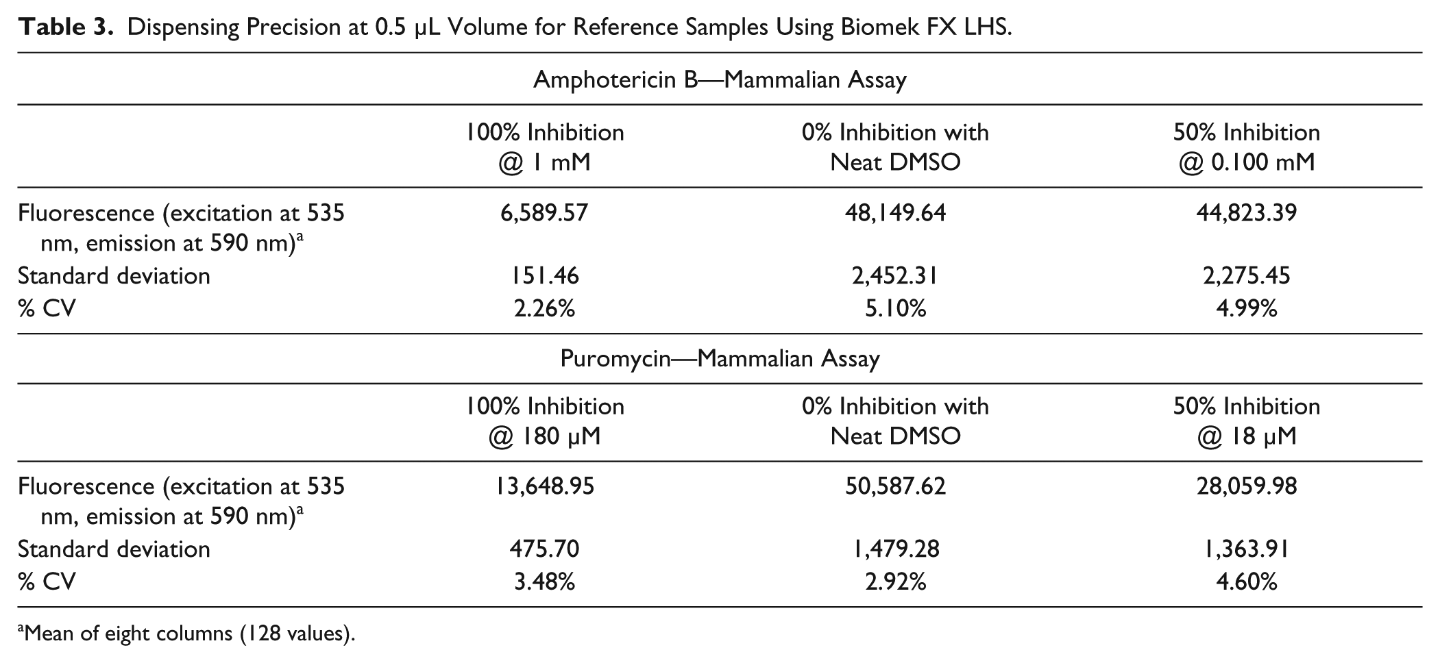
Mean of eight columns (128 values).
Validation Study
Two sets of samples were taken for the validation study. The first set contained 45 samples ( Table 4 ) and the second set contained 19 samples ( Table 5 ). The samples were prepared for screening in the cytotoxicity assay by two methods referred to as condition 1 and condition 2 respectively.
Validation Study 1: IC50 Values (100 μM Downward—10-Point Concentration) under Conditions 1 and 2.
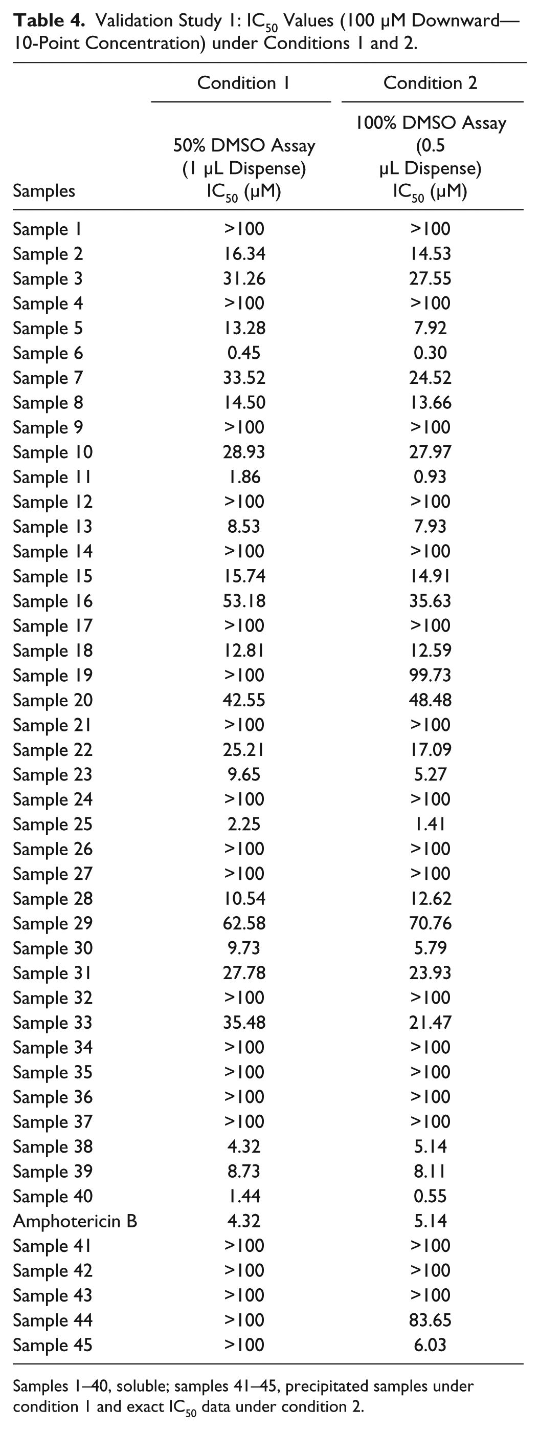
Samples 1–40, soluble; samples 41–45, precipitated samples under condition 1 and exact IC50 data under condition 2.
Validation Study 2: IC50 Values (100 µM Downward—10-Point Concentration) under Conditions 1 and 2.
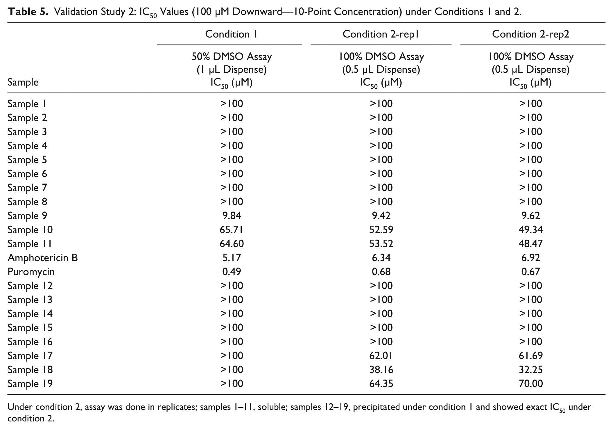
Under condition 2, assay was done in replicates; samples 1–11, soluble; samples 12–19, precipitated under condition 1 and showed exact IC50 under condition 2.
The IC50 values obtained for the validation set under the two conditions were evaluated.
In condition 1 (as described for compound plate preparation) the samples were serially diluted using 50% DMSO in water. Some of the sample solutions in the study were precipitated. Visual inspection of the plates was done to mark the wells that showed precipitation. One microliter from each well was transferred into the assay plate by the copy plate method on LHS as per condition 1 ( Fig. 4a ).
Flowchart showing plate preparation steps under (
In the condition 2 ( Fig. 4b ) the samples were serially diluted using neat DMSO. Compound stock (40 µL, 10 mM) was dispensed in columns 2 and 14. Reference compounds were added at N14 and O14. In the well N14 reference (R1) amphotericin B (10 mM in DMSO) was added, and in the well O14 reference (R2) puromycin (1.8 mM in water) was added. The compounds and references were added using a calibrated manual pipette. Neat DMSO (20 µL) was added in columns 3–12 and columns 14–23 using LHS. A twofold serial dilution was performed on LHS from columns 2 to 11 and from columns 14 to 23. The concentration ranged from 10 down to 0.0195 mM in the serially diluted intermediate plate. However, no precipitation was observed in the intermediate plate. From the serially diluted intermediate plate, 0.5 µL from each well was transferred into the assay plate by the copy plate method using 0.5 µL of the optimized technique using Axygen tips with a 384 pipetting head on Biomek FX. The assay plates prepared under the two conditions were tested in the A549 cytotoxicity assay. The final concentration of the samples and amphotericin B (R1) in the assay under the two conditions ranged from 100 down to 0.195 µM. The final concentration of puromycin (R2) in the assay under the two conditions ranged from 0.035 to 18 µM. All samples and references were with a DMSO level maintained at 1%.
Results and Discussion
The mammalian cytotoxicity assay has a DMSO tolerance of ≤1%. In order to achieve the required DMSO levels, the intermediate plates were serially diluted in 50% DMSO/water.
However, it was observed that about 10% to 25% of the samples precipitated. The IC50 data generated from such samples would therefore be unreliable.
In order to address the above issue and avoid the step of dilution with water, the alternative would be to serially dilute the plate with neat DMSO and copy 0.5 µL to maintain the DMSO concentration in the assay buffer with the required start sample concentration.
The Biomek FX LHS was optimized for dispensing 0.5 µL. The volume calibration data using tartrazine as given in Table 1 and Figure 3 and eosin as given in Table 2 indicated that the LHS is capable of dispensing low volumes with acceptable accuracy (108%) and precision (<6.5%). The percent CV for the inhibition data for amphotericin B and puromycin was in the acceptable range, as shown in Table 3 .
The validation study included samples that were soluble and also those that were precipitated in 50% DMSO/water. Samples with IC50 data >100 µM are the samples with minimal or no cytotoxicity liability. The samples flagged for precipitation under condition 1 showed IC50 values of >100 µM. Minimal or no toxicity for such samples could be due to very low concentration of the sample in the soluble form. The IC50 values from such samples would be inaccurate and could affect project progression at a later stage.
In the validation study1 done with 45 samples (
Table 4
) under two conditions in parallel, the following observations were made: 40 out of 45 samples (
Table 4
) were soluble and the IC50 data generated under the two conditions were not significantly different (within twofold variation). The low-volume dispensing therefore had no effect on the data quality.
Five out of the 45 samples ( Table 4 ) were found to be precipitated under condition 1. The IC50 values for three out of the five samples tested remained unchanged, indicating there were no toxicity issues. However, sample 45, which exhibited an IC50 of >100 µM under condition 1 (precipitated), showed a value of 6.03 µM under condition 2 (soluble), indicating it was toxic (IC50 curve, Fig. 5a,b ). This modified plate preparation approach enabled project teams to have reliable and consistent cytotoxicity data.
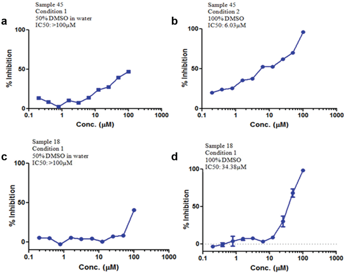
Validation study 1: IC50 curve for sample 45 under (
To further substantiate the observations made, validation study 2 with a set of 19 samples was run in duplicate under condition 2. The IC50 values were reproducible, and some of the precipitated samples with >100 µM showed definite IC50 values ( Table 5 ). The IC50 curve of a representative sample 18 that precipitated under condition 1 and the IC50 curve of the same sample under condition 2 have a definite IC50 value of 34.38 µM ( Fig. 5c,d ).
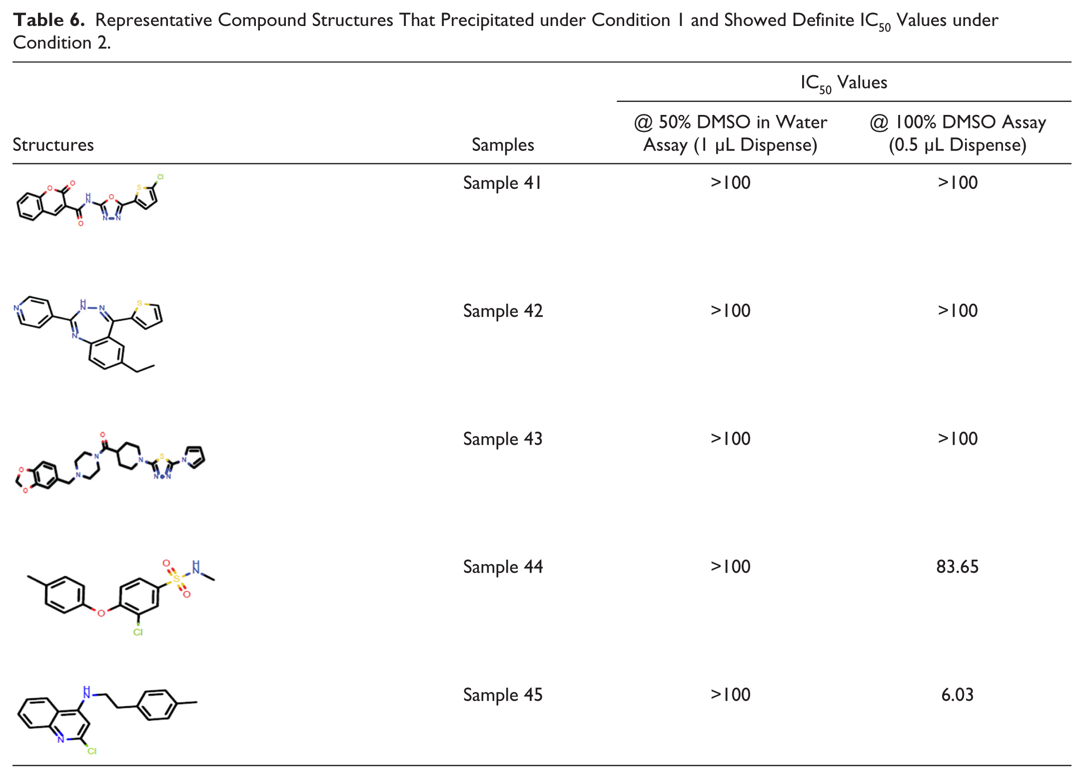
Representative compound structures shown in Table 6 are the samples that precipitated under condition 1 and showed a definite IC50 value under condition 2.
Representative Compound Structures That Precipitated under Condition 1 and Showed Definite IC50 Values under Condition 2.
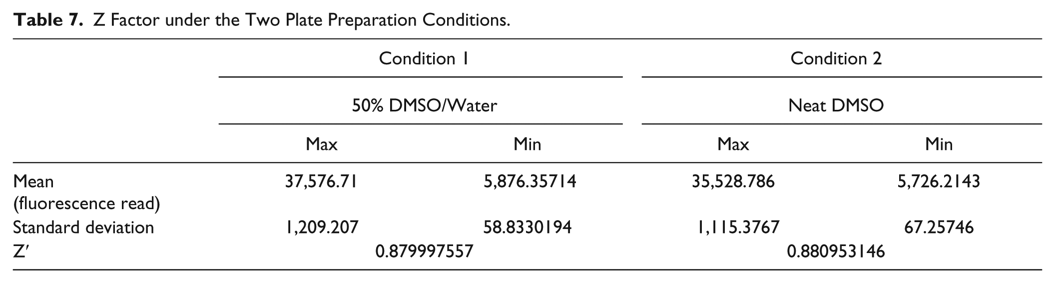
The reference samples amphotericin B and puromycin showed expected IC50 values under both conditions ( Fig. 6a–d ). The validation sets screened against the mammalian A549 assay had a Z factor of ~0.8, as shown in Table 7 . The data from the validation study indicated that the Biomek FX LHS was amenable for dispensing volumes of <1 µL, which enabled the team to address the precipitation issues in the mammalian cytotoxicity assay.
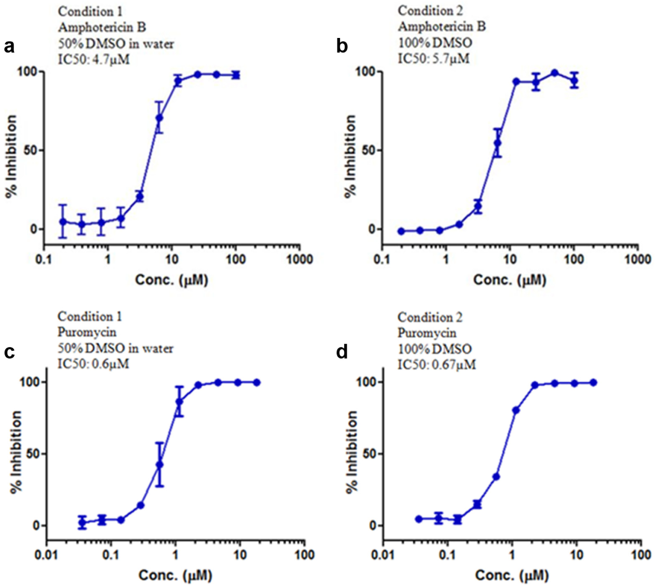
IC50 curve for amphotericin B: IC50 value ranges from 4.5 to 7 µM under both (
Z Factor under the Two Plate Preparation Conditions.
The consistency and reliability of the data generated from the validation study facilitated the CM team moving over to dispensing 0.5 µL using the Biomek FX LHS for the mammalian cytotoxicity assay on a routine basis. This change resulted in enhanced plate preparation efficiency coupled with generation of reliable cytotoxicity data for the samples tested.
Conclusions
All project compounds synthesized were subjected to mammalian cytotoxicity assay in the first wave of the test cascade. Compounds with potentially less or no toxicity issues passed the criteria and moved forward to the next cascade without any solubility flags. But for compounds that had issues with precipitation during plate preparation, the IC50 data were not reliable and were flagged. Optimization of the Biomek FX LHS for dispensing 0.5 µL with precision and accuracy and modification of the compound plate preparation method using neat DMSO translated into generation of quality data from the cytotoxicity assay.
Based on the results obtained from this study, the Biomek FX LHS is capable of dispensing 0.5 µL volumes within a statistically acceptable range of accuracy and precision. The modified compound plate preparation method has circumvented the problem of compound precipitation in the intermediate plate, thereby generating accurate, dependable, and consistent cytotoxicity data for project compounds. Elimination of the water addition step and avoidance of the visual inspection of the intermediate plates for tracking precipitation in condition 2 have resulted in enhanced efficiencies for the CM team. The modified method was able to generate reliable IC50 data for about 10% to 25% of the compounds that were flagged due to precipitation in the intermediate plate under condition 1 (in 50% DMSO in water). The low-volume optimization to address the precipitation issues in the mammalian cytotoxicity assay was therefore a value addition exercise both from the CM and the assay perspective. Roll out of this method on a routine basis has resulted in generation of quality data for all the projects at Bangalore. This has enabled project teams progress scaffolds with minimal or no cytoxicity liability in the drug discovery cascade.
Footnotes
Acknowledgements
The authors thank the microbiology team for the validation study done in the mammalian cytotoxicity assay and Dr. Dwarakanath Prahlad and Parvinder Kaur for helpful comments and suggestions.
Declaration of Conflicting Interests
The authors declared no potential conflict of interest with respect to the research, authorship, and/or publication of this article. The entire work was carried out at AstraZeneca as a part of internal projects.
Funding
The authors disclosed receipt of the following financial support for the research, authorship, and/or publication of this article: The work was supported by AstraZeneca only. No external funders have any role to play.