
Abstract
Three-dimensional culture systems bridge the experimental gap between in vivo and in vitro physiology. However, nonstandardized formation and limited downstream adaptability of 3D cultures have hindered mainstream adoption of these systems for biological applications, especially for low- and moderate-throughput assays commonly used in biomedical research. Here we build on our recent development of a 384-well hanging drop plate for spheroid culture to design a complementary spheroid transfer and imaging (TRIM) plate. The low-aspect ratio wells of the TRIM plate facilitated high-fidelity, user-independent, contact-based collection of hanging drop spheroids. Using the TRIM plate, we demonstrated several downstream analyses, including bulk tissue collection for flow cytometry, high-resolution low working-distance immersion imaging, and timely reagent delivery for enzymatic studies. Low working-distance multiphoton imaging revealed a cell type–dependent, macroscopic spheroid structure. Unlike ovarian cancer spheroids, which formed loose, disk-shaped spheroids, human mammary fibroblasts formed tight, spherical, and nutrient-limited spheroids. Beyond the applications we describe here, we expect the hanging drop spheroid plate and complementary TRIM plate to facilitate analyses of spheroids across the spectrum of throughput, particularly for bulk collection of spheroids and high-content imaging.
Introduction
Many cell culture applications are moving toward more physiological 3D model systems to bridge the gap between nonphysiological 2D platforms and in vivo conditions. Mass transport of drugs, nutrients, and metabolites in these systems is more representative of profiles of parameters such as hypoxia and metabolism. Despite these advantages, previous technologies for spheroid cultures were not used extensively due to lack of throughput (e.g., microfluidics), limited spheroid uniformity (e.g., rotating flasks), and cumbersome nonstandardized handling and analysis. Challenges to standardization, implementation, and analysis of 3D tissue cultures have limited widespread adoption. 1 Comparison and standardization of spheroid formation systems is also necessary to reconcile observed differences in gene expression and respiratory status for spheroids formed in hanging as compared with microfluidic systems. 2
To address many of these issues, we developed a 384 hanging drop plate for high-throughput formation and analysis of individual and uniform spheroids.3,4 Our hanging drop system consists of 384 through-holes to facilitate formation of uniform spheroids and ready access to the medium via the through-hole to exchange media, add treatments, or add cells at defined time points. Other groups have developed systems for hanging drop formation that rely on gravity-enforced cell assembly. 5 The simplest but least stable strategy for hanging drop formation is to plate droplets on the underside of nonadherent culture dishes. 6 The company, InSphero (Schlieren, Switzerland), also developed a 96-well gravity-driven hanging drop system that uses a similar through-hole strategy as our 384-well plate.7,8 Many imaging-based analyses, however, require a spheroid to be transferred to a secondary dish for stable focusing. High-fidelity transfer of hanging drop spheroids to a fixed substrate for downstream analysis requires highly parallel and consistent pipetting, which is inefficient and time-consuming by hand and costly to automate. While the InSphero GravityTrap system enables transfer of 96-well array format hanging drop spheroids to a secondary 96-well plate substrate, it disallows immersion-based imaging and facile bulk collection.
Here we report the development of a transfer and imaging (TRIM) plate to expand the usefulness of the 384 hanging drop system and facilitate these previously difficult downstream analyses. We differentiate our system from the InSphero system based on increased throughput (384-vs. 96-well array format) and low aspect transfer wells that facilitate high-fidelity spheroid capture, immersion-based imaging, bulk spheroid collection, and timely enzyme-substrate kinetic studies.
Materials and Methods
Plate Design
We used SolidWorks (Dassault Systèmes SolidWorks Corp., Waltham, MA) to conceptualize the TRIM plate in three dimensions. We fabricated the TRIM plate in the material Accura 60 (3DSystems, Rock Hill, SC) using the SLA Viper si2 dual-resolution SLA system, which is conducted by the University of Michigan Medical Innovation Center. The dimensions of the 384 wells are a total depth of 1.25 mm, which serves as the optical working distance, and a total well diameter of 4 mm. The wells are formatted as a standard 384-well plate to match the 384 hanging drop plate with a 16-by-24 array spaced every 4.5 mm. The inner-wall dimensions of the original 384 hanging drop plate match the outer wall of the TRIM plate and serve as physical guides for high-fidelity matching between droplets and wells. We developed a multiradial well contour to facilitate settling of the spheroid in the center of the well while limiting spheroids sticking in corners due to surface tension (
Cells and Spheroid Cultures
We cultured all cells in Dulbecco’s modified Eagle’s medium (DMEM) supplemented with 10% fetal bovine serum and 1% antibiotic. We used several cell types to form spheroids, including MDA-MB-231 breast cancer cells (ATCC, Manassas, VA), HeyA8 ovarian cancer cells (gift of Gordon Mills, MD Anderson Cancer Center, Houston, TX), and a human mammary fibroblast cell line expressing green fluorescent protein (GFP). 9 We transduced HeyA8 ovarian cancer cells with a lentiviral vector for GFP and sorted for a population of stably transduced cells by flow cytometry. 10 We previously have described MDA-MB-231 cells stably expressing eqFP650, GFP, or firefly luciferase. 11 We formed and maintained spheroids in 25 µL of culture medium as we described previously using the 384 hanging drop plate.3,4
Contact-Based Spheroid Transfer and Collection
To transfer hanging drop spheroids, we aligned the TRIM plate based on designed plate guides and lowered the hanging drop plate to contact the TRIM plate (
Fig. 1A
–
D
). We allowed spheroids to settle into the transfer wells for 1 to 2 min to improve transfer efficiency. We evenly lifted and separated the hanging drop plate from the recipient TRIM plate for spheroid transfer. See
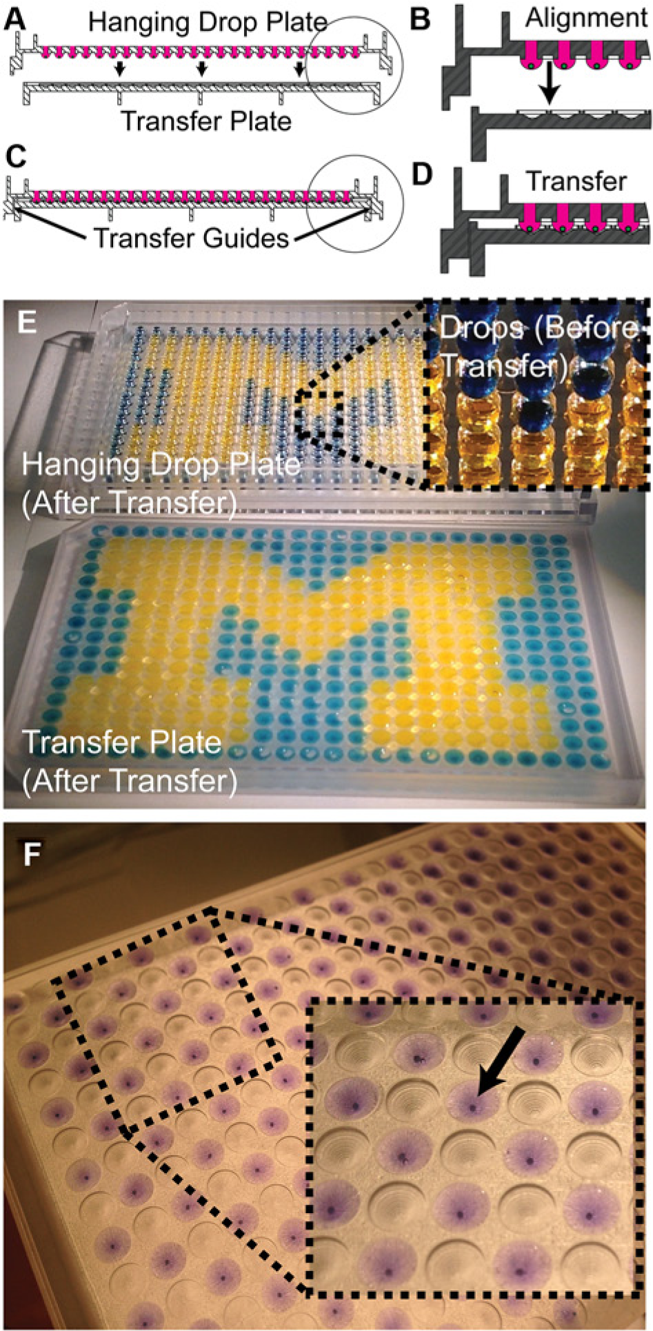
Transfer and imaging of 384 hanging drop spheroids. (
Spheroid Analysis and Imaging
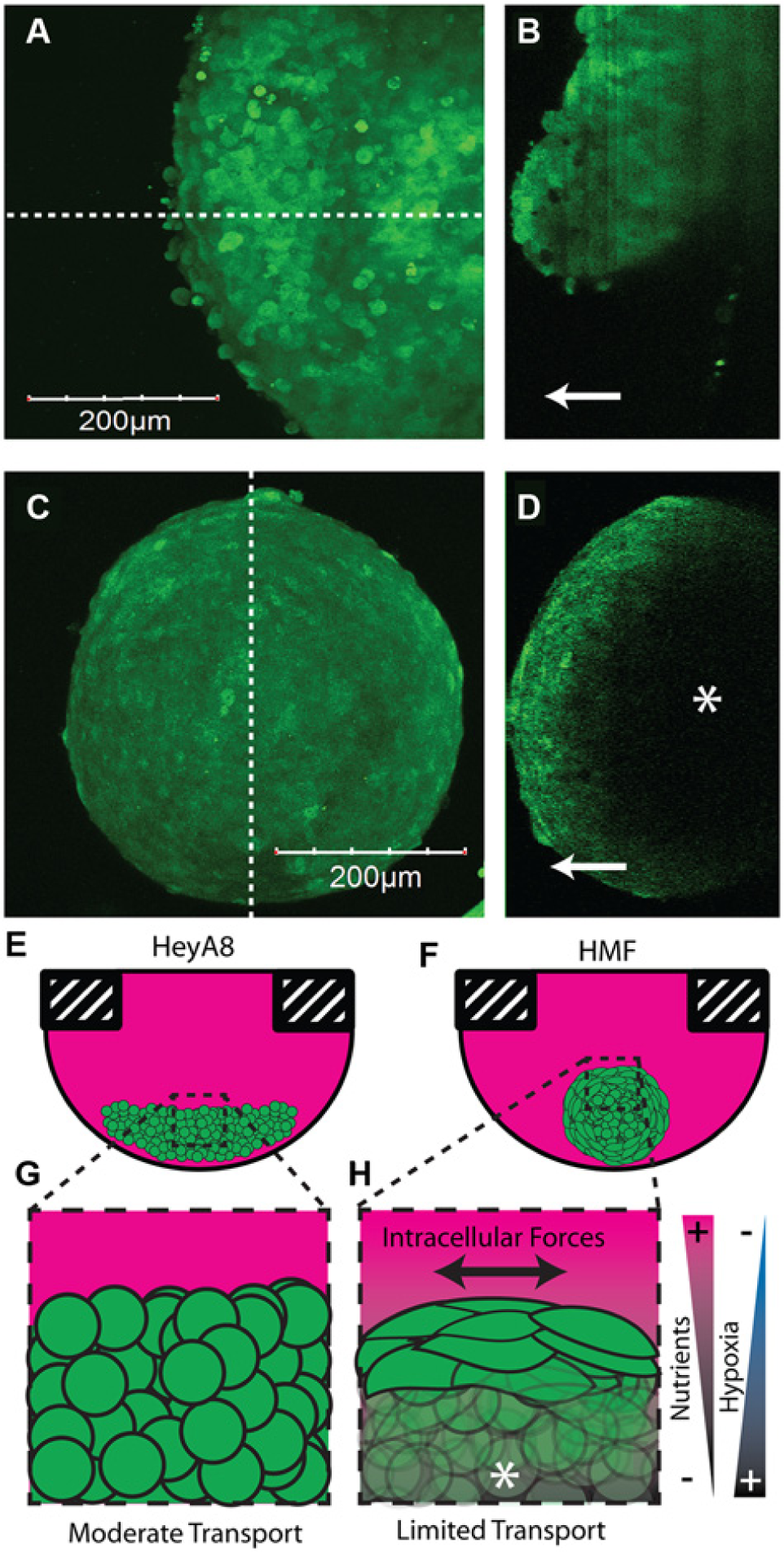
For two-photon imaging of spheroids, we used a 25× objective (XLPLN, NA: 1.05; Olympus, Center Valley, PA) and an upright confocal and two-photon microscope (Olympus MPE Twin) as we have described before. 12 We imaged individual HeyA8 spheroids and HMF spheroids both containing 15,000 cells per droplet. We very gently immersed the TRIM plate in growth medium after spheroid transfer for upright immersion microscopy. For imaging, we maintained resolution and z-step increment but varied the laser intensity between HMF and HeyA8 spheroids due to differences in their overall fluorescent intensity.
We collected 100 spheroids containing 10,000 cells total with different ratios (1:1 and 1:9) of 231 cells expressing either nuclear-GFP or FP650 for flow cytometry analysis ( Fig. 2 ). To collect spheroids, we transferred them to the TRIM plate and used ~50 mL of DMEM culture medium to wash spheroids from the TRIM plate into a 50-mL conical tube. We centrifuged the spheroids and resuspended the pellet in 0.25% trypsin with EDTA. Once spheroids were dissociated, we neutralized trypsin with full DMEM, centrifuged the sample, and resuspended the cells in phosphate-buffered saline (PBS) for flow cytometry analysis. Flow cytometry was done using a Becton Dickinson (Franklin Lakes, NJ) FACS DiVa Flow Cytometer. We set the sort quadrants with an excitation at 488 and 633 nm based on fluorescence-negative MDA-MB-231 cells ( Fig. 2B ). The quadrant statistics were determined with the Becton Dickinson software.

Spheroid collection for analysis by flow cytometry. (
For bioluminescence imaging, we seeded 154 spheroids (every other well in 384 wells) containing 10,000 cells per drop total at a ratio of 9:1 HMF cells and MDA-MB-231 cells expressing firefly luciferase. We plated water in the outer wells to minimize evaporation. Prior to transfer of spheroids to the TRIM plate, we added 3 µL of 15 mg/mL luciferin diluted 4× in phosphate-buffered saline to each corresponding well on the TRIM plate. Contact between the spheroid droplet and the preplated luciferin caused simultaneous mixing of all transferred droplets. We quantified the number of wells with detectable bioluminescence of firefly luciferase as we have described before (
We stained HMF spheroids with trypan blue after 3 to 4 days of culture to increase contrast by staining for dead cells. To stain the cells, we added 2 µL of 4× dilution of trypan blue in PBS to each droplet for 4 h prior to transfer. We exchanged 10 µL of media twice to remove the residual trypan blue in each droplet. After transfer, we imaged the TRIM plate with a white light back light using a 5-MP camera ( Fig. 1F ).
Results and Discussion
Spheroid Transfer and Imaging
The TRIM plate structure is complementary to the 384 hanging drop plate designed by our group with designs to enable (1) gentle, high-fidelity, contact-based drop and spheroid transfer; (2) bulk spheroid collection; and (3) high-resolution imaging with short optical working-distance objectives (
Figs. 1
,
2
, and
3
, respectively). The low aspect ratio of our plate is markedly different from the InSphero GravityTrap system, which disallows immersion imaging and bulk collection.
7
We designed low-aspect ratio wells less than 2 mm in depth, which is less than the height of the hanging 25-µL droplets for contact-based drop transfer and low working-distance immersion imaging (
Fig. 1D
). A visual demonstration with dyed droplets illustrates the simple high-fidelity transfer process using 384 droplets (
Fig. 1E
,
F
and
Three-dimensional imaging with the transfer and imaging (TRIM) plate. (
We demonstrate 100% spheroid capture efficiency (154/154) based on the number of bioluminescent spheroids that transferred to TRIM plate wells containing the preplated enzymatic substrate, luciferin (
One challenge with analysis of adherence-independent cultures such as spheroids is to physically but reversibly immobilize the sample to minimize movement and facilitate organized downstream analysis. We highlight these challenges particularly for immersion imaging, where sample stability is at a premium and imaging in the hanging drop plate is impossible. Transfer of spheroids to a rigid substrate enhanced stability and allowed for immersion imaging. Although transfer to standard culture dishes is possible, movement due to flow in standard culture formats disrupts imaging. To improve this, we implemented a multicurvature well contour (
Spheroid Collection and Analysis
Standard 2D cell culture platforms are amenable to subcellular, single-cell, and population analysis. While 3D and high-resolution imaging applications discussed above address the two former analysis scales, population analysis for population-, DNA-, RNA-, protein-, and metabolite-level assays requires high efficiency recovery of many cells, which is difficult in most 3D culture formats. We used the TRIM plate to collect bulk spheroids simultaneously for dissociation and analysis using flow cytometry. After transfer of spheroids, we released them by tilting the TRIM plate, directing flow from a pipette down into the wells, and collecting the overflow in a 50-mL conical tube ( Fig. 2A ). Following enzymatic dissociation of the spheroids with trypsin, we analyzed spheroids containing 1:9 and 1:1 ratios of 231 cells expressing either GFP or FP650 ( Fig. 2C , D ). We note that the FP650 cells were not homogeneously expressing, causing them to show up in both bottom quadrants of the flow cytometry plot. However, these are still intensity shifted for FP650 relative to the negative control ( Fig. 2C – E ). Our flow cytometry analysis closely matched the proportion of 1:1 and 1:9 for GFP to FP650 by summing the bottom quadrants for the FP650 cells and top quadrants for the GFP cells ( Fig. 2E , F ).
High-Resolution 3D Imaging
Since the spheroid settles to the lower curvature and is relatively fixed, we demonstrate imaging of the 3D volume of large spheroids using two-photon microscopy ( Fig. 3A – D ) for HeyA8 ovarian cancer ( Fig. 3A , B ) and HMF spheroids, both expressing GFP. We note the hemispherical disk shape of the ovarian cancer “spheroid” to be striking ( Fig. 3B ), as the perception of a spheroid is understandably spherical. The HMF cells formed a smaller, tight structure with largely spherical morphology. The density of the HMF spheroid also led to metabolic limitations in the center of the spheroid, which we speculate decreased protein synthesis and resulted in lower GFP intensity. Based on the morphology of the spheroids, the shape of the HeyA8 spheroid seems to be physically dictated by the droplet radius ( Fig. 3E , G ). Others have observed disk-like ovarian cancer spheroids that were formed in hanging droplets and cite the importance of defining the geometric spheroid morphology. 15 The strong intracellular forces between the HMF cells, on the other hand, lead to a tight spherical shape ( Fig. 3F , H ). Others have defined nutrient transfer limitations and hypoxia in 3D cell spheroids for structures more than ~100 to 200 µm in diameter. 16 However, nutrient limitations are very much cell type and spheroid shape dependent and must be characterized for new cells at the onset of a project. For defining nutrient limitations and hypoxia, the complementary hanging drop and TRIM plates enable 3D multiphoton image-based screening of many combinations of cell types and conditions in parallel.
Future Directions and Opportunities
Widespread adoption of 3D tissue culture is dependent on three aspects: ease of use, standardization, and scale. We previously developed a 384-well hanging drop plate to improve standardization of spheroid culture and spheroid uniformity. However, it is biased toward facilitating high-throughput analyses, such as robotic automation and standard plate reader assays. Efficient tools for recovery of spheroids will broaden applicability of hanging drop spheroids and encompass the full spectrum of throughput, from high-throughput analysis to low- and moderate-throughput analyses, such as high-resolution imaging and flow cytometry. We used the TRIM plate to demonstrate facile handling for high-resolution imaging of individual spheroids and flow cytometric analysis of bulk-collected spheroids. The TRIM plate improves compatibility of hanging drop spheroids with low- and moderate-throughput analyses that are bioassay staples. We believe these systems substantially improve the utility of spheroid cultures for biomedical research, bridging the gap between low-throughput in vivo models and standard 2D tissue culture systems.
Footnotes
Acknowledgements
We thank Sasha Cai Lesher-Perez for useful discussion of spheroid cultures and their analysis and handling. We also thank Michael Deininger, from the University of Michigan, Medical Innovation Center, for useful discussion of considerations for 3D prototyping.
Declaration of Conflicting Interests
The authors declared the following potential conflicts of interest with respect to the research, authorship, and/or publication of this article: S.T. has licensed the 384 hanging drop array technology to 3D Biomatrix and also has stock options.
Funding
The authors disclosed receipt of the following financial support for the research, authorship, and/or publication of this article: This work was supported by U.S. National Institutes of Health grants R01CA170198, R01CA136553, R01CA136829, R01CA142750, and P50CA093990. S.P.C. was supported on Advanced Proteome Informatics of Cancer Training Grant T32 CA140044.