
Abstract
Study design
Clinical study.
Objective
Our work was aimed at exploring the expression and clinical significance of microRNA-138-5p (miR-138-5p) and Transforming Growth Factor-beta 3 (TGF-β3) in peripheral blood of patients with ankylosing spondylitis (AS).
Methods
Forty-seven patients with AS were selected as the AS group, and the staging of the enrolled AS patients was based on the BASDAI score: <4 points were classified as the stable stage (stable group) and ≥4 points were classified as the active stage (active group). Forty-seven cases were selected from the same period of healthy physical examination in our hospital as the control group. miR-138-5p and TGF-β3 levels and disease activity factors in peripheral blood were measured in all patients.
Results
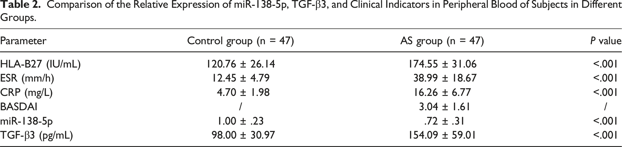
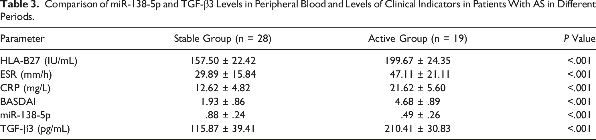
Compared to healthy subjects, reduced miR-138-5p levels and increased TGF-β3 levels were found in AS patient. Even more, level of miR-138-5p was decreased and level of TGF-β3 was found to be increased in active disease stage of AS in comparison to inactive disease. Correlation analysis disclosed that miR-138-5p expression in peripheral blood of AS patients was negatively correlated with TGF-β3, HLA-B27, ESR, CRP, and BASDAI; serum TGF-β3 was positively correlated with HLA-B27, ESR, CRP, and BASDAI. The ROC curve analysis disclosed that miR-138-5p and TGF-β3 had certain diagnostic value for AS, and the combined detection could improve the clinical diagnostic capability of this disease.
Conclusion
miR-138-5p and TGF-β3 in peripheral blood of AS patients are potential biological markers for the diagnosis of AS and are expected to be new clinical diagnostic indicators.
Keywords
Introduction
Ankylosing spondylitis (AS) is known as a chronic inflammatory autoimmune disease that primarily influences the spine, sacroiliac and peripheral joints and can result in significant pain, limitation of motion, and spinal deformity. 1 Several causative factors contribute to the development of AS, including infections, genetic susceptibility, and environmental triggers, particularly immune disorders that could induce the AS onset or be involved in its pathogenesis. 2 Current treatment modalities consist of tumour necrosis factor blockers and non-steroidal anti-inflammatory drugs, while therapeutic modalities targeting the interleukin (IL)-23/IL-17 pathway has recently been revealed to have significance in AS pathogenesis and be beneficial in diverse other inflammatory conditions. 3 To date, there are no criteria for the prompt diagnosis of AS, and the effective therapy methods remain largely undefined to prevent bone destruction and control ankylosis in AS patients. 4 As a result, identifying a potential hallmark is vital to monitor disease activity and seek favorable approaches to prevent and treat this type of irreversible skeletal disease.
MicroRNAs (miRNAs) controls the target mRNA functions and cellular processes, are present in a stable form in human plasma and appear as possible hallmarks for disease activity, pathogenesis, as well as prognosis. 5 Meanwhile, miRNAs represent a potentially powerful candidate to be perfect biomarkers in immune- or autoimmune-associated rheumatic diseases. 6 Identification of specific miRNA expression patterns in autoimmune diseases appears to hold promise not only as novel molecular diagnostic markers but also new therapeutic approaches for treating autoimmune diseases, such as AS. 5 Recently, miR-138-5p has been revealed to play crucial parts in diverse bone diseases, such as osteoarthritis (OA), 7 osteoporosis, 8 and osteosarcoma. 9 Especially, miR-138-5p suppression can enhance chondrocyte growth and migration, and restrain chondrocyte apoptosis and inflammatory response. 7 The transforming growth factor-β (TGF-β) family has gained considerable attention due to its property to induce mesenchymal stem cells toward chondrogenesis via the SMAD pathway activation and upregulation of chondrogenic genes. 10 The TGF-β gene comprises 3 isoforms, namely TGFB1, TGFB2, and TGFB3, which encode protein precursors of TGF-β1, TGF-β2 and TGF-β3, respectively. 11 Particularly, TGF‐β exerts functions inarticular cartilage growth, development, and maintenance 12 and TGF‐β is a critical immunomodulatory protein of B lymphocytes and the activation of TGF‐β results in functional immunomodulatory effects. 13 As reported, TGF-3β is most usually applied in conjunction with delivery systems to impede the apoptosis and hyper-trophy of chondrocytes. 14 Nevertheless, Few studies have directly investigated the relevance of miR-138-5p and TGF-β3 with AS. Therefore, our work was aimed at exploring the expression and clinical significance of miR-138-5p and TGF-β3 in peripheral blood of patients with AS.
Materials and Methods
Ethical Approval
This research was ratified by the Ethics Committee of our hospital (approval number: 20160518). Written informed consent from patients and consent from the hospital ethics committee were obtained for the collection of the above samples.
Study Subjects
According to the diagnostic criteria for the classification of AS recommended by the American College of Rheumatology in 2009, 47 patients with confirmed AS (AS group) admitted to the Department of Rheumatology and Immunology of our hospital were selected as study subjects using the random number method. The staging of the enrolled AS patients was based on the Bath Ankylosing Spondylitis Disease Activity Index (BASDAI) score: 15 <4 points were classified as stable stage (stable group, n = 28) and ≥4 points were classified as active stage (active group, n = 19). Forty-seven cases were randomly selected as the control group from the same period of healthy physical examination our hospital. Inclusion criteria: (1) patients with AS diagnosed by the above diagnostic criteria; (2) patients had positive serum HLA-B27 levels. Exclusion criteria: (1) history of infection in the last 3 months; (2) severe hypertension and diabetes mellitus; (3) history of malignancy; (4) combined spinal degenerative diseases. The subjects selected for the study were excluded from other autoimmune diseases and diseases of vital organ systems. Informed consent from patients and consent from the hospital ethics committee were obtained for the collection of the above samples.
Determination of miR-138-5p Relative Expression in Peripheral Blood
miR-138-5p relative expression in peripheral blood was evaluated by real-time fluorescence quantitative PCR method. Fasting elbow venous blood (5 mL) was harvested from all patients within 24 h of admission and from healthy controls on the day of physical examination, and the total RNAs were extracted using Trizol reagent (Invitrogen, Carlsbad, CA, USA). The ratio of absorbance value at 260 nm to 280 nm was estimated by applying UV spectrophotometer (Eppendorf, Hamburg, Germany), and the ratio of 1.8-2.0 was considered as qualified for purity. The acquired total RNA (1 μg) was taken and reverse transcribed into cDNA using All-in-OneTM miRNA First-Strand cDNA Synthesis Kit (GeneCopoeia, Guangzhou, China), and then was used to perform qRT-PCR using All-in-One miRNA qRT-PCR Detection Kit (GeneCopoeia). qRT-PCR was performed using ABI Prism 7500 system. The relative expression of miR-138-5p was calculated using the 2−ΔΔCt method with U6 as the internal reference. The primers were designed and synthesized by GenePharma (Shanghai, China): miR-138-5p: forward: 5’-AGCTGGTGTTGTGAATCAGGCCG-3’; U6: forward: 5’-GCTTCGGCAGCACATATACTAAAAT-3’; reverse: 5’-CGCTTCACGAATTTGCGTGTCAT-3’.
Measurement of TGF-β3 Expression in Peripheral Blood
TGF-β3 expression in peripheral blood was measured using the TGF-β3 enzyme linked immunosorbent assay (ELISA) kit (Shanghai Jinkang Bioengineering Co., Ltd, Shanghai, China) following the kit requirements.
Detection of Disease Activity Factors
HLA-B27 was tested by flow cytometry (Becton-Dickinson Pharmingen, San Diego, CA, USA), erythrocyte sedimentation rate (ESR) was measured by Weil's method, and C-reactive protein (CRP) was assessed by immunoturbidimetry (Beckman IMMAGE 800; Beckman, Fullerton, California, USA). Ultrasound was utilized to evaluate the lesions of peripheral joint attachment points in AS patients, and the ultrasound was performed using MyLab TM One ultrasound diagnostic instrument (Esaote, Italy).
Statistical Methods
SPSS 20.0 statistical software was adopted for statistical analysis. Measurement data were expressed as mean ± standard deviation and compared by the t test between two groups. Enumeration data were denoted as the number of cases and processed by chi-square test. Correlation analysis was implemented using Pearson test, and ROC curves were plotted for analyzing the diagnostic value of indicators for AS. P < 0.05 indicated a statistically significant difference.
Results
General Information Comparison
Comparison of General Information Between the Two Groups.

Comparison of the Expression of miR-138-5p, TGF-β3 and Clinical Markers Between the Control and AS Groups
Comparison of the Relative Expression of miR-138-5p, TGF-β3, and Clinical Indicators in Peripheral Blood of Subjects in Different Groups.
miR-138-5p and TGF-β3 Levels in Peripheral Blood and Levels of Clinical Indicators in Patients with AS in Different Periods
Comparison of miR-138-5p and TGF-β3 Levels in Peripheral Blood and Levels of Clinical Indicators in Patients With AS in Different Periods.
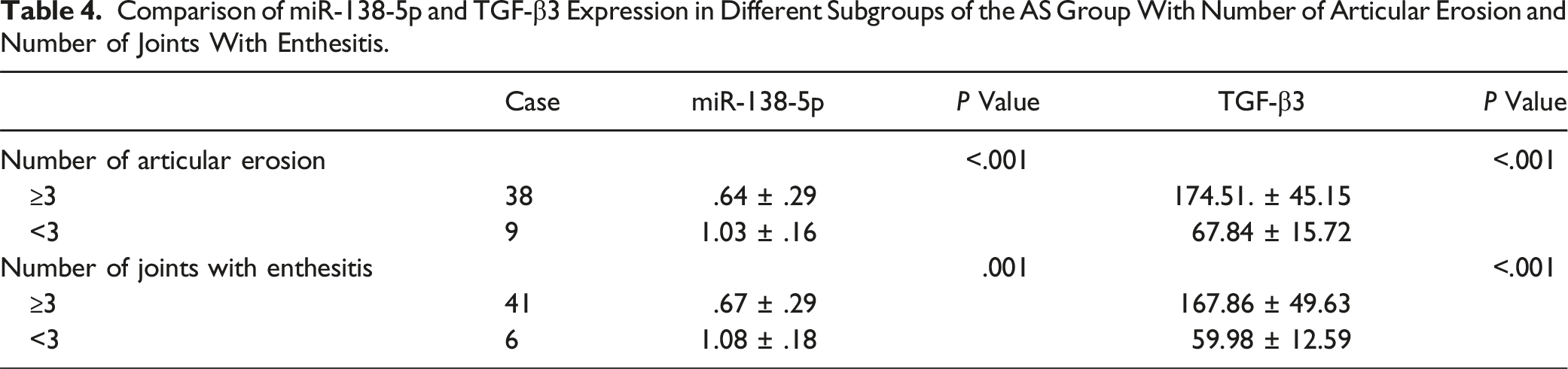
miR-138-5p and TGF-β3 Expression in Different Subgroups of the AS Group with Number of Articular Erosion and Number of Joints with Enthesitis
Comparison of miR-138-5p and TGF-β3 Expression in Different Subgroups of the AS Group With Number of Articular Erosion and Number of Joints With Enthesitis.
Correlation of miR-138-5p and TGF-β3 in Peripheral Blood of AS Patients
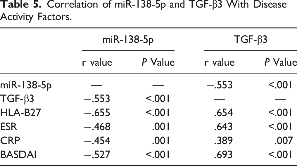
Pearson correlation analysis of miR-138-5p and TGF-β3 in peripheral blood of AS patients unveiled that the expression level of miR-138-5p in peripheral blood of AS patients was negatively correlated with TGF-β3 level (r = −.553, P < .001).
Correlation of miR-138-5p and TGF-β3 With Disease Activity Factors.
Diagnostic Values of miR-138-5p and TGF-β3 for AS
ROC Curve Analysis of miR-138-5p and TGF-β3.
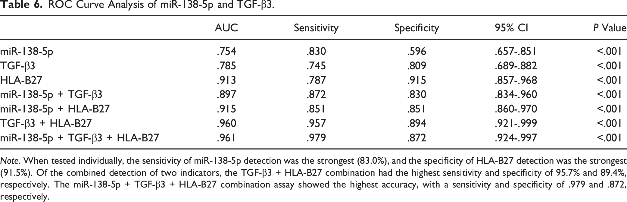
Note. When tested individually, the sensitivity of miR-138-5p detection was the strongest (83.0%), and the specificity of HLA-B27 detection was the strongest (91.5%). Of the combined detection of two indicators, the TGF-β3 + HLA-B27 combination had the highest sensitivity and specificity of 95.7% and 89.4%, respectively. The miR-138-5p + TGF-β3 + HLA-B27 combination assay showed the highest accuracy, with a sensitivity and specificity of .979 and .872, respectively.
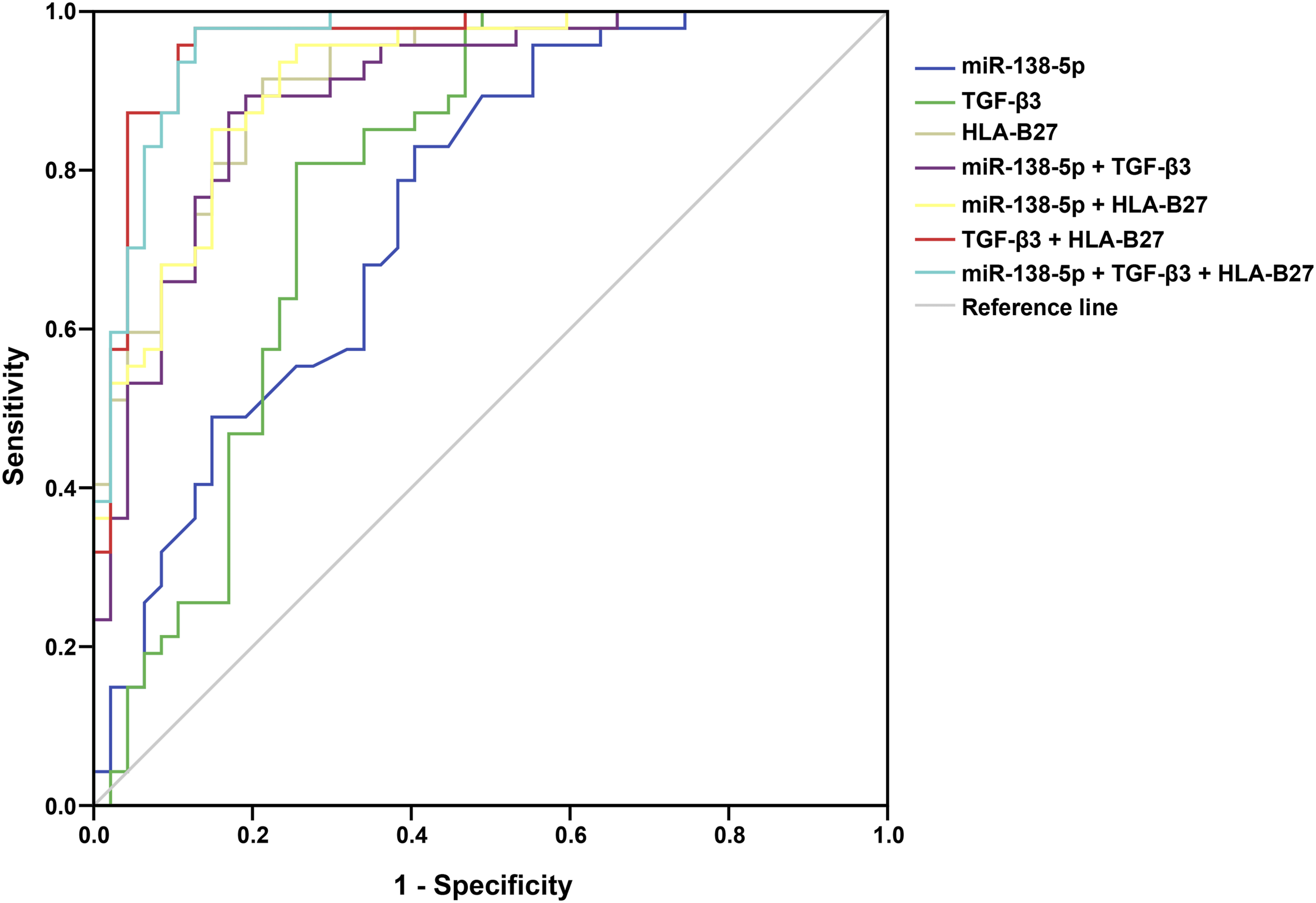
ROC curve analysis for the diagnostic values of miR-138-5p and TGF-β3 for AS. When tested individually, the sensitivity of miR-138-5p detection was the strongest (83.0%), and the specificity of HLA-B27 detection was the strongest (91.5%). Of the combined detection of two indicators, the TGF-β3 + HLA-B27 combination had the highest sensitivity and specificity of 95.7% and 89.4%, respectively. The miR-138-5p + TGF-β3 + HLA-B27 combination assay showed the highest accuracy, with a s-ensitivity and specificity of .979 and .872.
Discussion
AS is a chronic inflammatory joint disease that involves the entheses and the axial joints, causing functional impairments and inflammatory pain. 16 Long delay in diagnosis and inadequate response to existing therapeutics argue for a better recognition of disease pathogenesis. 17 Consequently, in our paper, we aimed to figure out the expression and clinical significance of miR-138-5p and TGF-β3 in peripheral blood of patients with AS. Taken together, this paper demonstrates that miR-138-5p and TGF-β3 in peripheral blood of AS patients are potential biological markers for the diagnosis of AS.
Recent evidence has shown that miRNAs exhibit distinctive expression profiles in cells of the adaptive and innate immune systems and exert functions in modulating cell development and function. 18 In the meantime, the mediation of miRNAs may play a crucial part in AS inflammatory drivers, which are implicated in the process of new bone formation and modulating the function of diverse immune cells. 19 A research has disclosed that miR-138-5p expression is reduced in OA, and depletion of miR-138-5p strengthens the proliferation and migration of human chondrocyte cell lines, while hindering their apoptosis and inflammatory reaction. 7 In our study, we found reduced miR-138-5p levels in peripheral blood of AS patients, and ROC curve suggested that miR-138-5p had a better value for the diagnosis of AS. Zhang et al., 20 have supported that miR-138-5p is downregulated in osteogenic differentiated human bone mesenchymal stem cells, and elevation of miR-138-5p decreases the expression levels of markers for osteodifferentiation, alkaline phosphatase activity, and Alizarin Red staining activity. 20 Another recent article has confirmed that miR-138 level is notably reduced in cartilage tissues of OA patients in contrast to those of normal controls, and reduced miR-138 advances the cartilage tissue destruction among OA patient. 21
TGF-β exerts a dual function in the immune response by either suppressing the generation and release of inflammatory mediators or strengthening the inflammatory response. 22 The role of TGF-β in AS pathogenesis has been clarified, particularly via modulating inflammatory response and bone metabolism. 23 In our study, we found elevated TGF-β3 levels in peripheral blood of AS patients, and ROC curve suggested that TGF-β3 had a better value for the diagnosis of AS. Similarly, TGF-β2 and -3 serum protein levels have been revealed to be significantly higher in OA patients, and serum TGF-β2 and TGF-β3 are positively linked to pain, functionality, as well as radiographic staging in OA patients. 24 In addition, correlation analysis of our study disclosed that miR-138-5p expression in peripheral blood of AS patients was negatively correlated with TGF-β3 level. Zhang et al 25 have stated that miR-138 is time-dependently reduced via TGF-β1 treatment, and TGFβ1-induced reduction of miR-138 leads to epithelial-mesenchymal transition in lung cancer cells. 25 Nevertheless, the relevance of miR-138-5p and TGF-β3 in AS remains to be unearthed.
Furthermore, our work also validated the correlation of miR-138-5p and TGF-β3 with disease activity factors (HLA-B27, ESR, and CRP) and BASDAI. Pearson analysis exhibited that peripheral blood miR-138-5p was negatively correlated with levels of disease activity factors HLA-B27, ESR, CRP, and BASDAI, and TGF-β3 was positively correlated with these parameters. AS has long been linked to inheritance of the HLA-B27, one of the strongest genetic relationships of all common human diseases. 26 All patients received laboratory tests and clinical assessments such as ESR, CRP, and BASDAI. 15 HLA-B27 is very sensitive while harbors a low specificity, whereas ESR and CRP possesses both low specificity and sensitivity. 27 As reported, inflammatory parameters such as ESR or CRP have a poor relevance with BASDAI and have a worse predictive power in longitudinal studies of AS patients. 28 However, the correlation of miR-138-5p and TGF-β3 with disease activity factors in AS patients needs further validation.
In summary, our work suggests that miR-138-5p and TGF-β3 in peripheral blood of AS patients are potential biological markers for the diagnosis of AS and are expected to be new clinical diagnostic indicators. Detection of miR-138-5p and TGF-β3 are ideal candidates to improve the early diagnosis of AS. Further studies including clinical data with more large size, as well as cell experiments, are warranted to verify the findings.
Footnotes
Declaration of conflicting interests
The author(s) declared no potential conflicts of interest with respect to the research, authorship, and/or publication of this article.
Funding
The author(s) received no financial support for the research, authorship, and/or publication of this article.
IRB statement
This research was ratified by the Ethics Committee of The Third Affiliated Hospital of Anhui Medical University (approval number: 20160518). Written informed consent from patients and consent from the hospital ethics committee were obtained for the collection of the above samples.