
Abstract
Study Design:
Basic science.
Objective:
To compare the effects of a neuropeptide Y1 receptor antagonist (NPY-1RA) to estrogen on maintaining vertebral bone microarchitecture and disc height in a rat model of menopause.
Methods:
This study was an institutional animal care approved randomized control study with 104 ovariectomized rats and 32 intact control animals. Comparison of disc height, trabecular bone, body weights, circulating levels of NPY and estrogen, and distribution of Y1 receptors in the intervertebral disc in an established rodent osteoporotic model were made at baseline and after 2, 4, and 8 weeks after receiving either an implant containing estrogen or an antagonist to the neuropeptide Y1 receptor. Data was compared statistically using One-way analysis of variance.
Results:
Circulating levels of estrogen increased and NPY decreased following estrogen replacement, with values comparable to ovary-intact animals. NPY-1RA-treated animals had low estrogen and high NPY circulating levels and were similar to ovariectomized control rats. Both NPY-1RA and estrogen administration were able reduce, menopause associated weight gain. NPY-1RA appeared to restore bone formation and maintain disc height, while estrogen replacement prevented further bone loss.
Conclusion:
NPY-1RA in osteoporotic rats activates osteoblast production of bone and decreased marrow and body fat more effectively than estrogen replacement when delivered in similar concentrations. Annulus cells had NPY receptors, which may play a role in disc nutrition, extracellular matrix production, and pain signaling cascades.
Introduction
Osteoporosis is a worldwide health problem that affects 200 million women and accounts for >17 billion dollars in direct care cost. 1 Osteoporosis occurs primarily in women, with post-menopausal women having the highest risk. Clinical studies have shown that vertebral fractures account for 20% of postmenopausal fractures, and women diagnosed with a vertebral fracture have an increased risk of sustaining additional vertebral fractures and for morbidity and mortality.2-5 Osteoporosis is characterized by low bone mass and deterioration of microarchitectural structure of the bone. Osteoblasts and osteoclasts have receptors for estrogen, and estrogen deficiency is a major risk factor for inducing bone loss. 6 Age-related osteoporosis is accompanied by an increase in marrow adipose tissue, implying an adipogenic process may contribute to bone loss. 7
While lower estrogen levels certainly correlate with decreased bone density, this hormone may not be solely responsible for osteoporosis. The regulation of bone remodeling has classically been associated with hormones, mechanical load, and the production of local bone factors. 8 In addition, recent reports provide evidence that bone growth is under the influence of both central and neural control.9-12 Clinical studies show that blockade of specific sympathetic adrenergic receptors results in increased bone density.13-15 Neuropeptide fibers and neuropeptide Y (NPY) receptors have been linked to bone density and may have a more direct responsibility in the development of osteoporosis. NPY is secreted by the hypothalamus, has circulating levels inversely proportional to estrogen, and is inversely related to bone mineral density. NPY receptors have been found on osteoblasts, osteoclasts, annulus cells, blood vessels, platelets, adipocytes, and macrophage cells.16-19 Exogenous administration of NPY is associated with decreased bone mass,12,20 while NPY knockout (germline and hypothalamic Y1) animals show increased bone densities.
Decrease in disc height is strongly correlated with estrogen withdrawal at menopause.21,22 Estrogen is important because it has a significant impact on maintaining hydrophilic glycosaminoglycans, water content, collagen, and elastin of the intervertebral discs. This suggests that the metabolic changes related to disc degeneration are under endocrine control.23,24 NPY has been shown to be associated with cells in the outer annulus and in the vascular buds, but its role in the intervertebral disc during menopause has not been fully defined. In this study, we hypothesize that local sustained release of a selective neuropeptide Y1 receptor antagonist (NPY-1RA) will delay the progression of intervertebral endplate calcification in an ovariectomized rat model of osteoporosis comparable to estrogen.
Materials and Methods
Experimental Design
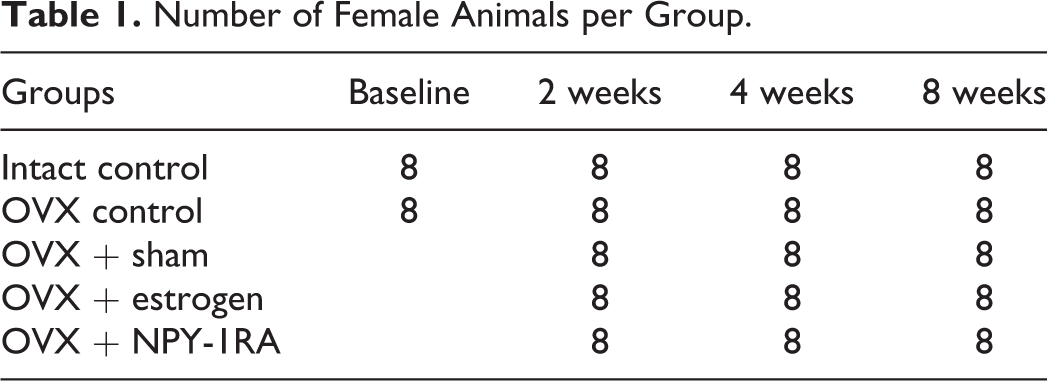
Following approval from the University of Mississippi Medical Center Institutional Animal Care and Use Committee, a total of 104 ovariectomized (OVX) 3-month-old Sprague-Dawley rats were divided into 4 groups along with 32 intact control rats were used in the study (Table 1). Five weeks postovariectomy, 8 rats from the OVX control group and 8 rats from the intact control group were euthanized to obtain baseline values for blood and bone density, and OVX animals in groups 2 to 4 underwent surgical implantation of a tricalcium phosphate (TCP) delivery device. At 2, 4, and 8 weeks postimplant treatment, 8 rats per group were euthanized. Blood levels of estrogen and NPY were analyzed; organs and L4-5 vertebrae were collected; histological evaluations were performed; and the data was compared and evaluated for statistical differences.
Number of Female Animals per Group.
TCP Preparation and Drug Delivery
The microcrystals of calcium phosphate were prepared by following standard laboratory protocol. 25 Briefly, microcrystals of TCP powder were prepared by mixing 70% calcium nitrate with 100 mL of ammonium hydroxide. The resulting precipitate was collected by centrifugation and washed 3 times with distilled water. The precipitate was resuspended in 3% ammonium sulfate and dried at 75 °C. 26 The TCP microcrystals (<38 µm) were calcined at 1200 °C, sieved then sintered at 1200 °C for 36 hours. TCP (0.5 g) material alone or combined with estrogen or NPY-1RA (Sigma, St. Louis, MO) were cold pressed into capsules capable of delivering 5 ng/d.
Surgical Approach
The animals were given an intraperitoneal (IP) injection of a ketamine/xylazine. The area around L4-5 was shaved, scrubbed, and draped for surgery. A 1-inch longitudinal right paramedian incision was made, and the external oblique muscle was exposed and a 0.5 mm × 0.4 mm sterile TCP implant was implanted adjacent to the spine and the fascia was reapproximated with interrupted 3-0 Vicryl, and the skin was closed with wound clips. Carprofen (5 mg/kg) was given subcutaneously (SC) just prior to surgery and once daily for 3 days. The wounds were checked daily and the staples were removed at day 10.
Necropsy
At 2, 4, and 8 weeks postsurgery, the animals were sacrificed. Blood, vital organs, and spine were collected and processed for biochemical or histopathological analysis.
Blood Analysis
Neuropeptide Y
A commercially available colorimetric competitive enzyme- linked immunosorbent assay (ELISA) was used to obtain quantitative measurements of NPY in serum (Abnova, Walnut Hills, CA). Standards or serum were pipetted into NPY-coated wells and the manufacturer instructions were followed. The intensity of the color of the samples was compared with a standard curve and the amount of NPY in the sample was determined.
Estradiol (E2)
A commercially available ELISA kit was used to obtain quantitative concentrations of estradiol (ALPCO). Briefly, standards, or rat serum were dispensed in duplicate into the microtiter wells followed by an additional 50 µL incubation buffer. The plate was incubated for 120 minutes, followed by the addition of 50 µL of an enzyme conjugate for an additional 60 minutes. The contents of the well were discarded, and the wells were washed 4 times and 200 µL of substrate solution was added for 30 minutes. Following the incubation, 50 µL of stop solution was added and the absorbance was determined at 450 nm. The specimens and controls were run concurrently with the standards, and the concentration of E2 was calculated from the standard curve.
Histology
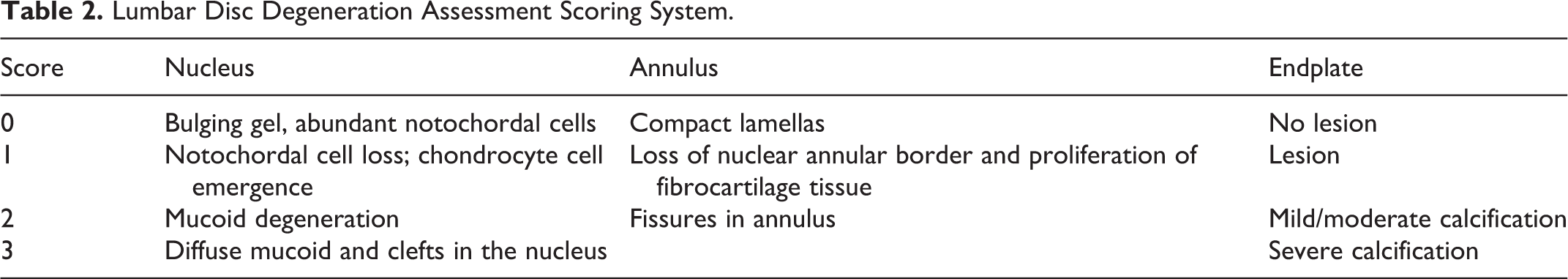
The L4-5 disc samples were embedded in paraffin, following decalcification in CalEx II solution (Fisher Scientific, Pittsburgh, PA). Each specimen was cut sagittally into sections of 10 µm in thickness and stained with hematoxylin and eosin. Disc degeneration was graded by 4 independent observers using a scale similar to that reported by Wang et al. 27 The scoring system takes into account changes in the nucleus pulposa, annulus, and cartilage endplate. The scores increase according to the degree of degeneration observed in each of the tissues (nucleus pulposa 0-3, annulus 0-2, and cartilage endplate 0-3) (Table 2). The degenerative discs were graded on a scale of I to IV as follows: I—normal (score of 0-1), II—mild (score 2-3), III—moderate (score 4-5), and IV—severe (score >5). The 4 observers were blinded to the treatment groups, and the slides were independently reviewed. A second review was repeated 2 weeks later by each reviewer on the same set of slides that were randomly ordered. Agreement and interrater reliability were determined.
Lumbar Disc Degeneration Assessment Scoring System.
Disc Height Measurements
L4-5 disc height was measured as the distance between the L4 and L5 endplates. Images were taken using a 4× objective and 10× ocular lens for a total magnification of 40×. NIH ImageJ software was calibrated using a standard micrometer calibration slide (Fisher Scientific, Pittsburgh, PA), and the lengths between the endplates (5 measurements per disc and 8 animals per group) were determined in millimeters.
Micro-Computed Tomography Analysis
A SkyScan 1172 micro-computed tomography (micro-CT) scanner with a 10-megapixel camera (Skyscan, Aartselaar, Belgium) was used for high-resolution analysis of the endplate thickness and underlying trabecular bone in naïve control, OVX control, and treated groups. The scanning parameters were as follows: accelerating voltage of 49 kV, current of 200 μA, exposure time of 474 ms per frame, Al 0.5 mm filter, rotation step at 0.700° (180° rotation), and image pixel size of 34.04 μm. After scanning, the x-ray projections were reconstructed by thresholding grayscale images with values that discriminated between bone, marrow, and disc tissue to form a 3-dimensional model, which was saved as a stack of BMP-type files using NRecon software (Skyscan, Aartselaar, Belgium). Beam hardening correction of 60% and ring artifact correction of 3 were used for the reconstruction. Structural indices were calculated using the CTAn software (Skyscan, Aartselaar, Belgium).
Statistical Analysis
Data was analyzed using SigmaPlot (Systat Software, San Jose, CA). The descriptive data is reported as mean ± standard deviation. For baseline data between 2 groups, the Student t test was used and for comparison of multiple treatment groups, One-way analysis of variance (ANOVA) was used to determine differences in the mean values. Student Newman-Keuls post hoc tests were used when differences were found for parametric data, and Kruskal-Wallis tests were used for nonparametric data.
Results
Baseline Evaluation
Five weeks post-OVX, animals were 30% larger than their intact control litter mates (P < .05) (Table 3), and their circulating estrogen levels were reduced from 39 to 9 pg/mL (P < .05), while NPY levels increased from 0.14 to 0.18 pg/mL (P > .05).
Baseline Values for Body Weight and Circulating Estradiol Levels at 5 Weeks Postovariectomy.a
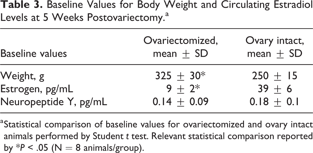
a Statistical comparison of baseline values for ovariectomized and ovary intact animals performed by Student t test. Relevant statistical comparison reported by *P < .05 (N = 8 animals/group).
Post Estrogen and NPY-1RA Delivery
Ovariectomized animals were significantly larger than intact control animals for the duration of the study (Table 4 and Figure 1). However, body weights of the OVX + estrogen and OVX + NPY-1RA-treated animals were significantly less when compared with OVX control and OVX + sham implant groups (Table 4). At necropsy it was noted that the animals receiving sustained delivery of estrogen and NPY-1RA had less central body fat compared with the OVX + sham and OVX control.
Body Weights Following Implantation of Empty TCP Sham, Estrogen, or NPY-1RA Into Ovariectomized Sprague-Dawley rats at 2, 4, and 8 Weeks.
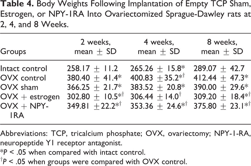
Abbreviations: TCP, tricalcium phosphate; OVX, ovariectomy; NPY-1-RA, neuropeptide Y1 receptor antagonist.
*P < .05 when compared with intact control.
†P < .05 when groups were compared with OVX control.
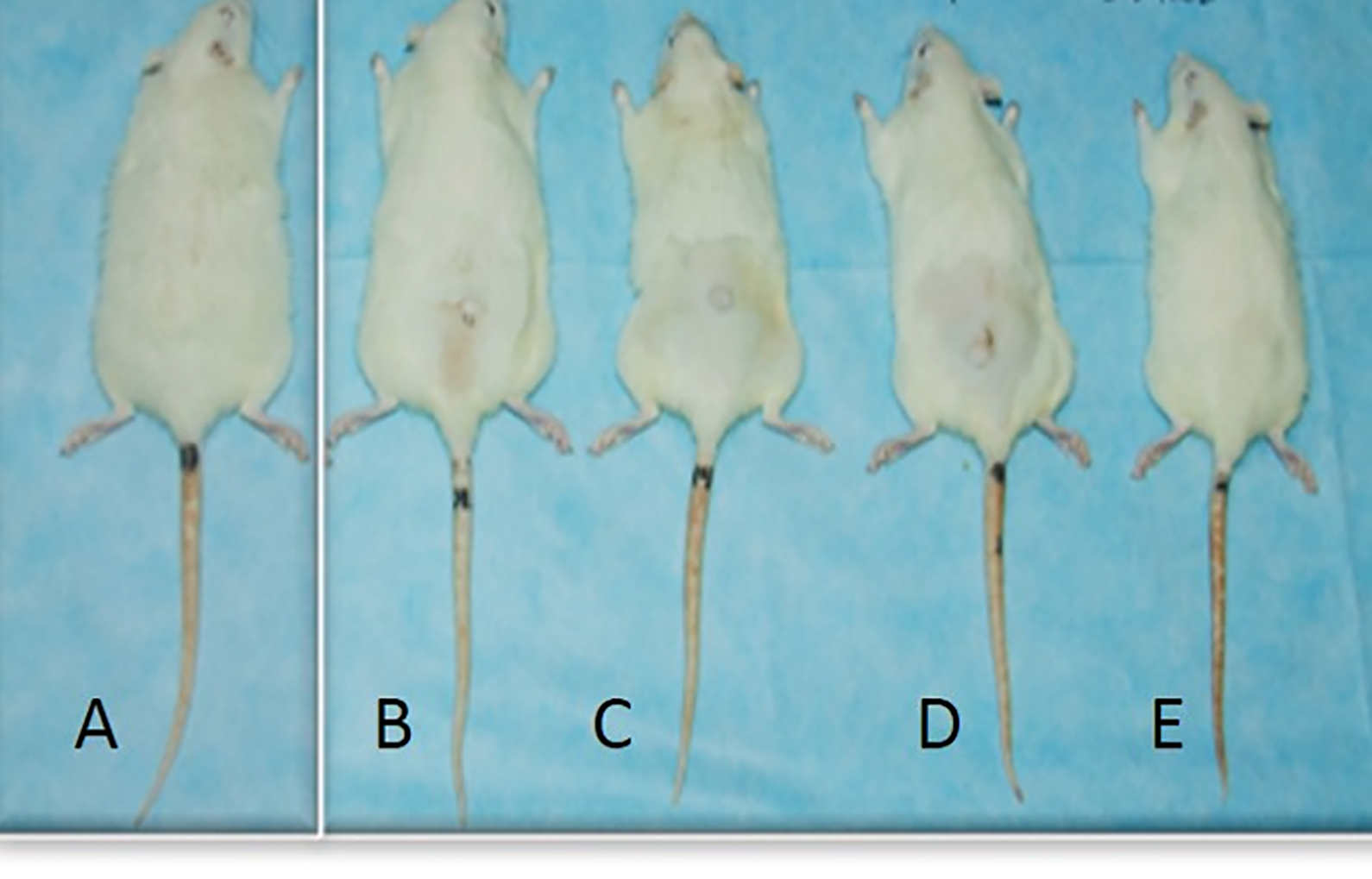
Representative changes in body size following administration of compounds after 2 weeks of tricalcium phosphate (TCP) implantation of sham or TCP drug carrier. Note: A (Ovx), B (Sham), C (Estrogen), D (NPY-1RA), E (Control).
Blood Analysis
Estradiol Levels
Intact control animals had estrogen levels ranging from 38 to 43 pg/mL for the duration of the study. Animals in OVX control, OVX + sham, and OVX + NPY-1RA groups had estrogen levels measuring from 5 to 9 pg/mL for the duration of the study. Animals receiving a TCP implant containing estradiol had estrogen levels ranging from 60 to 90 pg/mL after 2 weeks, followed by estrogen levels ranging from 40 to 45 pg/mL for the duration of the study (Figure 2).
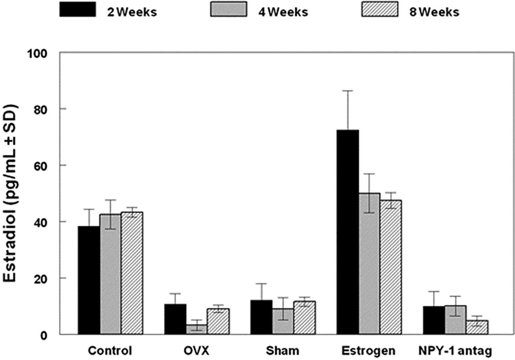
Serum levels of estradiol beginning 2 weeks postimplantation of tricalcium phosphate (TCP) delivery devices. Data is represented as mean ± SD (n = 8 per group). Enzyme-linked immunosorbent assay (ELISA) was run in triplicate for each sample (*indicates significant difference from the control at each time point).
Circulating NPY Levels
Animals in the intact control and OVX + estrogen groups had NPY levels that were significantly lower than animals in the OVX control, OVX + sham, and OVX + NPY-1RA groups (Figure 3).
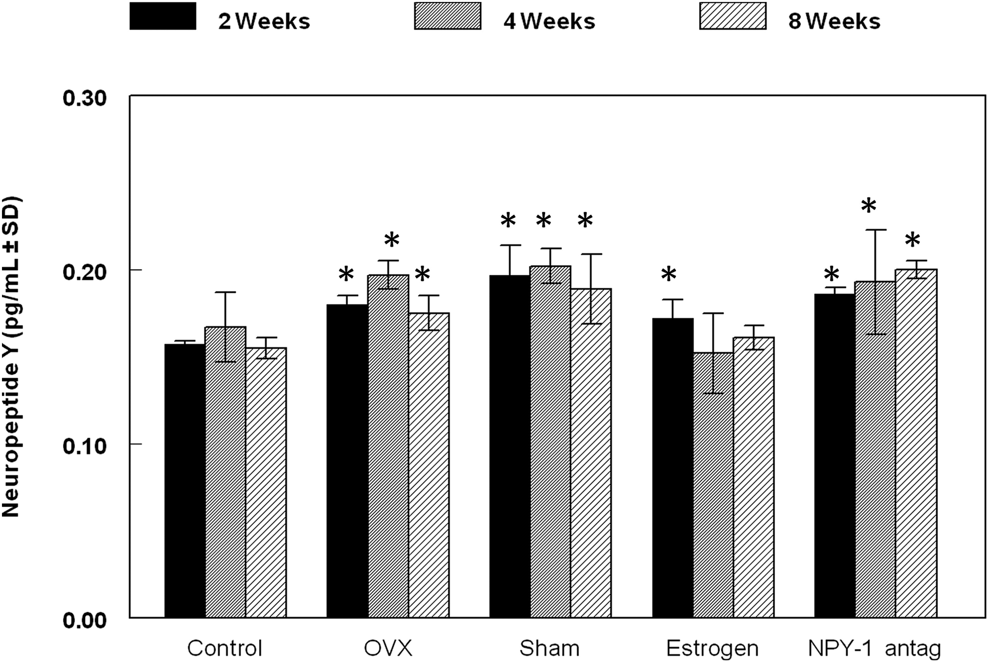
Serum levels of neuropeptide (NPY) beginning 2 weeks postimplantation of tricalcium phosphate (TCP) delivery devices. Data is represented as mean ± SD (n= 8 per group). Enzyme-linked immunosorbent assay (ELISA) was run in triplicate for each sample (* indicates significant difference from the control at each time point).
Disc Height
Disc height changes were evident 5 weeks post-OVX and were significantly different in the OVX control and OVX + sham animals compared with intact control 8 weeks post drug delivery surgery (Table 5). Disc degeneration and calcification of endplates was observed in all OVX groups (Figure 4). Animals receiving sustained delivery of estrogen or NPY-1RA had smaller regions of calcification and lower lumbar disc assessment scores (Table 5). Inter- and intrarater agreement between the 4 independent reviewers in disc assessment showed moderate (intraclass correlation coefficient [ICC] = 0.49) to strong (ICC = 0.76) interclass correlation (kappa ≥ 0.65). We considered ICC and kappa >0.75 as excellent, 0.45-0.75 as moderate, and scores less than 0.4 were considered poor or no agreement.
Hounsfield Units (HU), Mean ± SD (n = 3 Animals per Group).a
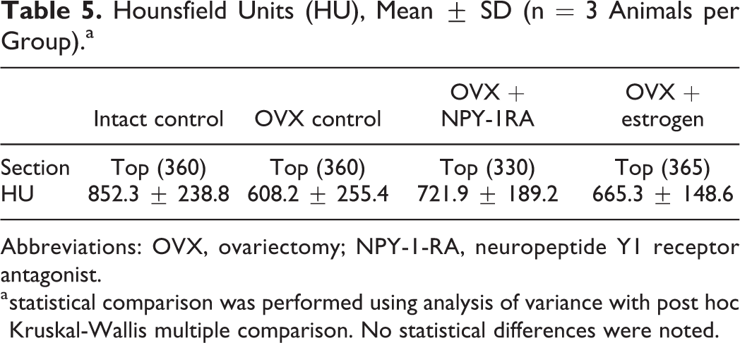
Abbreviations: OVX, ovariectomy; NPY-1-RA, neuropeptide Y1 receptor antagonist.
a statistical comparison was performed using analysis of variance with post hoc Kruskal-Wallis multiple comparison. No statistical differences were noted.
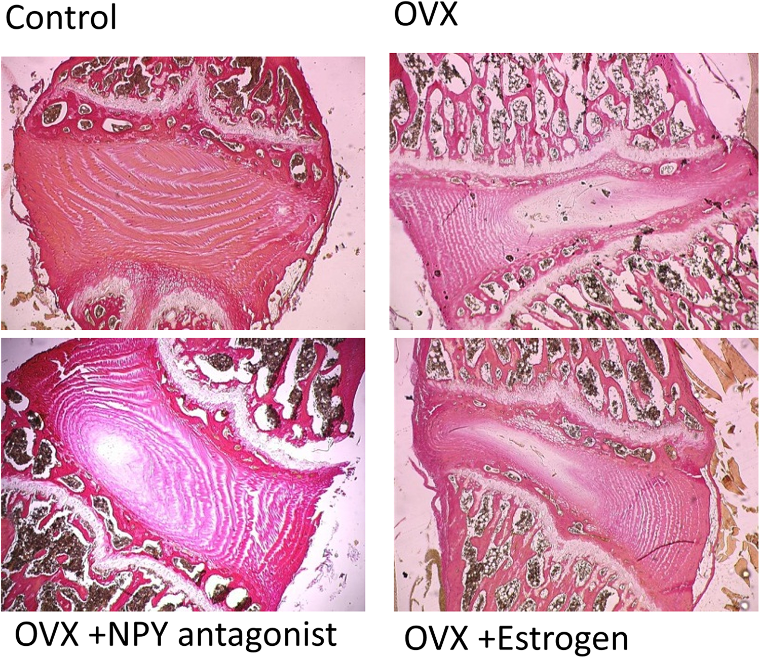
Histological illustration van Gieson staining of the L5-6 segments of the lumbar spine at 2 months postsurgery. Images were captured using 2× objective for a total magnification of 20×.
The intervertebral discs in the control group appeared normal. The nucleus pulposus contained abundant notochordal cells surrounded by large zones of extracellular matrix; the cartilage endplates were hyaline cartilage composed of chondrocytes; and the fibrocartilage of the annulus were arranged in lamellae (Figures 4 and 5).
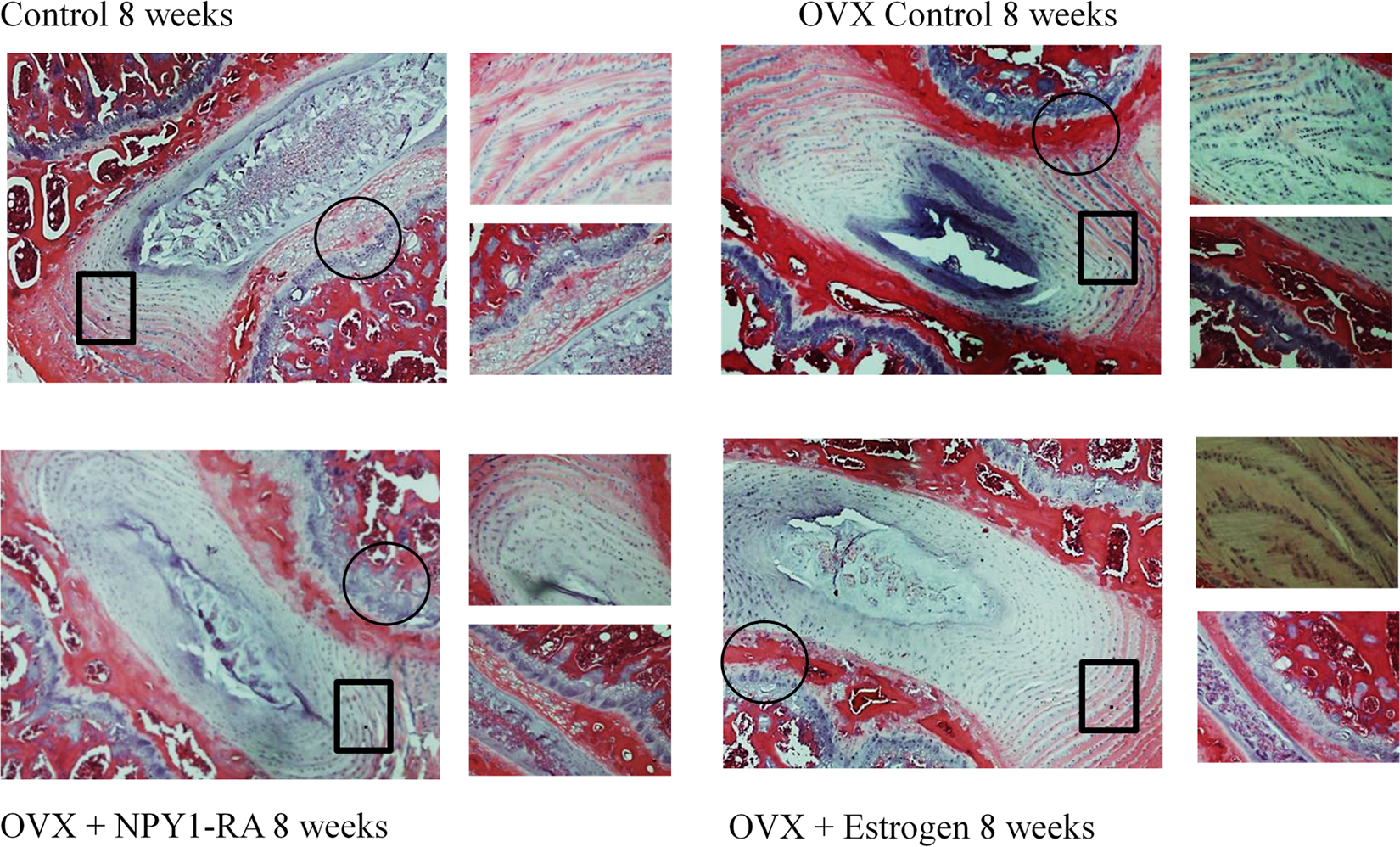
Images of the discs were taken at 10X. The black box is represented at 40X view in the top panel to the right, and the black circle is magnified (40X) in the bottom panel to the right.
In the OVX control and OVX + sham implant group, the discs showed degenerative changes where the nucleus pulposus appeared reduced and composed of relatively few clustered doublets of chondrocyte-like cells. Mucoid degeneration could also be seen eroding the nucleus pulposus, with clefts forming within them, and an increased number of small chondrocytes appeared in the inner layer of the annulus.
Bony tissues that contained bone marrow, hematopoietic lineage cells, and mineralized bone became more obvious in the deep zone of the middle cartilage endplate (Figure 5). In OVX + estrogen and OVX + NPY1-RA there were no significant differences in the number of the notochordal cells, with only a few chondrocyte-like cells appearing in the nucleus pulposus and some small chondrocytes in the inner layer of the annulus.
Mucoid degeneration was not observed in most samples in the OVX + NPY-1RA group, and there was less bony tissue forming in the middle cartilage endplate (Figures 4 and 5).
Immunostaining for NPY 1 Receptor in Disc
The presence of NPY Y1 receptors was located on the annulus cells (Figure 6) in all groups. There did not appear to be differences in the number of positively stained cells between the groups. However, the quality of the staining in the OVX groups was recognizably different and found in areas with significant calcifications in addition to the annulus. NPY-1 receptor staining was also present in the end plate and around the vascular buds in all groups.
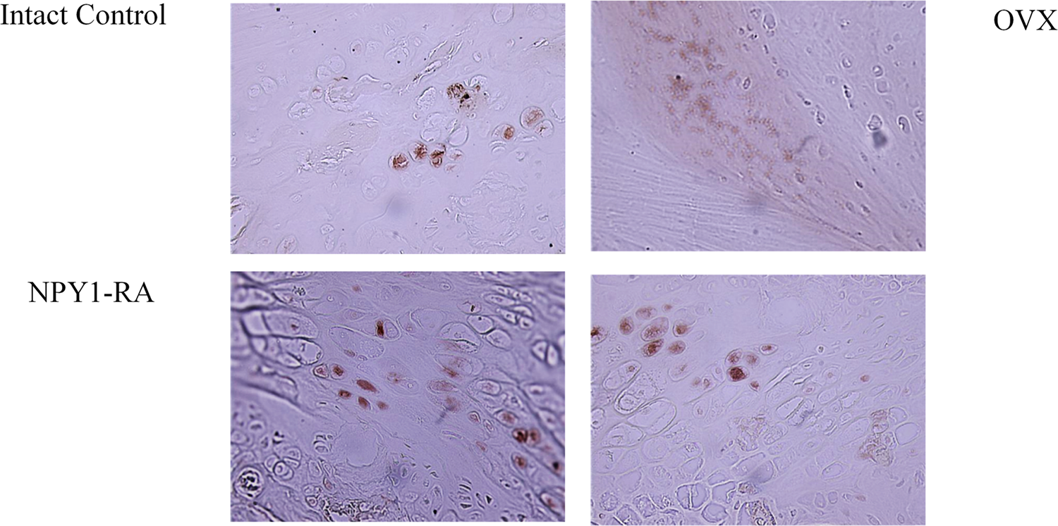
Photomicrograph representation of immunostaining using antibody against neuropeptide Y-1 (NPY-1) receptor in the disc. Positive staining is indicated by the dark brown deposition of DAB (3,3′-diaminobenzidine).
Micro-CT Bone Density
Three-dimensional micro-CT reconstructions demonstrated differences between intact control, OVX control, OVX + estrogen and OVX + NPY-1RA-treated animals (Figure 7). Calculation of Hounsfield units (HU) for each group showed the ovary intact control animals had the highest HU (852.3 ± 238.8) followed by NPY-1RA (721.9 ± 189.2) and estrogen (665.3 ± 148.6). Both treated groups had HU values that were higher than the OVX animals without treatment (608.2 ± 255.4).
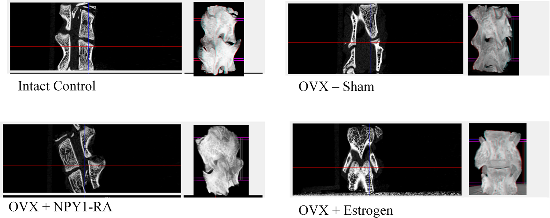
Representative micro-computed tomography (micro-CT) reconstruction of the L4/L5 region.
Discussion
Menopausal women have a tendency to undergo accelerated bone loss and disc degeneration.21-23 Replacement estrogen therapy has been widely used for the treatment of postmenopausal osteoporosis. Estrogen does not increase bone formation but improves the biomechanical properties of the vertebrae and protects against microstructural changes that lead to disc degeneration. Although no single animal model accurately mimics post-menopausal osteoporosis, the OVX rat model shows a pattern of bone loss, increased bone turnover rates similar to humans, and is suitable for the evaluation of estrogen replacement and potential therapeutic agents. 28 In our study, estrogen replacement was not able to reverse bone loss to premenopausal values, but it was able to maintain the existing bone, which is in agreement with human studies and with the OVX rat literature.27-29 Neuropeptide Y is a complex neuropeptide that is involved with blood flow, satiety, metabolism, immune function, and reproduction.16-18 It is unclear the role NPY plays at menopause; however, the diverse effects it has on the body may explain the changes in body composition and bone loss at menopause. In this study, circulating levels of plasma NPY were increased following estrogen loss and returned toward control levels when estrogen replacement was provided. Blockade of the Y1 receptor had no effect on circulating plasma NPY or estrogen levels, but it did have a significant effect on bone formation. The data suggests that estrogen alone may not be fully responsible for bone maintenance. Both estrogen and Y1 receptors are located on the bone, but only the blockade of the Y1 receptor resulted in bone formation. This is consistent with findings in the literature for loss of NPY-1 receptor. There are several sources of NPY production in the periphery. It is produced by sympathetic nerves, 30 the adrenal medulla, 31 vascular smooth muscle, osteoblast, and osteocytes.16,32 Neuropeptide Y production by the osteoblasts or osteocytes can regulate bone mass in both an autocrine and paracrine manner via the Y1 receptor.16,32,33 Isolated calvarial osteoblast studies show addition of NPY decreased bone cell numbers along with the expression of osteocalcin and decreased mineralization.34-36 Osteoblast cells isolated from Y1 receptor knockout mice showed no effect on bone cell numbers or cellular maturation. 16 Germline deletion of Y1 receptors leads to increased proliferation and differentiation of mesenchymal progenitor cells. 37
Weakening of the trabecular bone may influence the shape and support of the vertebrae leading to decreases in disc height. Ding et al 38 have shown that estrogen withdrawal in rats leads to cartilage endplate calcification and disruption of the structural integrity of the intervertebral disc. 39 In our study, supplementation of either estrogen or NPY-1RA improved disc height when compared with ovariectomized animals without treatment. A decrease in bone mineral density reduces bone loading strength, which subsequently affects the disc metabolism. Deng et al 29 used a sensitive magnetic resonance imaging technique and determined impaired disc diffusion in ovariectomized rats.
Diffusion of nutrients into the disc is extremely important for maintaining the annulus and nucleus. Intervertebral disc height depends on a proportionate distribution of collagen types, elastin, hydrophilic glycosaminoglycans, and water.40-42 It has been shown that premenopausal women have intervertebral discs that contain higher concentrations of types II, IV, and IX collagen along with collagen I than women at menopause. At menopause, types I, III, and VI predominate along with significant decreases in elastin and glycosaminoglycans, which alters the hydration of the tissue.40,43,44 In humans, estrogen has been shown to significantly increase the synthesis of sulphated glycosaminoglycans, 45 and women with post-menopausal osteoporosis had significant increases in urinary excretion of calcium and glycosaminoglycan. 46 In our study, ovariectomized animals receiving either 5 ng/mL/d estrogen or NPY-1RA showed lower lumbar disc scores and smaller regions of calcification in the endplate when compared to ovariectomized animals without treatment. Additional studies are needed to assess the glycosaminoglycan content of the discs. Our study is the first to show an effect of blocking Y1 receptors and maintenance of the disc height. It is possible NPY may decrease glycosaminoglycan production or increase metalloproteases whereas blockade of the receptor preserves the degradation or improves the synthesis of the extracellular matrix. Work by Salo et al 47 supports our theory regarding the role of NPY on the extracellular matrix. They found that NPY stimulation of ligament cells increased inflammatory mediators in vitro while significantly lowering mRNA levels for growth factors and matrix molecules. In addition, recent findings demonstrate rabbit annulus fibrous cells express NPY 1 receptors and under strain, the cells are capable of producing NPY and in the presence of inflammatory products (IL-1), the production of NPY increases, as does metalloproteases. 48
Estrogen receptors (ERα and ERβ) are found in the nucleus pulposus of both male and female animals, with less density in severely degenerated discs.49-51 Currently, there is minimal information regarding the function or specific action of the estrogen receptors located within the nucleus pulposus. The mechanism(s) for NPY on bone building and disc maintenance also have not been clearly established. Interestingly, we have found Y1 receptors in the annulus of the disc, on the chondrocytes, and around the vascular buds in the endplate. This information is critical for understanding a possible mechanism for movement of nutrients into the disc. Stimulation of the Y1 receptors on blood vessels results in constriction, and antagonism of the receptors improves blood flow, which can improve oxygen and nutrient flow into the disc. The increased supply of nutrients and oxygen will contribute to maintaining spinal structural integrity and suppressing cartilage endplate calcification, thereby supporting the overall function of intervertebral discs. Neuropeptide Y nerve fibers have been implicated as a player in both nociceptive and neuropathic pain, and the location of the Y1 receptors on annulus cells suggest a possible role for NPY in discogenic pain. Sowa et al 52 have demonstrated an increase in circulating NPY levels in patients with low back pain.
The results of our study suggest a clear role for the Y1 receptor in both bone and disc maintenance. Contribution of the Y1 receptor for bone loss is well established; however, more research is needed to determine the role of NPY and the Y1 receptor within the disc.
In summary, our data demonstrates that NPY-1RA in osteoporotic rats activates osteoblast production of bone and decreases marrow and body fat more effectively than the gold standard estrogen replacement when delivered in similar concentrations. This study also shows the presence of the receptor on the annulus cells, which may play a crucial role in disc nutrition, extracellular matrix production, and pain signaling cascades.
Footnotes
Declaration of Conflicting Interests
The author(s) declared the following potential conflicts of interest with respect to the research, authorship, and/or publication of this article: MT received funding for this project from CPP FFOB 2010 closed call. RM is president of the AO Spine Foundation and is a consultant for Rehab, Inc and Zavation. HB is executive director for the Mississippi Academy of Sciences and is vice president for Rocky Mountain Bioengineering Symposium.
Funding
The author(s) disclosed receipt of the following financial support for the research, authorship, and/or publication of this article: MT received funding for this project from CPP FFOB 2010 closed call.