
Abstract
The present study determined the effects of electroacupuncture on interstitial cells of Cajal and investigated whether changes in the stem cell factor pathway were involved. Animals were assigned to normal, diabetic, diabetic plus sham stimulation, diabetic plus low-frequency stimulation, and diabetic plus high-frequency stimulation groups. Electroacupuncture was performed daily for 8 weeks. In vitro contractility of colonic muscle strips were studied. Expression of c-kit (the marker of interstitial cells of Cajal) and stem cell factor were measured. The results showed that (1) contraction of colonic muscle strips was significantly elevated in low- and high-frequency stimulation groups and (2) in contrast to the diabetic group, the expressions of c-kit and stem cell factor were markedly increased in the low- and high-frequency stimulation groups. These results indicate that both low- and high-frequency stimulation can promote the contractility of colonic muscle strips partially through increasing the number of interstitial cells of Cajal, and these effects could be mediated by an elevated endogenous stem cell factor.
Disorders of gastrointestinal motility are known to occur frequently in diabetic patients, including gastroparesis, constipation, and diarrhea, 1 and affect blood glucose control and the quality of life in patients with diabetes mellitus. The mechanism of gastrointestinal dysmotility has been associated with pathological changes in interstitial cells of Cajal. 2 In patients with diabetes, the phenomena of cellular degradation and decrease in the number of interstitial cells of Cajal affect the whole gastrointestinal tract.
Electroacupuncture, a modification of acupuncture, which stimulates acupoints with electrical current instead of manual manipulation, is a potential method of treating gastrointestinal motility disorders. 3 Although extensive studies have investigated the effect of electroacupuncture on gastric motility,4–6 relatively little information exists to address its effect on the colon. Electroacupuncture at the Zusanli (ST-36) acupoint has been reported to accelerate colonic motility in conscious rats. 7 Our previous study has found that electroacupuncture at ST-36 can promote distal colonic contractility through the cholinergic pathway. 8 Jin et al 9 suggested that electroacupuncture is effective in the treatment of constipation. Recently, it has been documented that electroacupuncture can increase the density of interstitial cells of Cajal 10 and upregulate the expression of colonic c-kit (the marker of interstitial cells of Cajal) in slow-transit constipated rats. 11 However, the potential role of electroacupuncture in the interstitial cells of Cajal in diabetic rats have scarcely been investigated.
Interstitial cells of Cajal express c-kit protein (a receptor tyrosine kinase integral to interstitial cells of Cajal), which is the receptor for the stem cell factor. The function and survival of interstitial cells of Cajal depend on the activation of c-kit by the stem cell factor. The stem cell factor includes 2 isoforms: the soluble stem cell factor and membrane-bound stem cell factor. The binding of c-kit and stem cell factor can activate tyrosine kinase and a series of downstream signaling events that have been known to be essential for the maintenance of interstitial cells of Cajal phenotypes and differentiation and proliferation. 12 Reduced expression of the stem cell factor has been reported in the colon of diabetic animals. 13 Recently, it has been reported that electroacupuncture could elevate stem cell factor levels.10,14 However, it is unknown whether the regulative role of electroacupuncture on interstitial cells of Cajal is associated with the stem cell factor pathway.
The aims of our study were to evaluate the effect of electroacupuncture at ST-36 on interstitial cells of Cajal in the colon of diabetic rats and to investigate whether the stem cell factor signaling pathway was involved in the regulative effect of electroacupuncture on interstitial cells of Cajal.
Materials and Methods
Animals
A total of 50 adult male Sprague-Dawley rats (250-350 g) were used in the present study. Rats were purchased from the Experiment Animal Center of Tongji Medical College, Huazhong University of Science and Technology, Wuhan, China. They were housed under normal laboratory conditions (12/12-hour light-dark cycle, 18°C-22°C) and were given water ad libitum. The study was started after 1 week, when the rats were adapted to the environment. All animals in our study were treated strictly in accordance with the National Institutes of Health Guide for the Care and Use of Laboratory Animals, and the work was approved by the ethics committee of laboratory animals.
Experimental Protocols
Animals were randomly divided into 5 groups: normal control group, diabetic group, diabetic plus sham stimulation group (only acupuncture but no electrical current, 30 minutes), diabetic plus low-frequency stimulation group (10 Hz, 1-3 mA, 30 minutes), diabetic plus high-frequency stimulation group (100 Hz, 1-3 mA, 30 minutes). Diabetes was induced by intraperitoneal injection with streptozotocin (Alexis Biochemicals, San Diego, CA) 60 mg kg−1 prepared in citrate buffer (pH 4.5, Sigma, St Louis, MO, USA). Age-matched controls were injected with citrate buffer at the same dose as the diabetic group. The food and water supply was sufficient for free intake by animals during the experimental period. Blood glucose and body weight were measured before and 1 week after injection as well as prior to obtaining specimens. Animals were considered diabetic if the random blood glucose 1 week after streptozotocin injection was higher than 16.7 mmol/L.
Electroacupuncture was performed at ST-36 daily for 8 weeks. 8 At the end of 8 weeks, animals were killed, and specimens of the distal colon were obtained from each rat. Each specimen collected from the animals was divided into 4 pieces of similar size, which were then either immediately immersed in Kreb’s solution used for the contractility study, or snap frozen in liquid nitrogen and stored at −80°C for real-time polymerase chain reaction (PCR) and Western blot analyses, or placed into 4% paraformaldehyde for immunohistochemistry analysis.
Measurement and Analysis of the Contractile Activity of Colonic Muscle Strips
In vitro experiments were performed on the distal colon. Circular muscle strips measuring 7 × 2 × 1 mm3 were cut from the distal colon. 15 Each strip was carefully tied at both ends by threads and immediately mounted on the circular axis in a 25-mL organ bath filled with warmed Kreb’s solution with the following composition (mmol/L): 119 NaCl, 4.7 KCl, 1.2 MgSO4, 1.2 K2HPO4, 2.5 CaCl2, 25 NaHCO3, and 11.1 D-glucose. The organ bath was maintained at 37°C and continuously gassed with 5% CO2 and 95% oxygen to maintain a pH of 7.4. The upper part of the thread was connected to isometric force transducers (Fort-10, WPI, USA), which were connected to a chart recorder (MP-100 system) to record contractile activity concurrently. Data were analyzed by Acknowledge 3.7.1 (Biopac Systems Inc, USA).
Experiments began following equilibration for at least 1 hour in an organ bath. Cumulative concentrations of acetylcholine (10−7, 10−6, 10−5, 10−4, and 10−3 mol L−1), a cholinergic receptor agonist, were consecutively applied to each muscle strip at 5-minute intervals. The concentration–response curve for acetylcholine on spontaneous contractile activity of colonic circular muscle strips was generated.
Then, we evaluated the contractions of muscle strips with interstitial cells of Cajal removed, by methylene blue incubation and intensive illumination. 16 Muscle strips were incubated in Kreb’s solution containing 50 μmol/L methylene blue at 37°C and gassed with 95% O2 and 5% CO2 for 40 minutes in the dark. Then, the muscle strips were immediately exposed to light (50 mW/cm2) for 5 minutes after methylene blue staining. The contractions of muscle strips with interstitial cells of Cajal were measured as mentioned above for the common muscle strips.
Concentration–response contractile curves of the muscle strips before and after removal of interstitial cells of Cajal were compared. The variation in percentage of mean area under the curve measured every 5 minutes was calculated as percentage variation, reflecting changes of effect–concentration curves before and after adding each concentration of acetylcholine. The disparity in percentage variation between common colonic muscle strips and muscle strips with interstitial cells of Cajal removed, at each concentration of acetylcholine, were calculated as percentage decrease reflecting changes of interstitial cells of Cajal at each concentration in different groups. In this manner, percentage decrease could be calculated as follows: Percentage decrease = Percentage variationbefore ICC removed − Percentage variationafter ICC removed. (Here, ICC refers to interstitial cells of Cajal.)
Expressions of c-kit in Colon Tissue by Immunohistochemistry Analysis
The immunohistochemical study was carried out under the following conditions using antikit antibody (1:200, Santa Cruz Biotechnology, Inc, Santa Cruz, CA) and antigoat Histostain-plus Kit (Jing Mei Biotech, Shenzhen, China), which included secondary antibodies for immunohistochemistry and HRP-linked streptavidin.
Distal colon tissue samples were fixed by 4% paraformaldehyde, then processed for embedding and cut into sections. Paraffin tissue sections of 5 μm were deparaffinized in xylene and hydrated in a graded solution of ethanol. After endogenous peroxidase activity was quenched with 3% hydrogen peroxide for 15 minutes, the specimens were microwaved (at 750 W) for 5 minutes and were treated with normal rabbit serum in phosphate-buffered saline for 30 minutes to diminish the nonspecific binding and then incubated with primary antibody (c-kit, 1:200, Santa Cruz Biotechnology, Inc) in a moist chamber overnight at 4°C. After washing 3 times with phosphate-buffered saline (pH = 7.2), incubation was carried out with secondary antibodies (rabbit antigoat IgG) for 30 minutes at 37°C. Later, the specimens were washed 3 times and incubated in a solution containing diaminobenzidine and H2O2. The slides were washed again, counterstained, and then dehydrated. Specificity of the antibody was confirmed by negative control in the absence of primary antibody treatment. 17 Two observers evaluated the slides using an Olympus FV500 optical microscope (Olympus, Tokyo, Japan). Positive immunostaining was evaluated at a magnification of 400.
The mRNA Expression of c-kit and Membrane-Bound Stem Cell Factor in Colon Tissue by Real-Time PCRs
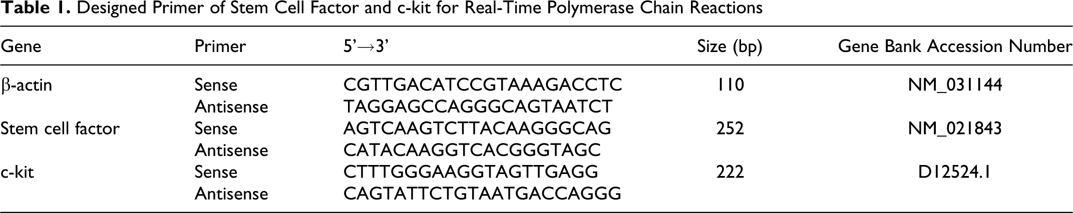
Total RNA was extracted using the TRIzol Reagent (Invitrogen Life Technologies, 15596-026, Carlsbad, CA), and RNA samples were reverse transcribed according to the instructions of the manufacturer (ReverTra Ace-α, FSK-100). The specific primers used in our study are listed in Table 1 . PCRs were performed as described by the manufacturer using the Toyobo Thunderbird SYBR Green polymerase chain reactions Mix (QPS-201). The reaction was performed in 25 μL total volume containing 12.5 μL SYBR green/enzyme reaction mix, 2.5 μM of primer, and 2 μL cDNA. The PCR conditions were 95°C for 1 minute, followed by 40 cycles at 95°C for 15 s, 58°C for 20 s, and 72°C for 20 s. Real-time detections were performed on an ABI-StepOne Detection System (Applied Biosystems, Carlsbad, CA). Melting curve analysis was performed by increasing the temperature by 1°C from 72°C to 95°C and measuring the fluorescence at every temperature point for a period of 20 s. Potential contaminations were monitored using nontemplate controls. The transcript level of each specific gene was normalized to the β-actin amplification. The 2−ΔΔCT method was used to quantify the relative change of gene expression.
Designed Primer of Stem Cell Factor and c-kit for Real-Time Polymerase Chain Reactions
Expression of c-kit and Membrane-Bound Stem Cell Factor in Colon Tissue by Western Blot Analysis
Distal colon specimens of about 0.5 g were ground into the cell suspension with a blade. The suspension was centrifuged (Eppendorf 5403) at 12 000 g for 10 minutes at 4°C, and protein concentrations were measured by the Bradford method. Then, equivalents of 50 μg of extracted proteins were separated using 10% sodium dodecylsulfate polyacrylamide gel electrophoresis and transferred to a PVDF (polyvinylidene fluoride) membrane (Millipore, Bedford, MA). The PVDF membranes were blocked in 5% nonfat milk for 1 hour, followed by incubation with primary antibodies against c-kit (1:200, Santa Cruz Biotechnology, Inc) and stem cell factor (1:400, Abcam, Cambridge, UK) at 37°C for 1 hour and then at 4°C overnight. Rabbit antirat actin (1:400, Santa Cruz Biotechnology, Inc) served as the internal control. This was followed by incubation with horseradish peroxidase-linked secondary antibody (1:6000, horseradish peroxidase-linked antigoat or horseradish peroxidase-linked antirabbit, Jing Mei Biotech, Shenzhen, China) for 1 hour at room temperature. Detection of protein was achieved with enhanced chemiluminescence (ECL reagents, Amersham Pharmacia Biotech, Piscataway, NJ), and the blot was subjected to autoradiography.
Detection of Soluble Stem Cell Factor in Serum by Enzyme-Linked Immunosorbent Assay
Blood samples amounting to 1 to 2 mL were taken from each animal. The concentration of soluble stem cell factor in serum was measured using the enzyme-linked immunosorbent assay kit (CSB-E04720r, Cusabio Biotech Co, Ltd.) according to the manufacturer’s instructions. The standard curve of soluble stem cell factor was obtained by spectrophotometrically measuring the standard with known concentrations at a wavelength of 450 ± 2 nm, and the concentration of soluble stem cell factor in each sample was determined by comparing the optical density (OD) of the samples with the standard curve.
Measurement and Statistical Analysis
All data were expressed as mean values ± standard deviation. Differences among multiple groups were evaluated by 1-way analysis of variance (ANOVA). Contractility of muscle strips were analyzed using 2-way ANOVA with Bonferroni’s post hoc test. Pearson’s correlations and linear regressions were calculated to determine the relationship between c-kit protein expression and other experimental variables, and P < .05 was taken as a statistically significant difference. Statistical analysis was performed by using SPSS 17.0 (SPSS Inc, Chicago, IL).
Results
Body Weight and Blood Glucose Levels
No differences were noted in the baseline body weight among the 5 groups (Figure 1A ). At the end of 8 weeks, the body weight of untreated diabetic rats did not increase, whereas the high-frequency stimulation group gained weight during the study period (P = .000). In the sham and low-frequency stimulation groups, body weight was not significantly altered compared with the untreated diabetic group (P = .283 and .270).

Body weight and blood glucose in each group: A. No differences were noted in the baseline body weight among the 5 groups. At the end of 8 weeks, the high-frequency electroacupuncture-treated (HEA) group gained weight during the study period compared with the diabetic (DM) group (P = .000). B. All the diabetic rats displayed markedly increased blood glucose levels. In the sham, low-frequency electroacupuncture-treated (SEA, LEA), and HEA groups, blood glucose levels did not show any significant changes compared with those of untreated diabetic rats (P > .05).
Meanwhile, no differences were noted in the baseline blood glucose among the 5 groups. All the diabetic rats in our study displayed markedly increased blood glucose levels. At the end of 8 weeks, the nonfasting blood glucose concentration in the untreated diabetic rats was nearly 5 times higher than that of the controls (P = .000). In the sham and low- and high-frequency stimulation groups, blood glucose levels did not show any significant changes compared with those of untreated diabetic rats (P = .087, .063, and .252, respectively; Figure 1B).
The Contractile Activity of Colonic Muscle Strips
The concentration–response contraction obtained for acetylcholine before and after removal of interstitial cells of Cajal are shown in Figure 2A . When the concentration of acetylcholine was 10−3mol/L, colonic muscle strips contracted sufficiently enough, and the percentage variation before removal of interstitial cells of Cajal was markedly decreased in the untreated diabetic group compared with controls (43.0 ± 9.6 vs 177.7 ± 11.5, P = .000), but it was elevated in the low- and high-frequency stimulation groups (99.1 ± 9.7 vs 43.0 ± 9.6, P = .000; 106.1 ± 11.6 vs 43.0 ± 9.6, P = .000). Also, there was no significant difference between the sham and untreated diabetic groups (47.8 ± 4.7 vs 43.0 ± 9.6, P = .403).

Contraction of colonic muscle strips in vitro: A. Contraction of colonic muscle strips in each group. B. When the concentration of acetylcholine (Ach) was 10−3 mol/L, the percentage decrease between before and after interstitial cells of Cajal were removed was significantly increased in low- and high-frequency electroacupuncture treated (LEA and HEA) groups compared with the diabetic (DM) group.
To determine whether restoration of impaired interstitial cells of Cajal was involved in the elevated contractility of the electroacupuncture-pretreated group, the percentage decrease between common muscle strips and muscle strips with interstitial cells of Cajal removed were used to reflect the changes in the interstitial cells of Cajal. As shown in Figure 2B, the percentage decrease was significantly lower in the untreated diabetic group than in the control group (P = .000), but it was greater in the low- and high-frequency stimulation groups compared with the untreated diabetic group at 10−3 mol/L of acetylcholine (P = .010 and .028). There was no significant change in the sham stimulation group compared with the untreated diabetic group (P = .999).
Expression of c-kit by Immunohistochemistry
Figure 3 shows the immunohistochemical staining of c-kit, with a large number of c-kit-positive cells present in colon tissue in normal rats. c-kit Expression was markedly decreased in the untreated diabetic group. There was no significant change in the sham stimulation group. However, in the low- and high-frequency stimulation groups, the expression of c-kit was increased.

Localization of interstitial cells of Cajal in colon tissue: interstitial cells of Cajal are indicated with black arrows. In untreated diabetic rats and diabetic rats treated with sham stimulation, positive cells were decreased markedly. The treatment with low- and high-frequency electroacupuncture stimulation (LEA and HEA) increased the number of interstitial cells of Cajal compared with the diabetic (DM) group.
The mRNA Expression of c-kit and Membrane-Bound Stem Cell Factor in Colon Tissue by Real-Time PCRs
The mRNA expression of c-kit and membrane-bound stem cell factor in colon tissue are shown in Figure 4 . Real-time PCR studies suggested that the mRNA levels of c-kit and membrane-bound stem cell factor in colon tissue were decreased markedly in the untreated diabetic group compared with the control group (P = .002 and .000). Significant increases can be observed in the low-frequency (P = .044 and .032) and high-frequency stimulation groups (P = .049 and .000) compared with the untreated diabetic group. However, there was no difference between the sham stimulation and the untreated diabetic groups (P = .930 and 1.000).

mRNA expressions of c-kit (A) and membrane-bound stem cell factor (M-SCF) (B) in the distal colon. Significant increases were observed in low- and high-frequency electroacupuncture-treated (LEA and HEA) groups compared with the diabetic (DM) group. However, there was no significant change between sham stimulation (SEA) and DM groups
Expression of c-kit and Membrane-Bound Stem Cell Factor in Colon Tissue by Western Blot Analysis
As shown in Figure 5 , the protein expression of c-kit in the untreated diabetic group was markedly decreased compared with that in the control group (P = .000). However, c-kit protein expression was significantly increased in the low- and high-frequency stimulation groups compared with the untreated diabetic group (P = .000 and .000) and nearly recovered to control values in the high-frequency stimulation group. There was no difference between the sham stimulation and untreated diabetic groups (P = .222). Similar results were obtained in the expression of membrane-bound stem cell factor.

Western blot analysis of c-kit and membrane-bound stem cell factor (M-SCF) in the distal colon: The protein expression of M-SCF (A, E) and c-kit (B, D) were decreased in the diabetic (DM) group. However, they were significantly increased in the low- and high-frequency electroacupuncture-treated (LEA and HEA) groups. There were no differences between the sham stimulation (SEA) and DM groups
Soluble Stem Cell Factor in Sera by Enzyme-Linked Immunosorbent Assay
Besides membrane-bound stem cell factor in colon tissue, we also evaluated the levels of soluble stem cell factor to determine the changes in soluble stem cell factor levels in serum. In parallel to the expression of membrane-bound stem cell factor protein in colon tissue, soluble stem cell factor concentration in serum also displayed similar changes. In contrast to the control group, soluble stem cell factor was decreased significantly in the untreated diabetic group. However, diabetic rats treated with low- and high-frequency stimulation had increased soluble stem cell factor levels compared with the untreated diabetic group (29.6 ± 6.9 vs 13.3 ± 4.9 pg mL−1, P = .000; 25.4 ± 8.9 vs 13.3 ± 4.9 pg mL−1, P = .001). No differences were noted between the sham stimulation and untreated diabetic groups (15.7 ± 4.4 vs 13.3 ± 4.9 pg mL−1, P = .475).
Correlation and Regression Analysis
To detect the association between soluble stem cell factor, membrane-bound stem cell factor, and c-kit, Pearson’s correlations and linear regression were evaluated between c-kit protein expression, soluble stem cell factor, and membrane-bound stem cell factor in the 5 groups. The correlations between soluble stem cell factor, membrane-bound stem cell factor, and c-kit protein expression were as follows: soluble stem cell factor, r = 0.577 (P = .000); membrane-bound stem cell factor, r = 0.972 (P = .000). In the linear regression analysis, soluble stem cell factor and membrane-bound stem cell factor were entered as dependent variables (R 2 = 0.949), and the result suggested that only the membrane-bound stem cell factor had significant correlation with c-kit protein expression (P = .000). The regression equation was Y = −0.062 + 0.003X 1 + 0.968X 2 (X 1 is the soluble stem cell factor and X 2 the membrane-bound stem cell factor). In a further stepwise regression analysis, it seemed that the membrane-bound stem cell factor was the only independent effect of the potential determinant on the c-kit protein expression (R 2 = 0.945; P = .000); no significant correlations were observed between c-kit and soluble stem cell factor (P = .148), with the regression equation being Y = −0.024 + 1.007X 2 (Figure 6 ).

Correlations between c-kit and membrane-bound stem cell factor (M-SCF): There was a significant and high, positive correlation between c-kit and M-SCF levels (R 2 = 0.9454; P = .000). Each point represents the value from each individual rat.
Discussion
In the current study, we demonstrated that electroacupuncture at ST-36 could stimulate the contractility of colonic muscle strips and restore impaired interstitial cells of Cajal in the colon of diabetic rats; this is accompanied by upregulation of the stem cell factor both in the colon tissue and in serum.
There are some studies that reported stimulating effects of electroacupuncture on colonic motility in vivo.7,8 However, the effects of electroacupuncture on colonic muscle strip in vitro and the precise mechanisms underlying electroacupuncture on colonic motility have scarcely been investigated. The hypothesis that impaired interstitial cells of Cajal could be restored by the process of electroacupuncture has been proposed on the basis of previous studies.10,11 In line with this hypothesis, removal of interstitial cells of Cajal by methylene blue and exposure to light was used in our study. 16 The effects of methylene blue and light exposure in muscle strips were specific only to interstitial cells of Cajal, without affecting the enteric nerve system and smooth muscle cells.16,18 In our study, the contractility of common muscle strips in the untreated diabetic group was markedly decreased compared with controls, but it was elevated in the low- and high-frequency stimulation groups. Moreover, when interstitial cells of Cajal were removed, the percentage decrease was significantly lower in the untreated diabetic group than in controls, but it was greater in the low- and high-frequency stimulation groups compared with the untreated diabetic group. This phenomenon implies that pretreatment with electroacupuncture, not only high-frequency but also low-frequency treatment, had a restoring effect on impaired interstitial cells of Cajal, which may be attributed to the stimulatory effect on the contractility of colonic muscle strips.
Decrease in the number of interstitial cells of Cajal in diabetes has been reported in animal models19–21 as well as in people with diabetes. 22 The model of diabetes in our study was established successfully. To provide sufficient assessment of the interstitial cells of Cajal in the colon, immunohistochemistry, real-time PCRs, and Western blot were performed. In the current study, Western blot showed an 80% loss of interstitial cells of Cajal in the colon of diabetic rats compared with the control group, which is consistent with previous findings. 19 Our study also found that the expression of c-kit increased significantly in low- and high-frequency-stimulation-treated diabetic rats but was not altered in the sham stimulation group, suggesting that electroacupuncture, both low and high frequency, has the capability to increase the expression of c-kit in diabetic rats. This was consistent with the previous results.10,11
In addition to investigating the effect of electroacupuncture on interstitial cells of Cajal, the experiment was also designed to study the possible mechanisms involved. The stem cell factor is essential for maintaining the function and survival of interstitial cells of Cajal. 23 Together with injury to the interstitial cells of Cajal, diabetic rats also showed a reduced expression of stem cell factor in both serum and colon tissue in our study. These findings were consistent with those of previous studies.13,24,25 Other studies have reported that the abnormalities of interstitial cells of Cajal can attributed to a deficiency in the endogenous stem cell factor. 25 The exogenous stem cell factor can partially reverse the pathological changes in interstitial cells of Cajal.23,25 The role of the stem cell factor in the development of interstitial cells of Cajal has been studied extensively in vivo and in vitro.25,26
Moreover, it was reported that electroacupuncture could elevate the soluble stem cell factor level, 14 but there is no study of electroacupuncture on the membrane-bound stem cell factor. Little information about stem cell factor levels in electroacupuncture-treated diabetic animal and human tissue is available. In the present study, we demonstrated that both soluble stem cell factor and membrane-bound stem cell factor were increased significantly in low- and high-frequency-stimulation-treated diabetic rats, indicating that the increased expression of stem cell factor might be involved in the mechanism of electroacupuncture on colonic contractility and interstitial cells of Cajal.
In our study, we noticed that the changes in both soluble stem cell factor and membrane-bound stem cell factor paralleled the c-kit protein expression. Although both had significant correlations with c-kit, regression analyses indicated that membrane-bound stem cell factor was the only independent effect of the potential determinant to c-kit protein expression, and there was a significant positive correlation between them. This suggests that the higher the membrane-bound stem cell factor level was, the more c-kit was expressed in colon tissue. Our results are consistent with the observation that membrane-bound stem cell factor is a more effective agonist for c-kit receptor compared with soluble stem cell factor and preferentially stimulates the c-kit receptor.26,27
Weight loss and hyperglycemia were the 2 main parameters that characterized the streptozotocin-induced diabetic rats. 28 Li et al 29 reported that electroacupuncture could increase the appetite, sleep, and body weight. In the current study, diabetic rats treated with high-frequency stimulation exhibited an increase in body weight. Blood glucose levels after treatment with different modalities of electroacupuncture were not significantly lower than those of untreated diabetic rats, and they were still hyperglycemic. Our results indicate that electroacupuncture can increase the body weight of diabetic rats but has no effect on blood glucose.
In conclusion, electroacupuncture at ST-36 can promote the contractility of colonic muscle strips partially through increasing the number of interstitial cells of Cajal in diabetic rats, and these effects may be mediated by elevated endogenous membrane-bound stem cell factor levels but are not related to blood glucose levels.
Footnotes
Acknowledgments
The authors would like to thank Dr Roger Brumback and Dr Barbara Gilligan for their editorial support.
JX was involved in planning and conducting the study, acquisition of data, interpreting data, and drafting the manuscript. YC was involved in conducting the study. SL was involved in planning and interpreting data, drafting and revising the manuscript, and procuring the financial support. XH interpreted the data. The final draft was approved by all authors.
The authors declared no potential conflicts of interest with respect to the research, authorship, and/or publication of this article.
The study was supported by a grant from the National Natural Science Foundation of China (project No.30670775) and project of the National Key Technologies R&D Program in the 11th Five Plan period (2007BAI04B01).
All animals in our study were treated strictly in accordance with the National Institutes of Health Guide for the Care and Use of Laboratory Animals, and the work was approved by the ethics committee of Laboratory Animals.