
Abstract
Introduction
Cryosurgery is recognized as a treatment option for various types of oral lesions. Although cryosurgery is less invasive and easier to perform than surgical treatments, adverse events, such as stomatitis and scarring can occur if the freezing is excessive. There are few studies regarding the effects of cryosurgery on the surrounding soft tissues. Thus, this study investigated the extent of tissue destruction and healing progress in tongues of mice who underwent cryosurgery.
Methods
Eight-week-old male BALB/c mice were used. An instrument cooled with liquid nitrogen was lightly touched on the right side of the tongue for 5 s, and a second test was performed 10 s later. Histological evaluation was performed 3, 7, and 14 days after cryosurgery. Blood vessels were evaluated with India ink at 1, 3, 7, 14, and 21 days after cryosurgery.
Results
Destruction of the soft tissue spread to the left side of the tongue after 3 days. At 7 days, it was confirmed that the muscle tissue was in the process of repair and was completely repaired at 14 days. Although blood vessels were not confirmed at 3 days, they were visible after seven days and were confirmed at 21 days all over the tongue.
Discussion and Conclusion
These results indicated that the tissue destruction caused by cryosurgery was extensive and suggest that the duration and frequency of freezing should be minimized for clinical use.
Lay Summary
Cryosurgery is a treatment method for various types of oral lesions. Freezing the lesion causes the tissue to collapse, resulting in its disappearance. Although cryosurgery is less invasive and easier to perform than surgical treatments, adverse events, such as stomatitis and scarring can occur if the freezing is excessive. This study investigated the extent of tissue destruction and healing progress in tongues of mice who underwent cryosurgery.
The right side of mice tongues were frozen by an instrument cooled with liquid nitrogen for 5 s, and a second test was performed 10 s later. The tissue destruction was evaluated at 3, 7, and 14 days after freezing. Blood vessels were evaluated with India ink at 1, 3, 7, 14, and 21 days after freezing. Tissue destruction spread to the left side of the tongue after 3 days. At 7 days, it was confirmed that the muscle tissue was in the process of repair and was completely repaired at 14 days. Blood vessel repair was confirmed at 21 days in the throughout tongue. These results indicated that the tissue destruction caused by cryosurgery was large and suggest that the duration and frequency of freezing should be minimized for clinical use.
Introduction
Cryosurgery has been recognized as a treatment option for various tumor lesions, including oral lesions, since the 1980s.1–3 Currently, it is widely applied to the entire body, and is used to effectively treat skin diseases, kidney cancer, breast cancer, liver tumors, and uterine fibroids.4–8 Cryosurgery is less invasive and easier than surgical treatments. 9 Basic cryosurgery research has determined that an osmotic pressure gradient is generated because the thawing speed differs between the inside and outside of the cell and the loss of intracellular water causes cell necrosis. 10 There are few studies regarding the effects of cryosurgery on the surrounding soft tissue, although it is considered that the peri-lesion tissue is also damaged and destroyed by freezing at the site where cryosurgery is applied. If the vascular system, especially the drainage system, which is necessary for tissue repair, is damaged, congestion and edema occur, and the healing process is affected.
In the field of oral surgery, there are many opportunities to perform resection and excision for mucous cysts and vascular lesions.9,11 When performing surgical procedures, there is a possibility of scar formation, deformation, and paresthesia due to neuropathy associated with resection.9,11,12 Cryotherapy is mildly invasive and produces a strong cold sensation during treatment. For oral diseases, a single treatment consists of freezing for 30 s and is applied two to three times to cover the entire lesion. 9 In most cases, the procedure can be safely performed without bleeding or the need for local anesthesia. Therefore, cryosurgery, which is a less invasive and simple treatment, is effective for older patients taking anticoagulants who are at a risk of bleeding after surgical treatment and infants and young children who cannot undergo surgical resection under local anesthesia.9,13 However, excessive freezing is associated with severe stomatitis and scarring. 9 If the extent of the tissue affected by cryosurgery can be predicted, serious side effects can be prevented. Therefore, this study investigated the extent of tissue destruction and the healing process caused by cryosurgery in the tongues of mice.
Materials and methods
Animals
Eight-week-old male BALB/c mice (Japan SLC, Inc.) were used. The study protocol followed the principles of laboratory animal care and national laws and was approved by the Animal Research Committee of Iwate Medical University (approval number: 02-021). Temperature and humidity were managed at the experimental facilities and the animals were used after one week of preliminary breeding. After experimentation, the animals were carefully observed for any harmful phenomena, such as wound compartment infection, decreased appetite, and death.
Cryosurgery on mouse tongue
A dental instrument with a spherical tip of 3 mm used for amalgam filling was used to cool the tissues (Figure 1(a)). The tips were cooled to −196.8 °C with liquid nitrogen. The experimental mice were anesthetized by isoflurane inhalation and the tongue width of each mouse was approximately 5 mm. The tongue was pulled forward using tweezers, and the cooled tip of the dentistry tool was lightly pressed until half of the tip was buried at the right tongue margin surface, 7 mm from the tip of the tongue (Figure 1(b)). The freezing time was set to 5 s, and freezing was performed twice. There was an interval of 10 s between consecutive treatments. The experimental mice were fixed at 3, 7, and 14 days after cryosurgery for histological evaluation and at ,1, 3, 7, 14, and 21 days after cryosurgery for evaluation of the vascular system. The same experiment was performed five times.

(a) The instrument for cooling the mouse tongue margin. The diameter of the tip is 3 mm. (b) Illustration of the treatment and evaluation areas. The right side of tongue margin was treated, and the tip of the tongue region, anterior to the frozen area, and the frozen area were used for evaluations.
Histological evaluation
The mice were euthanized by administering carbon dioxide and fixed with 5% phosphate-buffered paraformaldehyde (pH 7.4) via perfusion through the aorta. The tongues were resected, dehydrated in a graded series of ethanol solutions, and embedded in paraffin. Serial sections with a width of 5 μm were prepared using a microtome (Reichert-Jung HN 40; Cambridge Instruments GmbH, Nussloch, Germany). All sections were stained with hematoxylin and eosin, and photographs were taken using a photomicroscope (VHX-5000 Digital Microscope; KEYENCE, Osaka, Japan). The tips of the tongue region, anterior to the frozen area, and the frozen section were used for evaluation (Figure 1(b)). A toluidine blue-stained section of an untreated tongue was used as a control.
Evaluation of blood vessels
Gelatin-containing India ink was then perfused. After cooling, the tongues were resected. The samples were dehydrated in a graded series of ethanol solutions and embedded in paraffin. Serial sections with a width of 5 μm were prepared using a microtome (Reichert-Jung HN 40; Cambridge Instruments GmbH, Nussloch, Germany). All sections were stained with hematoxylin and eosin, and photographs were taken using a microscope (VHX-5000 Digital Microscope; KEYENCE, Osaka, Japan). The tips of the tongue region, anterior to the frozen area, and the frozen section were used for evaluation (Figure 2).

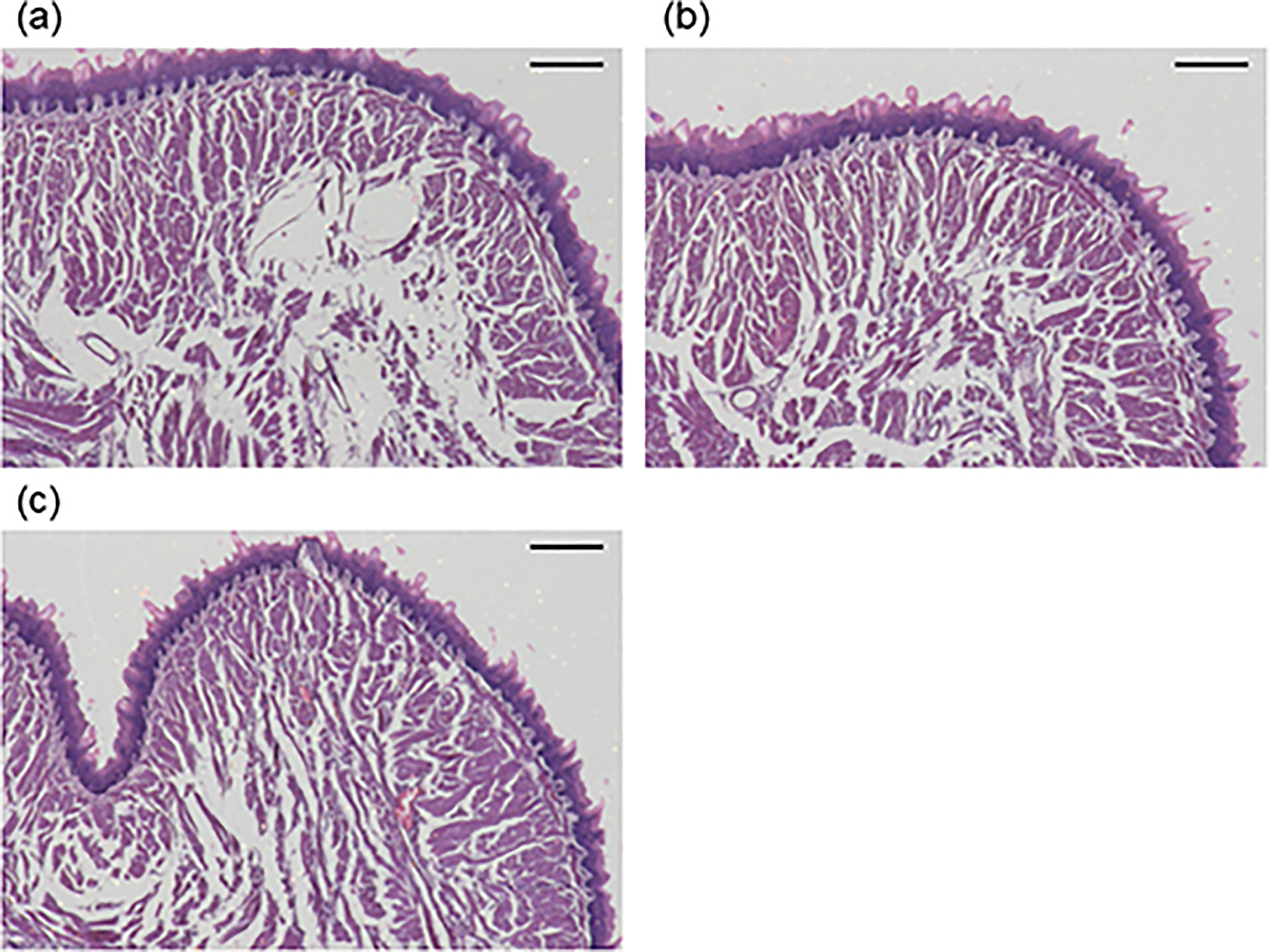
Overview of histological sections of tip of tongue area. Hematoxylin and eosin staining (a) 3, (b) 7, and (c) 14 days after cryosurgery. Scale bars = 500 μm. Tissue destruction is observed at 3 and 7 days. The muscle tissues are repaired after 14 days.
Results
Histological examination
Figures 2–4 present the overview of histological sections of the tongue tip, anterior to the frozen section, and the frozen section, respectively. Three days after cryosurgery, the destruction of soft tissue extended beyond the submucosal longitudinal tongue muscle to the vertical tongue muscle and transverse tongue muscle. Destruction of the tongue muscles spread to the left side and tissue destruction was confirmed in the frozen area and at the tip of the tongue. The collapsed muscle tissues were in the process of being repaired at 7 days. The muscle tissues were repaired after 14 days. The area of the tongue slice expanded after 3 days but subsequently decreased and recovered after 14 days. Figure 5 shows the magnified tongue image of the anterior to the frozen section: at 3 days (Figure 5(a)), tissue destruction accompanied by muscle tissue dissolution was observed; at 7 days (Figure 5(b)), new muscle fibers were observed and they were combined; and regeneration of muscle tissue could be confirmed at 14 days (Figure 5(c)). Similar findings were observed for each group of five mice.

Overview of histological sections of anterior to the frozen section. Hematoxylin and eosin staining (a) 3, (b) 7, and (c) 14 days after cryosurgery. Scale bars = 500 μm. Tissue destruction is observed throughout the section at 3 and 7 days. The muscle tissues are repaired after 14 days.

Overview of histological sections of the frozen section. Hematoxylin and eosin staining (a) 3, (b) 7, and (c) 14 days after cryosurgery. Scale bars = 500 μm. Tissue destruction is observed throughout the section at 3 days and 7 days. The healing process is observed at 14 days. The muscle tissues are repaired after 14 days. (d) A histological section of a control tongue that did not undergo cryosurgery. Toluidine blue staining was used.
Magnified images of the anterior to the frozen section. Scale bars = 200 μm. (a) At 3 days, muscle tissue dissolution is observed. (b) At 7 days, new muscle fibers are observed. (c) At 14 days, muscle tissue regeneration is observed.
Evaluation of blood vessels
Figure 6 shows the overview of histological sections with India ink at the tip of the tongue. India ink could not be confirmed after 14 days but on day 21, the India ink was detected. Figure 7 presents the overview of the histological sections with India ink anterior to the frozen area. Slight ink was detected on day 1; however, no ink was detected on days 3, 7, and 14. On day 21, the ink was observed on the opposite side of the treated region. Figure 8 shows the overview of the histological sections of the section frozen with India ink. India ink was observed until day 21; however, it was minimal on day 3. Overall, the ink was detected after 21 days. Figure 9 shows the magnified images of the anterior to frozen section at 1, 3, 7, 14, and 21 days. Although the presence of micro vessels could be confirmed at 1, 7, 14, and 21 days, they could not be confirmed at 3 days. Similar findings were observed for each group of five mice.

Overview of histological sections with India ink of the tip of tongue area (a) 1, (b) 3, (c) 7, (d) 14, and (e) 21 days after cryosurgery. Scale bars = 500 μm. The black circles indicate India ink. The ink is not detected until 14 days.

Overview of histological sections with India ink of the anterior to frozen section (a) 1, (b) 3, (c) 7, (d) 14, and (e) 21 days after cryosurgery. Bars = 500 μm. The black circles indicate India ink. The ink is detected in the opposite side of treated region at 21 days.

Overview of histological sections with India ink of the frozen area (a) 1 (b) 3, (c) 7, (d) 14, and (e) 21 days after cryosurgery. Bars = 500 μm. The black circles indicate India ink. At 3 days, it is detected in a small area but at 21 days, the ink is detected throughout the tongue.

Magnified image of histological sections with India ink of the anterior to frozen section (a) 1, (b) 3, (c) 7, (d) 14, and (e) 21 days after cryosurgery. Bars = 200 μm. The black circles indicate India ink. At 3 days, micro vessels are not detected, but are observed on days 7, 14, and 21.
Discussion
In this study, freezing was performed for a total of 10 s. Since no stomatitis or scarring was observed, it was considered that the degree of freezing was not excessive. However, because the tissue collapsed over a wide area, even a short period of freezing may be effective. In the current clinical practice of oral surgery, cryosurgery is performed by repeating the treatment for approximately 30 s several times;9,11 however it is considered that shortening the treatment time and reducing the number of repetitions would be just as effective. Histological evaluation showed strong tissue destruction; therefore, excessive cryosurgery may result in scarring even if the lesions disappear. In fact, in cases of intraoral mucous cysts treated with cryosurgery, strong scarring of the surrounding tissues is observed in pathological specimens that were removed because of poor healing despite repeated treatments. 9 Swelling occurred at each part of the tongue on days 1 and 3. Edema is considered to be caused by the destruction of the vascular system, including blood vessels, and muscle tissue. India ink was used to assess blood vessel integrity and was not confirmed until day 14 at the regions anterior to the frozen section. However, it was detected in the frozen tissue after 21 days. The effects of freezing may extend to the opposite side of the frozen area, and that blood flow to the anterior periphery is blocked. India ink was detected in the frozen section after 3 days; however, it was minimal. The tissue was considered to be the most disintegrated on day 3, similar to that shown in the histological examination. Furthermore, because India ink was observed only on the opposite side of the anterior site at 21 days, healing on the treated side still might have been incomplete in the peripheral region. In addition, the frozen area healed within 21 days, suggesting that the effects of freezing persist for up to 14 days.
In cryosurgery for mucous cysts, the tip of the probe is pressed hard so that freezing penetrates deep into the tissue. However, the results of this study indicated that the freezing range is wide, even when contact with the frozen tip is light. The minor salivary glands responsible for the formation of mucous cysts are located submucosally. Therefore, it was concluded that there is no need to press hard when treating mucous cysts. Cryosurgery was performed on all vascular lesions, including hemangiomas. The results of this study determined that the destruction of blood vessels by freezing was sufficient, and that recovery took a long time. If the main blood vessel of the vascular lesion is identified, freezing only the main blood vessel may have a sufficient effect and cause the lesion to regress, even if it is large. Thus, side effects such as swelling, and stomatitis may be reduced.
The limitation of this study was that a quantitative evaluation was not performed because the tissue expansion due to edema was large and it was not possible to accurately measure the depth of the tissue destruction. It may be possible to quantitatively evaluate the extent of soft tissue collapse using the buccal mucosa of large animals in future studies.
In conclusion, the current study evaluated tissue destruction caused by cryosurgery in mice tongues and showed that the tissue destruction caused by cryosurgery was substantial. Therefore, in clinical practice, a short cryosurgery time for lesions and a minimum number of treatments might be desirable. In addition, the results demonstrated that cryosurgery can be effective for at least 21 days.
Footnotes
Declaration of conflicting interests
The author(s) declared no potential conflicts of interest with respect to the research, authorship, and/or publication of this article.
Funding
The author(s) received no financial support for the research, authorship, and/or publication of this article.
How to cite this article
Tadashi K, Atsushi O, Isao H, Hiroyuki Y and Akira F. Histological evaluation of tissue destruction in mouse tongues caused by cryosurgery. Scars, Burns & Healing, Volume 10, 2024. DOI: 10.1177/20595131241230398.