
Abstract
To discuss the influence on proliferation and apoptosis of human intestinal epithelial cells by Helicobacter pylori (Hp). CCK-8 method and flow cytometry to test the influence on proliferation and apoptosis of intestinal epithelial cells by Hp and cell cycle distribution. Immunocytochemistry, qRT-PCR, and WB to analyze the expression and activation of ACVR1. Functional studies of TNBS-induced IBD on mice and DSS-induced IBD on C57BL/6J mice with HP used for intervention were also performed. Hp facilitated the proliferation of intestinal epithelial cells with the breeding ratios on the first and third day are 5.24% ± 3.26% and 34.18% ± 6.68% respectively. ACVR1 is activated and the expression of anti-apoptosis protein Bcl-2 and Bcl-XL is up-regulated. In vivo, TNBS-induced and Hp-induced exhibited inflammatory lesions of intestine, ACVR1 and Bcl-2 higher expressions as compared to wild-type mice. Hp is likely to play a very important role in the proliferation, apoptosis, and malignant transformation of intestinal epithelial cells through ACVR1 pathway.
Keywords
Introduction
Intestinal epithelial cells can sense microorganisms and respond to their stimuli to enhance their barrier function, it is also involved in regulating adaptive immune responses from tolerance to antipathogens.1–3 However, if the environmental stability of epithelial or immune cells is broken, microbial colonization can cause infection and inflammation. The stability of the internal environment depends on the multiple functions of intestinal epithelial cells. It includes the physical barrier of symbiotic bacteria and microbial signal integration. Therefore, intestinal epithelial cells are one of the most important mediators of gut homeostasis. 4 Establish an immune environment that allows symbiotic bacteria to colonize. Especially in infants and young children, the intestinal function has not yet been established. 5 The abnormal activation of the immune system by symbiotic bacteria is considered to cause inflammatory bowel disease (IBD).5,6
Helicobacter pylori (Hp) is generally accepted to be the major etiologic agent for gastritis and peptic ulcer disease, and it has been recognized that it closely correlates with the development of gastric cancer and lymphoma.7,8 However, in recent years strong evidence through animal models and human samples exist to indicate that Hp is possibly responsible for Cohn’s disease.9,10
This experiment is designed to study the influence on proliferation and apoptosis of intestinal epithelial cells and expression of activin A receptor type 1(ACVR1) by Hp for the further discussion whether Hp is related to Cohn’s disease. This provides a new clue for the pathogenesis of HP in some autoimmune diseases.11–13 This study was originally undertaken to find out if colonization of the upper gastrointestinal tract by H pylori might also play a part in the pathogenesis of chronic ulcerative diseases further down the gastrointestinal tract, in particular Crohn’s disease and ulcerative colitis. Materials and methods
Hp and intestinal epithelial cells
Hp international standard strains NCTC11637 (CagA+, VacA+) are introduced from Institute of Infectious Diseases in Chinese Prevention Academy of Science. Prepare the fresh Hp suspensions of live bacilli, use the spectrophotometer to adjust to 1 OD (1 × 10⁸ CFU/ml), and make the two-fold gradient dilution in the DMEM culture solution with no serum medium, the MOI(Multiplicity of infection)value, namely the ratios of bacteria to cell are 8:1–0.25:1.
Intestinal epithelial cells are cultured in the DEME culture solution with 10% fetal bovine serum (FBS) (containing 10% calf serum, penicillin 200 IU/ml, and streptomycin 50 µg/ml), and located in the 37°C, 5%CO2, and 95% humidity for culture, reproducing once per 3–4 days. Cells utilized in the experiment are all in the logarithmic growth phase.
Detecting cellular proliferation by CCK-8 colorimetry
Intestinal epithelial cells are inoculated in 96-well cell plates at a concentration of 0.05 × 10⁵/ml, 100 µl/well, after synchronization step, inject fresh Hp suspensions of live bacilli 100 µl/well, the MOI (multiplicity of infection) value, namely the ratios of bacteria to cell are 8:1–0.25:1. There are three repeated experiments in every working concentration, culturing for 1 and 3 days respectively. The cell group disposed of by the cell culture mediums with no bacilli and no serum is the control group. Adopt CCK-8 (kits were brought from Shanghai Sangon Biological Engineering Technology And Service Co., Ltd.) to determine the Proliferation Index (PI), and calculate the cell PI according to the formula below.
PI = (experimental group − control group)/control group × 100%. Observe the changes of cell form in the group with the most evident proliferation under the common inverted microscope.
RT-PCR semi quantitative
Cell Treatment and Grouping are the same as above, and make the general RNA extraction according to the Trizol specification. The first strand of cDNA was synthesized according to the specification of AffinityScript™ Multiple Temperature Reverse Transcriptase. Real-time reverses transcription quantitative polymerase chain reaction (RT-PCR) was carried out on a LightCycler (Roche Diagnostics GmbH, Mannheim, Germany) using the LC-Fast Start DNA Master SYBR Green I kit as recommended by the manufacturer and primers for ACVR1 and GAPDH (Promega, Mannheim, German).
TNBS induction of colitis
Briefly, rats were slightly anesthetized with ether following a 24 h fast, and then a medical-grade polyurethane canal for enteral feeding (external diameter 2 mm) was inserted into the anus and the tip was advanced to about 8 cm from the edge of anus. 1 ml TNBS (150 mg/kg, Sigma-Aldrich Company Ltd., Spain) dissolved in 50% ethanol was instilled into the colon through the cannula. Following the instillation of the TNBS/ethanol, the animals were maintained in a head down position for a few minutes to prevent leakage of the intracolonic instillation. C57BL/6 mice were euthanized within 3–7 days after the TNBS enteral administration, 0.5 cm intestinal tissue was taken from the proximal colon and the distal colon for histopathological sections. This study was carried out in accordance with the recommendations of Medical ethics committee of the Fujian Medical University.
DSS induction of colitis with HP intervention
Specific-pathogen-free female C57/BL6 mice aged 8–10 weeks were obtained from Shanghai Bio model Organism Science & Technology Development Co., Ltd. To induce acute colitis, 3% dextran sodium sulphate (DSS; MP Biomedicals, Solon, Ohio, USA) was given 2 h after the oral administration of respective endotoxin-free Hp (0.05 × 10⁵/ml) on day 0. DSS was given in the drinking water for 7 days and changed every 2 days to ensure purity. On day 7, DSS water was switched to regular drinking water, and a recovery period of 7 days was observed. The mice were killed on day 14.
Intracellular signaling mechanisms
Proteins were separated on 4%–12% Tris-glycine gels and transferred to nitrocellulose membranes. Membranes were probed with antibodies directed against ACVR1 (ABclonal, A10363), BCL-2 (ABclonal, A10965), BCL-XL (Cell Signaling Technology, 8965), and Gapdh (ABclonal, AC002). Immunofluorescence confocal microscopy was also undertaken to determine the correlation of ACVR1 and BCL-2. BCL-2 was detected with rabbit anti-human antibody (SantaCruz, USA) and labeled with a goat anti-rabbit secondary antibody conjugated to Cy3. ACVR1 was detected with a rabbit anti-human antibody (Wanlei, Shenyang, China) and labeled with a goat anti-rabbit secondary antibody conjugated to FITC. Nuclei were stained using 4′,6-diamidine-2-phenylidole (DAPI).
Statistics
Statistical differences between groups were assessed by the nonparametric Mann–Whitney U-test for two groups and Kruskal–Wallis test for more than two groups. Bonferroni–Dunn’s correction method was applied for post hoc multiple pair-wise comparisons. Spearman’s rank correlation coefficient estimated the degree of association between two variables. Significance was calculated at p < 0.05 by GraphPad Prism 5 (La Jolla, CA).
Results
Cell proliferation
At MOI (concentration ratio of bacteria to cell) of 0.5:1, 0.25:1, Hp could facilitate the proliferation of intestinal epithelial cells, among which the breeding ratios on the first and third day are 5.24% ± 3.26% and 34.18% ± 6.68% respectively, compared with the group at MOI of 0.25:1, having statistical significance, p = 0.001 (Figure 1(a)). Meanwhile, the G2/M ratio showed higher among Hp suspensions groups, indicating that Hp could cause S phase cell cycle arrest (Fig.1B).

Effect of Helicobacter pylori on cell proliferation and cell cycle: (a) cell proliferation detection by CCK-8 (mean ± SD, n = 3) and (b) effect of HP on intestinal epithelial cells cycle distribution.
Hp promotes irritable bowel syndrome and high expression ACVR1
Histopathological examinations showed a large number of inflammatory cells infiltrate the intestinal mucosa in the colitis mouse model (Figure 2(a)), and lymphoid follicles appeared (Figure 2(b)). Moreover, ACVR1 expression was significantly higher in colitis mice which was reflected by immunohistochemistry (Figure 2(c)) and RT-qPCR (Figure 2(d)).

Hp promotes irritable bowel syndrome and high expression ACVR1: (a)Hp-bearing mice, IBD (100×), (b)Lymphatic follicular hyperplasia in IBD mice, (c)ACVR1 was significantly higher expression in IBD mice which reflected by immunohistochemistry, and (d)ACVR1 was significantly higher expression in IBD mice reflected by quantitative real-time RT-PCR.
Hp activates the ACVR1 and the expression of anti-apoptosis protein Bcl-2 and Bcl-XL
To examine whether Hp effect on ACVR1 activates anti-apoptosis protein Bcl-2 and Bcl-XL, we performed a Western blot on the cell lysates from intestinal epithelial cells infected Helicobacter pylori in different concentrations, which indicated that Hp induced an increase in ACVR1. Similarly, the expression of anti-apoptosis protein Bcl-2 and Bcl-XL under Hp intervension was significantly higher than that of control (Figure 3). It showed Bcl-2 in the endothelial and smooth muscle layers from Hp and TNBS induction of IBD (Figure 4).
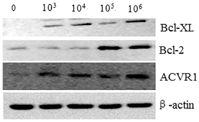
WB results of ACVR1, Bcl-2, and Bcl-XL.

Co-localization of ACVR1 and Bcl-2.
Discussion
Over half of the world’s population is colonized with Helicobacter pylori (Hp), which plays a key factor in the etiology of various gastrointestinal diseases, ranging from chronic active gastritis without clinical symptoms to peptic ulceration, gastric adenocarcinoma, and autoimmune disease. One of the major mechanisms proposed for IBD is that the mucosal immune system mounts an inappropriate response against normal luminal constituents such as bacteria, components of the diet or other ingested material.14,15
This experiment proved in vitro that Hp infection could facilitate the proliferation to host cells vigorously; meanwhile, cell cycle analysis showed that the bacteria could also promote the development of cell cycle after acting on cells, moreover may participate in the facilitating proliferation of cells. With the development of infection, cell proliferation was increased while apoptosis was reduced, thus instabilities of cell group were enhanced, resulting in the virulent transformation of mucous epithelium.
This experiment subsequently proceeds from the cell image that the proliferation of cells in vitro is quite obvious after Hp infection, selects ACVR1 gene index, studies the expression changes of that transcription factor in host cell after Hp infection, and provides a new clue for exploring the Hp mechanism of IBD. IFA revealed that the anti-apoptosis protein Bcl-2 activity of ACVR1 in cell could be activated with Hp. Meanwhile, immunocytochemical results for the expression of ACVR1 protein also showed that the protein expression in the group with Hp was increased significantly. All these indicated that activation and up-regulated expression of ACVR1 possibly played a very important role in the proliferation and anti-apoptosis process of intestinal epithelial cells with Hp, as for the mechanism activating ACVR1 by Hp infection, some studies reported that the relative antigen CagA of cytotoxin could up-regulate the expression of ACVR1.16-18
Bcl-2 family is the gene family regulating the cell apoptosis on which the most attention is paid, including Bcl-2 and Bcl-XL, which as the earliest apoptotic inhibitors are one of the inducible apoptotic inhibitors of ACVR1. 14 Studies find out that the activation of ACVR1 could enhance the expression of anti-apoptotic genes in the Bcl-2 family, thus having the anti-apoptosis effect. 15 Therefore, we presume that proliferation, apoptosis, cell cycle and a series of changes in intestinal epithelial cells as a result of ACVR1 activation induced by Hp, possibly associated with the expression conditions of anti-apoptosis protein Bcl-2 and Bcl-XL.
There are some defects in this study, including a small number of experimental animals and it had better use TNBS or DSS model to verify the impact of HP on colitis.
Conclusion
As stated previously, while Hp is influencing the proliferation and apoptosis of intestinal epithelial cells, the expression of Bcl-2 and Bcl-XL is likely to be regulated by ACVR1. This experiment proves the activation of transduction pathway of Hp-ACVR1-anti-apoptosis protein Bcl-2 and Bcl-XL, and provides valuable experimental materials and proof for the further discussion on the Hp effect in the development of IBD.
Footnotes
Author’s note
Jingwen Wang is now affiliated with Department of internal medicine, Tiantai People’s Hospital Of Zhejiang Province, Taizhou, China.
Declaration of conflicting interests
The author(s) declared no potential conflicts of interest with respect to the research, authorship, and/or publication of this article.
Funding
The author(s) disclosed receipt of the following financial support for the research, authorship, and/or publication of this article: This work was supported by the Fujian Natural Science Foundation (no. 2020J01655).
Ethical approval
Ethical approval for this study was obtained from The Medical ethics committee of the Fujian Maternal and Child Health Care Hospital (038).
Animal welfare
The present study followed international, national, and/or institutional guidelines for humane animal treatment and complied with relevant legislation.