
Abstract
The fruit and root of Clausena lansium are remedy for bronchitis in oriental traditional medicine. Extracts from Clausena lansium are reported to have antioxidant, anti-inflammatory, and anticancer activities. This study is designed to investigate whether the ethanol extract of Clausena lansium (Lour.) Skeels could inhibit the lipopolysaccharides (LPS)-induced inflammatory in RAW 264.7 macrophages and the underlining mechanisms. Enzyme-linked immunosorbent assay (ELISA) and reverse transcription polymerase chain reaction (RT-PCR) are used to detect the secretion of tumor necrosis factor alpha (TNF-α) protein and expression of TNF-α mRNA, respectively. RT-PCR and Western blotting are used to determine the mRNA and protein levels of TLR4, MYD88, and TRAF6, respectively. The extract of Clausena lansium suppressed the expression and secretion of TNF-α, which is released by RAW 264.7 cells after LPS induction. Meanwhile, the expression of TLR4, MYD88, and TRAF6 are decreased by extracts from Clausena lansium. Meanwhile, NBP2-29328, an inhibitor of MYD88, synergistically enhances the inhibiting effect of extract from Clausena lansium. The results imply that ethanol extract from Clausena lansium attenuate macrophage-mediated inflammation and TLR4/MYD88/TRAF6 pathway is its potential target.
Introduction
Clausena lansium (Lour.) Skeels is a fruit tree widely distributed in southern China. Its leaves, roots, and seeds have been used as a folk herb for the treatment of a cohort of inflammatory diseases such as cough, asthma, dermatological disease, viral hepatitis, gastro-intestinal inflammation, and ulcer. 1
Macrophages, the large-sized type of phagocytes, engulf and digest cellular debris, foreign substances, microbes, and cancer cells, and then play a critical role in nonspecific defense (innate immunity) and also help initiate specific defense mechanisms (adaptive immunity) by recruiting other immune cells such as lymphocytes. 2 However, exaggerated reaction of macrophages lead to tissue injury and deteriorate the inflammation. These macrophages have a strong pro-inflammatory signature characterized by the production of factors including tumor necrosis factor alpha (TNF-α), interleukin 1 beta (IL-1β), interleukin 6 (IL-6), and inducible nitric oxide synthase (iNOS). 3 TNF-α promotes the inflammatory response, which, in turn, causes many of the immune injuries, and TNF inhibitors can alleviate the inflammatory response. 4
TLR4 is a member of the toll-like receptor (TLR) family, which plays a fundamental role in pathogen recognition and activation of innate immunity. 5 They recognize pathogen-associated molecular patterns (PAMPs) that are expressed on infectious agents, one of which, with high affinity to TLR4, is lipopolysaccharides (LPS). Upon LPS recognition, conformational changes in the TLR4 initiate two major intracellular signaling pathways: MYD88-dependent pathway and MYD88-independent pathway. 6
In this study, we reported that ethanol extract from Clausena lansium decreased the release of TNF-α induced by LPS in macrophages and blocked the TLR4/MYD88/TRAF6 pathway.
Materials and methods
Materials
Fresh leaves of Clausena lansium were collected from Guilin, a southern city in China. Air-dried and finely powdered leaves of Clausena lansium were subsequently extracted with 70% EtOH (10 BV) under reflux for 8 h, and then extracted successively with petroleum ether, dichloromethane, and ethyl acetate. 1 The concentrate of ethyl acetate was stored at −20°C for further research. Enzyme-linked immunosorbent assay (ELISA) kit for TNF-α was purchased from Boster Biological Technology Ltd (Wuhan, China), LPS from Sigma-Aldrich (St. Louis, Missouri, USA), and Dulbecco’s Modified Eagle medium (DMEM) and fetal bovine serum (FBS) from Invitrogen (Carlsbad, CA, USA). Trizol, primers, reverse transcription polymerase chain reaction (RT-PCR) kit, and amplification kit were purchased from TIANGENG (Beijing, China). β-actin antibody was obtained from ZSGB-BIO (cat no. ZB2305; Beijing, China), TLR4 antibody from GeneTex (cat no. GTX113024; Irvine, CA, USA), and TRAF6 antibody from Abcam (cat no: ab33915; Cambridge, UK). MYD88 inhibitor, NBP2-29328, was purchased from Novus Biologicals (Littleton, CO, USA).
Cell culture
RAW 264.7 cells were obtained from ATCC (ATCC® TIB-71™, Beijing, China). These cells were cultured in DMEM, which was supplemented with 10% heat-inactivated FBS, 1% penicillin, and streptomycin. The cells were cultured at 37°C in a 5% CO2 atmosphere with 90% relative humidity.
RT-PCR analysis
RAW 264.7 cells were divided into three groups: (1) control group, (2) LPS treated group, and (3) LPS combined with 50 μg/mL extracts-treated group. First, all of the cells were treated with 50 μg/mL extracts from Clausena lansium for 1 h. Then, 1 ng/mL LPS was added for 48 h. Finally, cells were collected for detecting mRNAs of TLR4, MYD88, TRAF6, and TNF-α.
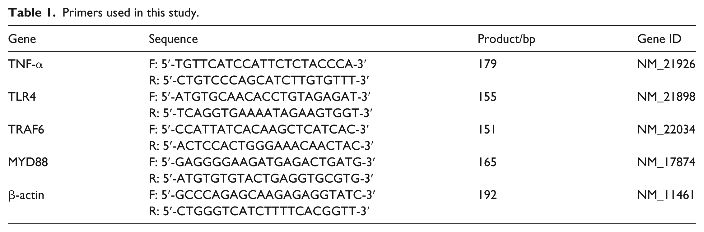
RT-PCR assay was briefly described as follows. First, total RNA was isolated from the cells using RNeasy mini kit (TIANGENG). Then, RNA was reversely transcribed to cDNA as per the manufacturer’s instruction. Polymerase chain reaction (PCR) amplification systems include 2 μL of cDNA, 1 μL of forward primer, 1 μL of reverse primer, 12.5 μL of 2 × Taq Plus Master Mix, and 8.5 μL of ddH2O. The PCR assays were performed as follows: 3 min at 94°C, 30 cycles at 94°C for 3 s, and then 60°C for 30 s and 72°C for 30 s. RT-PCR products were semiquantified by ImageJ software (National Institutes of Health). The primers used in this study are listed in Table 1.
Primers used in this study.
ELISA
To detect the induction effects on TNF-α secretion by LPS, 4 × 10⁶ RAW 264.7 cells per well were inoculated in 6-well plates and treated with 0.1, 1, 10, and 100 ng/mL LPS for 24 h before harvesting supernatants.
In order to detect inhibition effects on TNF-α secretion by the extracts from Clausena lansium and MYD88 inhibitor NBP2-29328, RAW 264.7 cells were divided into five groups: (1) control group, (2) 1 ng/mL LPS-treated group, (3) 1 ng/mL LPS + 5 mM NBP2-29328-treated group, (4) 1 ng/mL LPS + 50 μg/mL extracts-treated group, and (5) 1 ng/mL LPS + 5 mM NBP2-29328 + 50 μg/mL extracts-treated group. Cells were treated with extracts for 1 h, and then 1 ng/mL LPS was added. Subsequently, 5 mM NBP2-29328 was added 24 h before harvesting supernatants.
ELISA assays of TNF-α of the supernatants were according to the manufacturer’s instructions.
MTT analysis
To detect the cytotoxicity of extracts from Clausena lansium, RAW 264.7 cells (5 × 10⁴ cells per well) were incubated in 96-well plates. Cells were pre-treated with 25, 50, and 100 μg/mL extracts for 1 h, and then treated with 1 ng/mL LPS for 48 h. Subsequently, 20 μL of 5 mg/mL MTT (3-(4,5-dimethylthiazol-2-yl)-2,5-diphenyltetrazolium bromide) was incubated with the cells for 4 h. Finally, the solution was discarded, and 150 μL of dimethyl sulfoxide (DMSO) was added. The plates were oscillated for 10 min and immediately detected at 490 nm on a microplate reader (Molecular Devices, Santa Clara, CA, USA).
Western Blot assays
To observe the inhibiting effects of extracts from Clausena lansium on the expression of key components of TLR4/MYD88/TRAF6 pathway, RAW 264.7 cells were divided into five groups as aforementioned. The cells were lysed in 500 μL of radio immunoprecipitation assay (RIPA) lysis buffer (ThermoFisher Scientific, Rockford, IL, USA) with 1% phenylmethanesulfonyl fluoride (PMSF; Sigma, Santa Clara, CA, USA). The concentration of the protein was measured using Pierce™ BCA Protein assay kit (Thermo Fisher Scientific). Proteins were boiled for 10 min, segregated by sodium dodecyl sulfate-polyacrylamide gel electrophoresis, and transferred to polyvinylidene fluoride membrane. Subsequently, the protein was probed by the following antibodies: TLR4 (1:1000), TRAF6 (1:5000), and β-actin (1:500). Finally, the membrane was incubated with chemiluminescence reagent (Beyotime, Shanghai, China) for 1 min and exposed in X-ray films.
Statistical analysis
Statistical analysis was performed using Statistical Analysis System Software (SPSS 19.0, IBM, New York, USA). All the data were presented as mean ± SD. Statistical significance between multiple groups was assessed by performing analysis of variance (ANOVA). P < 0.05 was considered to be statistically significant.
Results
LPS induced the secretion of TNF-α in a time-dependent manner in RAW 264.7 cells
To determine the optimal concentration of LPS and to induce the differentiation of RAW 264.7 cells to macrophages and then secretion of TNF-α, RAW 264.7 cells were exposed to different concentrations of LPS (0.1,1,10, and 100 ng/mL) for 24 h. TNF-α reached a peak value when stimulated by 1 ng/mL of LPS (Figure 1(a)). So, 1 ng/mL of LPS was selected for the following experiments. Then, RAW 264.7 cells were exposed at different time points (4, 8, 12, 24, and 48 h), and the secretion of TNF-α increased in a time-dependent manner (Figure 1(b)). As RT-PCR results show, TNF-α mRNA overexpressed at early stage on stimulation of LPS at 4, 8, and 12 h (Figure 1(c)). Meanwhile, LPS induced the differentiation of RAW 264.7 cells to macrophages, which transformed from round or polygonous to fusiform or stellate with pseudopodia (Figure 1(d)).

LPS promoted the secretion of TNF-α and differentiation in RAW 26.7 cells: (a) ELISA results show that supernatnant TNF-α were increased after LPS stimulating for 24 h, (b) supernatnant TNF-α were increased by 1 ng/mL of LPS stimulation at different time points, (c) RT-PCR results indicated that LPS promoted the expression of TNF-α mRNA at early time points, and (d) morphology of RAW 264.6 cells transformed from round/oval to stellate after LPS stimulating for 24 h.
Extracts from Clausena lansium suppressed LPS-induced TNF-α secretion and TLR4/MYD88/TRAF6 pathway
To determine the cytotoxicity of Clausena lansium and LPS on RAW 264.7 cells, MTT assay was performed to detect the cell viability. Different concentrations (25, 50, 100 μg/mL) of extracts from Clausena lansium and 1 ng/mL LPS showed no significant influence on the cell viability of RAW 264.7 cells (Figure 2(a)). Treated with 25, 50, and l00 μg/mL extracts from Clausena lansium, TNF-α, which was drastically induced by LPS, decreased by 27.8%, 49.8%, and 80.6%, respectively (Figure 2(b)) in RAW 264.7 cells. Consistent with the results of ELISA, TNF-α mRNA also decreased (Figure 2(c)). The results clearly indicated that the extracts from Clausena lansium inhibited the expression and secretion of TNF-α, which was induced by LPS, in a dose-dependent manner. RT-PCR results showed that TLR4, MYD88, TRAF6, and TNF-α were all downregulated after being treated with 50 μg/mL of extracts (Figure 2(d)). The results indicated that the extracts from Clausena lansium could suppress TLR4/MYD88/TRAF6 signaling pathway and then TNF-α expression.

Extracts from Clausena lansium suppressed LPS-induced expression and secretion of TNF-α and TLR4/MYD88/TRAF6 pathway in RAW 264.7 cells: (a) LPS with or without different concentrations of the extracts from Clausena lansium show no significant inhibtion on the viability of RAW 264.7 cells by MTT assays, (b) extracts from Clausena lansium remarkably suppressed the LPS-induced secretion of TNF-α, (c) by RT-PCR, extracts from Clausena lansium downregulated the expression of TNF-α mRNA, and (d) extracts from Clausena lansium remarkably suppressed the LPS-induced upregulation of mRNA components of TLR4/MYD88/TRAF6 pathway and downstream effector of TNF-α.
MYD88 inhibitor NBP2-29328 and extracts from Clausena lansium synergistically suppressed secretion of TNF-α and expression of TRAF6 protein
To evaluate the inhibiting effects of the extracts from Clausena lansium and whether there is a synergistic effect between the extracts and MyD inhibitors, a traditional MyD inhibitor, NBP2-29328 is included in this study. Compared with the LPS group, secretory TNF-α decreased by the extracts, NBP2-29328, and the combination of both. Meanwhile, the extracts from Clausena lansium showed stronger inhibiting effect than NBP2-29328.
TLR4 is an upstream receptor of MYD88-dependent PAMPs-associated pathway. Upon LPS recognition, the signal passed to MYD88 and then to TRAF6. Western blot results showed that TLR4 decreased by 28.0%, 23.1%, and 30.1% when interfered by extracts from Clausena lansium, NBP2-29328, and combination of both, respectively (Figure 3(b)). While TRAF6 protein reduced by 58.1%, 56.6%, and 77.9%, respectively (Figure 3(c)), TNF-α decreased by 28.6%, 36.2%, and 58.2%, respectively (Figure 3(c)).

Extracts from Clausena lansium and MYD88 inhibitor NBP2-29328 synergistically inhibited the protein expression of TLR4/MYD88/TRAF6 pathway and downstream effector TNF-α in RAW 264.7 cells: (a) TLR4 and TRAF6 protein were downregulated by extracts from Clausena lansium, NBP2-29328 and combination of both, (b) the relative expression value of TLR4 protein, (c) the relative expression value of TRAF6 protein, and (d) supernatant TNF-α were reduced by extracts from Clausena lansium, NBP2-29328, and combination of both.
These data implied that extracts from Clausena lansium displayed suppressing effect on MYD88-dependent pathway. Furthermore, the combination of MYD88 inhibitor NBP2-29328 and the extracts from Clausena lansium synergistically inhibited MYD88-dependent PAMPs-associated pathway with drastically reduced TRAF6, the downstream molecular of MYD88, and pro-inflammatory factor TNF-α.
Discussion
Clausena lansium (Lour.) Skeels is a fruit tree distributed in southern mainland China, Southeast Asia, and North America. 7 Its leaves and roots have been used as a folk herb for the treatment of inflammatory diseases. 8 The extracts from Clausena lansium, such as lansiumamide B and SB-204900, could inhibit inflammation through reducing histamine release, IL-6 secretion, and downregulation of cyclooxygenase 2 (COX-2, an essential enzyme in synthesis of inflammatory mediators) expression in A23187-induced RBL-2H3 cells. 9
PAMPs are recognized by TLRs’ activate innate immune responses, protecting the host from infection. 10 Bacterial LPSs, a class of PAMPs, are specifically recognized by TLR4, a recognition receptor of the innate immune system.11,12 In this study, LPS (1 ng/mL) stimulation successfully constructed a inflammed cell model in RAW 264.7 cells, which showed increased expression and secretion of TNF-α (Figure 1(b) and (c)), a macrophage-like morphology with stretched pseudopods (Figure 1(d)), and also activated TLR4/MYD88/TRAF6 signaling pathway with upregulated TLR4, MYD88, TRAF6, and TNF-α (Figure 2).
LPS and extracts from Clausena lansium (100, 50, and 25 μg/mL) had no obvious influence on the viability of RAW 264.7 cells (Figure 2(a)). However, different concentrations of extracts significantly suppressed the secretion and mRNA expression of TNF-α, which were stimulated by LPS (Figure 2(b) and (c)). This implied that extracts from Clausena lansium can inhibit the inflammation of macrophage through inhibiting the expression of pro-inflammatory factor TNF-α.
To evaluate the inhibiting effects of extracts from Clausena lansium on TLR4/MYD88/TRAF6 signaling pathway, a polypeptide inhibitor of MYD88, NBP2-29328, was employed as a positive control. Both NBP2-29328 and the extracts decreased the expression of TRAF6 protein, a mediator downstream of MYD88, and showed a synergistic effect together (Figure 3).
Altogether, the extracts from Clausena lansium attenuate macrophage-mediated inflammation and TLR4/MYD88/TRAF6 pathway in macrophages, which suggest a potential value in the management of exaggerated inflammation.
Footnotes
Declaration of conflicting interests
The author(s) declared no potential conflicts of interest with respect to the research, authorship, and/or publication of this article.
Funding
The author(s) disclosed receipt of following financial support for the research, authorship, and/or publication of this article: The study was supported by the Guangxi Natural Science Foundation (grant nos. 2012GXNSFAA053079, 2018GXNSFAA281168), the Guangxi Administration of Traditional Chinese Medicine Research Project (grant no. GZKZ10-124), the Guilin Scientific Research and Technology Development Research Projects (grant nos. 20120121-1-17, 20140120-1-9, and 20170109-47), Guangxi Key Laboratory of Molecular Medicine in Liver Injury and Repair Research Projects (grant no. 15-140-46-14), the Beijing Medical and Health Foundation (grant no. YWJKJJHKYJJ-B184007), and the National Natural Science Foundation of China (grant nos. 81160066, 81260661, and 81860658).