
Abstract
Cognitive dysfunction resulting from the reduction of cerebral blood flow has been defined as “vascular cognitive impairment” (VCI) which has become the second cause of dementia only after Alzheimer’s disease (AD) and arouses great concerns. There is accumulating evidence that environmental enrichment (EE) can induce functional and anatomical alterations and then bring about overt improvement in memory and learning tasks in many injury paradigms, including ischemic brain injury. Moreover, necrostatin-1 (Nec-1), the special inhibitor of necroptosis, improved functional outcomes following ischemic brain injury and AD. The question of whether and what effect EE and EE + Nec-1 could bring about on cognitive performance and microenvironment and histopathological consequences in the mice suffering from VCI is still unclear. In this study, we investigated this question using the bilateral common carotid artery stenosis (BCAS) mouse model. A week after surgical operation for BCAS, mice were reared for 3 weeks either in standard housing condition or in an EE consisting of special cage filling with various stimulatory items. The results found that the mice in the BCAS + EE and BCAS + EE + Nec-1 groups showed significantly shorter latencies and distances to reach the platform in behavioral tests versus untreated mice at 4 weeks after BCAS surgery. However, three injured groups showed significant deficits compared with the sham group (P < 0.05). In addition, there were no differences between the EE-reared mice and EE + Nec-1-treated mice except in the level of expression of inflammation cytokines. Our results indicated that noninvasive environmental stimulation is beneficial in ameliorating cognitive deficits and inflammation response in mice following VCI and that Nec-1 enhanced the inhibitory effect of EE on inflammation response.
Introduction
The World Health Organization estimated that the number, 35.6 million people suffer from dementia in 2010, will increase to approximately 115.4 million by 2050. 1 More recently, there is a view accepted generally that vascular cognitive impairment (VCI) has become the second cause of dementia only after Alzheimer’s disease (AD). 2
Cognitive dysfunction due to the reduction of cerebral blood flow has been defined as “VCI”3–5 that encompasses the entire clinical spectrum from mild cognitive decline to more severe dementia2,5–8 and attracted great attention. It was reported that white matter (WM) damage is the most important component of VCI. 9 WM changes are mediated by a series of factors, including vascular dysfunction and inflammation, leakage of blood–brain barrier (BBB), damage to oligodendrocytes and microglial activation, and finally demyelination. 10 Reduction in the cerebral blood flow (CBF) leading to hypoperfusion is an early and characteristic phenomenon in the process of WM changes 10 and is associated with cognitive decline.11,12
There are a number of proposed animal models for VCI and vascular dementia, 13 and a widely accepted report is that the bilateral common carotid artery stenosis (BCAS) mouse model using microcoils is the best and most valid model for VCI. 14 There is still no known treatment once symptoms appear although some studies suggest that exercise may slow down cognitive decline duo to upregulation of endogenous protection. 15
Previous report demonstrated that mental, cognitive, social, and physical health interacted with leading an active life. 16 Furthermore, environmental enrichment (EE) can induce functional and anatomical alterations and then bring about overt improvements in memory and learning tasks in several injury paradigms of the brain. 17 Importantly, it has been demonstrated that the hippocampus, which is involved in many kinds of cognitive performance such as learning and memory tasks, 18 is one of the brain areas most susceptible to be affected by EE housing.
Inflammatory activation and sustained microvascular failure are the major mediators of stroke and cerebral ischemia, 19 which finally lead to VCI.3–5,10 And some previous studies indicated that EE alleviated inflammation responses in tumor necrosis factor (TNF)-induced acute inflammation mouse model 20 and that necrostatin-1 (Nec-1) improved cognitive function21,22 and ameliorates neuronal injury in both intracerebral hemorrhage mouse model 23 and ischemic brain injury. 24 So we hypothesized that EE will improve behavioral outcomes and reduce inflammation response in the BCAS mouse model of VCI and that combination administration with Nec-1 will enhance the effect of EE on VCI mice.
Results
Alterations of CBF in the experimental mice
The values of CBF in mice before and after surgical operation are demonstrated in Figure 1. The data suggested that the value of CBF reduced markedly by (63% ± 12%) at baseline 1 h after surgical operation for BCAS as compared with the preoperative baseline, and that the value of CBF recovered to (30% ± 9%) and (33% + 10%) in the BCAS + EE and BCAS + EE + Nec-1 groups versus the preoperative baseline, but no significant difference between the EE and EE + Nec-1 groups was observed between BCAS + EE and BCAS + EE + Nec-1 (*P < 0.05; Figure 1).
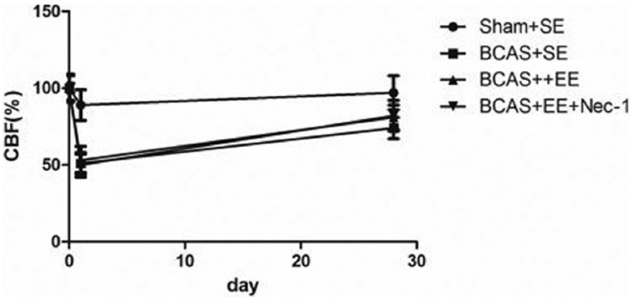
The value of CBF at three different time points.
Morris water maze test
During the first 5 days, we performed the visible platform trials on mice and measured two indicators (escape latency and distance to reach the platform), and no significant difference between the experimental animals was detected in escape latency. At the third, fourth, and fifth days, escape latency showed a significant difference between the sham and the three injured groups as well as between the BCAS + Standard environment (SE) and the two groups (BCAS + EE and BCAS + EE + Nec-1; *P < 0.05; Figure 2(a)). The data of escape distance suggested that the mice in the BCAS + SE group traveled the longest path to reach the platform. There was observed the same significant difference as the data of escape latency test (*P < 0.05; Figure 2(b)). At the sixth day, the performance of mice in the hidden platform (Morris water maze test (MWM)) was measured. Two indicators were recorded, including the time spent in the target quadrant and the number of times they crossed the platform site. The data on time in the target quadrant and the number of times crossed the platform showed that a significant difference between the BCAS + SE and BCAS + EE groups as well as between the BCAS + SE and BCAS + EE + Nec-1 groups were observed (*P < 0.05; Figure 2(c) and (d)).
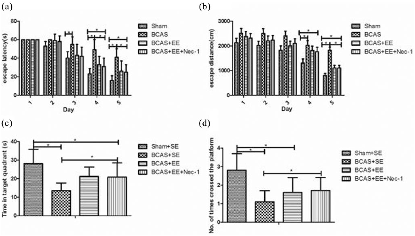
Behavioral performance in the Morris water maze test. (a) Escape latency in mice in MWM with visible platform. The data showed that escape latency decreased gradually with the flow of time. The time in both the BCAS + EE and BCAS + EE + Nec-1 groups was shorter than that in the BCAS + SE group (*P < 0.05), but there was no significant difference between the BCAS + EE and BCAS + EE + Nec-1 groups; (b) escape distance in mice in MWM with visible platform. On the last day of training, escape distance in both the BCAS + EE and BCAS + EE + Nec-1 groups decreased markedly compared with the BCAS + SE group (*P < 0.05); (c) time spent in the target quadrant. On the formal test day, a significant difference was observed in the time spent in the target quadrant without the platform between the Sham + SE group and the other three injured groups (*P < 0.05); (d) number of times the mice crossed the platform site. The same effect was observed as the other indicator, the time spent in the target quadrant (*P < 0.05).
Open field test
Open field test (OFT) was performed to assess the locomotor activity of mice. We recorded two indicators including the total distance traveled in the apparatus and the time spent in the center of the apparatus. The data showed that the total distance traveled in the apparatus in these three groups (BCAS + SE, BCAS + EE, and BCAS + EE + Nec-1 groups) was significantly longer than that in the sham group and that the BCAS group mice traveled longer distance than those in the BCAS + EE and BCAS + EE + Nec-1 groups (*P < 0.05; Figure 3(a)). The time spent in the center of the apparatus in the BCAS + SE group mice was significantly greater than that in the other three groups. In addition, mice in both the BCAS + EE and BCAS + EE + Nec-1 groups spent shorter time than in those in the BCAS + SE group (*P < 0.05; Figure 3(b)).
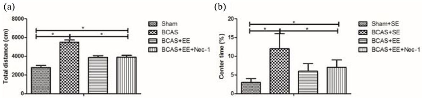
Behavioral performance in the open field test: (a) the total distance traveled in the apparatus in the Sham + SE, BCAS + SE, BCAS + EE, and BCAS + EE + Nec-1 groups (*P < 0.5) and (b) the time spent in the center of the apparatus in the four groups (*P < 0.05).
Histological alterations after different treatments in BCAS mice
There were significant changes in the cluster of differentiation 68 (CD68), ionized calcium-binding adapter molecule 1 (IBA1), and glial fibrillary acidic protein (GFAP) expressions. The top four images (Figure 4(a)–(d), 20×) were stained with CD68 antibody, a marker of activated microglia/macrophages; the middle four images (Figure 4(e)–(h), 20×) were stained with GFAP antibody, a marker of astrocyte; the bottom four images (Figure 4(i)–(l), 20×) were stained with IBA1, a marker of microglia. Figure 4(m) shows the statistical data from over 12 images. The data demonstrated that the expressions of CD68, IBA1, and GFAP in the BCAS mice were significantly stronger than those in sham mice (*P < 0.05), and that EE and EE + Nec-1 have the same significant therapeutic effect on the BCAS mice, but no superposition effect.
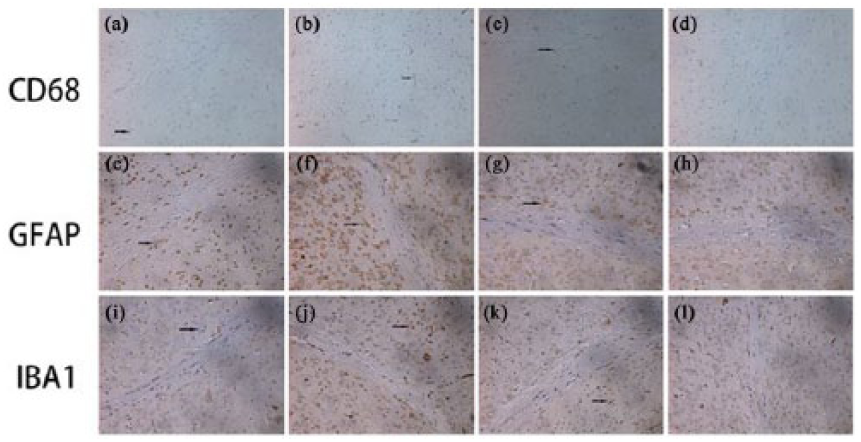
Immunohistochemical staining in the subregions of the hippocampus: (a)–(d) CD68 expression in the four groups of mice; (e)–(h) GFAP expression in the four groups of mice; and (i)–(l) IBA1 expression in the four groups of experimental mice. The arrows show the positive cells (dyed brown). The images are magnified 20×.
The alterations of receptor interacting protein 1, receptor interacting protein 3, and myelin basic protein from the hippocampus
To understand whether necroptosis signaling was involved in the chronic ischemic stroke induced by BCAS surgery, we examined the expression of receptor interacting protein 1 (RIP1), receptor interacting protein 3 (RIP3), and myelin basic protein (MBP) using Western blot. The kinase activities of RIP1 and RIP3 have been proposed to be required for necroptosis; our results indicated that the levels of RIP1, RIP3, and MBP in the BCAS mice were higher than those in the controls, as shown in Figure 5.

The expressions of proteins in the hippocampus, including RIP1, RIP3, and MBP, in the experimental mice in the Sham + SE, BCAS + SE, BCAS + EE, and BCAS + EE + Nec-1 groups, respectively.
The changes of inflammatory cytokines
The levels of some inflammatory cytokines were evaluated through enzyme-linked immunosorbent assay (ELISA) technology (Figure 6(a) and (b)). The data suggested that the levels of TNF-α and interleukin (IL)-1β protein in the mice from the BCAS + SE and Sham + SE groups were the strongest and weakest, respectively, and that the mice from the BCAS + EE group alleviated the levels of these inflammatory cytokines. There was an enhanced alleviated effect on these two inflammatory cytokines in the mice from the BCAS + EE + Nec-1 group (*P < 0.05).
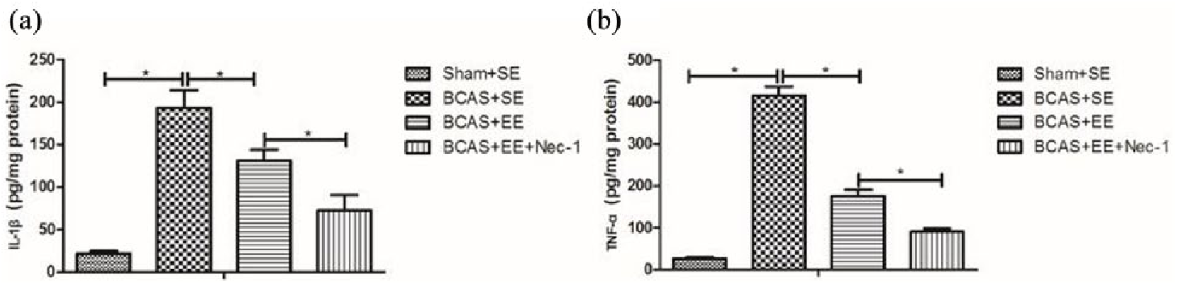
The changes of inflammatory cytokines in the hippocampal tissue measured by ELISA: (a) the level of TNF-α expression and (b) the level of IL-1β expression (*P < 0.05) compared to the Sham + SE group and the BCAS + SE group, respectively.
Discussion
In this study, we demonstrated that EE ameliorated cognitive function and alleviated inflammation responses in the VCI induced by the BCAS mouse model and that Nec-1 enhanced the effect of EE on VCI. Using microcoils, BCAS successfully induced moderate cerebral hypoperfusion and thus led to VCI. At 30 days after surgery, EE increased the value of CBF in BCAS mice, but there is no effect when combined with Nec-1 administration. In the other indices detected in this study, including behavioral performance, tissue staining, and protein measurement, the EE with Nec-1 combination can bring about the enhanced improvement.
EE has been shown to influence a number of brain parameters, including cell proliferation, 25 inflammatory cytokines, 20 and growth factor expression. 26 This study illustrates that the environmental factors and activity can alter functional outcomes, tissue inflammation, and cognitive recovery in VCI mice. As shown in Figure 2, VCI mice treated with EE and EE + Nec-1 spent shorter escape latency to find the platform in the MWM, which is associated with hippocampal function. 27 The same effects were observed in the other behavioral test after treatment with EE and EE + Nec-1, but there is no difference between the EE and EE + Nec-1 groups, such as OFT shown in Figure 3. A similar result was observed that EE reduced GFAP-, IAB1-, and CD68-positive cells, whereas this alteration was not enhanced by the administration of Nec-1, as shown in Figure 4. In this regard, the possible physiological mechanisms underlying the above effect have to be determined further in the future study. Necroptosis, that is, recently discovered programmed necrosis, is activated by TNF-α and/or Fas,24,28 which played important roles in a number of pathophysiological conditions such as ischemic stroke. 24 Accumulating evidence has shown that necroptosis is inhibited specifically by Nec-1.24,29 In this study, we found that the combination of EE with Nec-1 decreased the expressions of RIP1 and RIP3 and increased the expression of MBP in VCI mice. In addition, behavioral functions of mice were improved in the EE and EE + Nec-1 groups as compared with the untreated BCAS group. Necroptosis is a pathological process characterized by inflammatory responses. 22 Hence, we measured the level of inflammatory cytokines and confirmed that EE decreased the levels of TNF-α and IL-1β in the mouse model of VCI induced by BCAS; moreover, this effect was enhanced by the administration of Nec-1 in the VCI mice.
Our study has several limitations. First, the process of disease has a defined start in the study, whereas in fact it is insidious with slow onset in human. Second, the experimental mice were administrated for a relatively shorter period of time, that is, only 3 weeks. Third, the mice we used were young and male, but regardless of the gender the elderly are easy to suffer from VCI. In the future research work, we should investigate EE with or without Nec-1 in aged male and female animals and the therapeutic window and mechanism of treatment with EE + Nec-1. In addition, we have ongoing work to determine the influence and possible mechanism of neuroprotection by long-term EE + Nec-1 therapy.
In conclusion, our work suggests that treatment with EE and Nec-1 alleviated inflammation and improved cognitive function in a mouse model of VCI. Therefore, this study may provide a potential therapeutic target for the patients with VCI.
Materials and methods
Animals and experimental design
Male wild-type C57BL/6 mice were purchased from the Shanghai Laboratory Animal Center (SLAC), Chinese Academy of Sciences (20160966A284, 27 February 2016). Animals were housed in an animal breeding room under controlled temperature and provided with free access to food and water. Furthermore, all animals were maintained in accredited facility of altering 12-h light–dark cycle. Experiments were performed on 12-week-old mice (weighing 25–30 g).
Mice were matched in accordance with body weight and randomly assigned to one of the following four groups according to the random number table: (1) sham-operated group for the procedure of BCAS plus a standard housing condition (Sham + SE group), n = 16; (2) BCAS surgery housing in a standard environment (BCAS + SE group), n = 16; (3) BCAS housing in EE (BCAS + EE group), n = 16; and (4) BCAS housing in EE condition plus administration with Nec-1 (BCAS + EE + Nec-1 group), n = 16. The outcome measures were blinded.
Microcoil preparation and CBF measurement
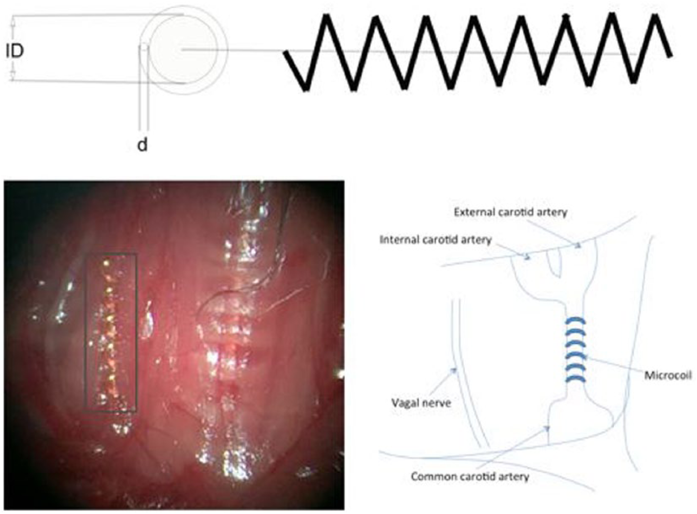
The microcoils used in this study were made of piano wire characterized by an inner diameter (ID) of 0.18 mm, a wire diameter (d) of 0.08 mm, a pitch of 0.50 mm, and the total length of 2.5 mm (Sawane Spring Co., Wuxi, China), as shown in Figure 7.
CBF was recorded through the exposed skull in the prone position by laser Doppler flowmetry (Omega Wave, Tokyo, Japan) 3 days before the surgery and 1 h after the surgery. The details were as follows: mice were deeply anesthetized with isoflurane (1.5%); the operation was performed as reported previously; 14 finally, the CBF values were expressed as the percentage of the baseline value.
Surgical operation on the mice
The surgery of BCAS was performed using microcoils as described previously. 14 Briefly, 12-week-old mice were deeply anesthetized with isoflurane (1.5%). And then the mouse was fixed in the supine position. Through a midline incision in the cervical part, common carotid arteries (CCAs) were exposed through the midline incision in the cervical part, and then the microcoils were gently twined around both arteries in turn (Figure 7). The body temperature was maintained at 37.0°C ± 0.5°C by a heating pad during the course of the surgery. Some mice underwent the sham operation, and only the same procedure was performed as the BCAS operation without using microcoils. After surgery, the animals were randomly divided into four groups, including the control, BCAS, BCAS + EE, and BCAS + EE + Nec-1 groups.
Drug treatment and housing condition
Mice were given 3.5 mg/kg Nec-1 (Calbiochem, San Diego, CA, USA) twice a week for 3 weeks after surgery. Nec-1 was dissolved in 10% methyl-beta-cyclodextrin solution (Sigma-Aldrich, St Louis, MO, USA) and administrated through intragastric administration at the time points indicated.
EE housing condition was described as follows: EE cage was a large plastic one (56 × 40 × 22 cm3) containing various items for mice to play and manipulate, such as plastic tunnels, wooden climbing frame, platforms, idling shelters, mini house, exercise wheel, and other novel objects. In order to keep novelty for mice, these items were rearranged or replaced completely and cleaned twice a week. At the same time, the mice from the BCAS + EE + Nec-1 group were administrated with Nec-1 while they were housed in EE condition.
Water maze testing
Water maze test was commenced after treatment for 3 weeks with EE or EE + Nec-1 according to the previous publication. 30 In brief, a cylindrical water tank was filled with water covering the platform. The temperature of water was kept at 22°C–25°C. And then edible white pigment was dissolved in water so that mice could not see the platform. The platform (10 cm diameter) was placed in the same position during the course of experiment. Mice were continuously trained for 5 days. The data were obtained and considered as the experimental values only when the mice found and climbed the hidden platform within 60 s and kept for 10 s. On the sixth day, mice were tested while the platform was removed, and the time spent on the platform quadrant and the number of time the mice crossed the platform site were recorded as the experimental values. The data were analyzed utilizing SMART-CS program (Panlab, Barcelona, Spain).
OFT
To evaluate a series of behaviors of mice, including exploratory, anxiety, and spontaneous locomotor activity, we performed an OFT for experimental animals, according to the procedure published elsewhere. 31 All mice were placed in the box one by one, an open field apparatus with a camera. The apparatus was cleaned thoroughly using 75% ethanol before the mouse was placed into it. Mouse could freely move around for 15 min in it. The total distance and pathway were recorded by the camera and determined.
Sample collection
After behavioral measurement, the experimental animals were sacrificed through anesthetization with 10% chloral hydrate. After decapitation, the hemispheres were harvested for further analysis.
Immunohistochemical staining
For the immunohistochemical examination, 3,3′-diaminobenzidine (DAB) staining was performed on the samples according to the procedure published elsewhere 32 with slight modifications. Briefly, the brains were removed and paraffin-embedded, and then 25-μm-thick sections were prepared. These sections were incubated in 0.3% H2O2 in phosphate-buffered saline (PBS) for 30 min and 0.3% Triton X-100 in PBS for 15 min in turn at room temperature. After a 30-min preincubation in 10% (wt%/vol%) bovine serum albumin (BSA), the sections were incubated overnight at 4°C with primary antibody (CD68, GFAP, and IBA1, 1:300; Santa Cruz Biotechnology, Inc., Dallas, Texas, USA). The following day, the sections were washed with PBS three times and treated with appropriate secondary antibodies (Vector Laboratories, Burlingame, CA, USA). The avidin–biotin complex (ABC) method was performed for immunoreactivity after rinsing in PBS, and then the sections were developed using DAB staining. Several fields were photographed in each section.
Western blot analysis
As previously described, 21 total protein extracts from the hippocampus were collected. Tissues were lysed in a homogenizer containing radioimmunoprecipitation assay (RIPA) buffer kept at 4°C (Beyotime Biotechnology, Shanghai, China). And then Western blotting was conducted to measure the expressions of proteins. Briefly, a total of 40 μg per protein sample was added to the gel well made of sodium dodecyl sulfate polyacrylamide gel electrophoresis (SDS-PAGE) and separated by 10% SDS-PAGE; after that, the proteins were transferred onto polyvinylidene difluoride (PVDF) membranes (Bio-Rad Laboratories, Hercules, CA, USA). And then the membranes were blocked with 3% BSA for 1 h and incubated with primary and secondary antibodies, respectively. The levels of RIP1, RIP3, and β-actin (Santa Cruz Biotechnology, Inc., USA.) were assayed. Finally, ImageJ software was used to quantify the proteins that were normalized with respect to the levels of β-actin as an internal control.
ELISA
Concentrations of IL-1β and TNF-α from the hippocampal homogenates were quantified using ELISA kits (Mouse IL-1 beta ELISA Kit and Mouse TNF-α ELISA Kit; RayBiotech, Norcross, GA, USA) according to the manufacturer’s protocol.
Statistical analysis
All results were expressed as mean ± standard deviation (SD). Differences between groups were analyzed by two-tailed t test. P < 0.05 was considered as significant difference. All graphs in this study were constructed using GraphPad Prism (version 5; GraphPad Software, San Diego, CA, USA).
Footnotes
Declaration of conflicting interests
The author(s) declared no potential conflicts of interest with respect to the research, authorship, and/or publication of this article.
Funding
The author(s) disclosed receipt of the following financial support for the research, authorship, and/or publication of this article: This study was supported by the Natural Science Foundation of China (NSFC; nos. 81672242) , the key projects of Shanghai Science and Technology on Biomedicine (nos. 17411953900) and Shanghai Municipal Commission of Health and Family Planning, Key developing disciplines (nos. 2015ZB0401).