
Abstract
In the study, we investigated the immune factors related to T helper 17 (Th17) cells and T regulatory (Treg) cells in spontaneous abortion mice. The expression of Th17 was analyzed by interleukin (IL)-6, IL-17A secretion, RAR-related orphan receptor γt (RORγt) expression, and proportion of CD4+IL-17+ cells. The levels of IL-10, transforming growth factor β (TGF-β), Foxp3, and CD4+Foxp3+ cells were presented the Treg expression. Higher embryo absorption rate was found in spontaneous abortion group than that in normal pregnancy group (P < 0.01). Compared with the normal pregnancy mice, spontaneous abortion mice showed higher levels of IL-6 and IL-17A and lower levels of IL-10 and TGF-β in serum and in decidua (P < 0.05). Furthermore, the expressions of Foxp3 and CD4+Foxp3+ cells were significantly decreased in spontaneous abortion mice than those in normal pregnancy mice (P < 0.05). However, the levels of RORγt and CD4+IL-17+ cells remarkably increased in spontaneous abortion mice (P < 0.05). The results reveal that Th17/Treg cells may play a vital role in immunoregulation during pregnancy.
Introduction
Spontaneous abortion is a common problem in clinics. Many reasons may be occurred in the processing of abortion, such as genetic, hormonal abnormalities, and tissue rejection. 1 Several researchers reported that autoimmune disorder was the major reason for abortion.
In pregnancy, T regulatory (Treg) cells play an important role in immunoregulation. The production of interleukin (IL)-10 and transforming growth factor β (TGF-β) would favor the maintenance of mammalian pregnancy, which are the secretions of Treg cells.2,3 Treg cells could activate the expression of Foxp3 in the nuclei. 4 CD4+CD25+Treg cells were described as a unique subpopulation of T cells, known playing physiology and pathophysiology of pregnancy.5,6 It is essential to know the changes of Treg cells in pregnancy and abortion.
T helper 17 (Th17) cells are another T cells, playing opposite actions to Treg cells. With the ability of producing IL-17 and IL-6, Th17 cells play a critical role in the development of autoimmunity and allergic reactions. 7 The IL-17 expression increased in placenta and in serum from women with intrahepatic cholestasis of pregnancy, and the differentiation of Th17/Treg could affect the pregnancy. 8 The differentiation of Th17 is controlled by a key transcription factor called RAR-related orphan receptor γt (RORγt), which can induce the secretion of IL-17 in naive T cells. 9 In humans, IL-6 was reported to be elevated at the onset of spontaneous abortion. It also found the numbers of IL-6 cells increased in placentas and deciduas of abortion mice. 10
In the study, we further investigated the changes of immune factors related to Th17 and Treg cells in pregnant and abortion mice to elucidate the immune function.
Materials and methods
Animals and tissues simples
Healthy female CBA/J, male DBA/2, and BALB/c mice which were 8-week-old were provided by the Institute of Laboratory Animal Sciences, Chinese Academy of Medical Sciences. All animals were kept in 23 ± 2°C under a 12-h dark/light cycle environment with free access to water and food. The spontaneous abortion model CBA/J × DBA/2 and the normal pregnancy model CBA/2 × BALB/c were established by mating CBA/J with DBA/2 mice and CBA/J with BALB/c mice, respectively. Then, two females and one male were fed in one cage separated from other mating groups. The date was treated as 0 day after pregnancy when vaginal plug was detected.
Blood was extracted from the heart of mice after anesthetized with 1% pentobarbital sodium (Sigma, Burlington, MA, USA) at 14 days after pregnancy. Mice were sacrificed and uterine tissues surrounding the embryo were collected. Spleen was taken and grinded for about 3 min to prepare the cell homogenate. The rate of embryo absorption (R) was calculated as following, R = Re/(Re + F). Re is the number of absorbed embryos, and F is the number of surviving embryos.
Animal care and experimental procedures were approved by the Institutional Animal Care and Use Committee at the Qingdao University.
Enzyme-linked immunosorbent assay
The levels of IL-6, IL-10, IL-17A, and TGF-β in serum and in decidua were measured by enzyme-linked immunosorbent assay (ELISA) kit (MAPKET, USA) according to the instructions. The values at 450 nm were recorded. Standard concentration and optical density (OD) values were used to establish the standard curve to calculate the concentration of each factor.
Reverse transcription-polymerase chain reaction
Total RNA was extracted from the decidua tissue using TRIzol kit (Invitrogen, Carlsbad, CA, USA). The purity of the decidua was calculated by calculating the absorbance value. Only the RNA samples with a260/a280 ratio between 1.8 and 2.0 were selected for further study. Reverse transcription were performed with a kit from Tiangen Biotech Co., Ltd (Beijing, China). SYBR Green master (Bio-Rad, Hercules, CA, USA) was used for polymerase chain reaction (PCR). The following primers were used: 5′-ACAGAAGCAGCGTCAGTACC-3′ (sense) and 5′-CGGGGTATTTTTGGCAAGGC-3′ (anti-sense) for Foxp3, 5′-TCACATCACCCCGTCATTGG-3′ (sense), and 5′-CTCCACGACTGCCCATCATT-3′ (anti-sense) for RORγt, 5′-GTCAAGGCTGAGAACGGGAA-3′ (sense) and 5′-AAATGAGCCCCAGCCTTCTC-3′ (anti-sense) for glyceraldehyde 3-phosphate dehydrogenase (GAPDH). The primers were all designed and synthesized by Shanghai Sangon Biotech Co., Ltd (Shanghai, China). The images were observed and photographed with gel imaging system (Bio-Rad).
Western blot
The protein expressions of Foxp3 and RORγt were detected by Western blot. The protein concentration was detected by bicinchoninic acid (BCA) method. And the total proteins were separated by sodium dodecyl sulfate polyacrylamide gel electrophoresis (SDS-PAGE) and transferred to polyvinylidene difluoride (PVDF) membrane (Merck, Darmstadt, Germany). The PVDF membrane was immersed in 5% milk powder in TBST buffer (20 mM Tris, 137 mM NaCl, pH 7.6, with 0.1% Tween 20) and blocked for 1 h. Then, the PVDF membrane was incubated with the corresponding primary antibody (1:1000; Abcam, Cambridge, UK) for 2 h at room temperature, and the corresponding horseradish peroxidase (HRP)-conjugated secondary antibody (1:2000; Abcam) was added and incubated for 1 h at room temperature. The enhanced chemiluminescence (ECL) system (Thermo Fisher Scientific, Waltham, MA, USA) was used to detect the signal on the specimen membrane. Relative expression levels of each protein were normalized to endogenous control GAPDH using Image J software (Thermo Fisher Scientific).
Flow cytometry
Cell homogenate was centrifuged and supernatant was removed. The cells were harvested and cultured in RPMI 1640 medium (Gibco, Gaithersburg, MD, USA) and then adjusted to a concentration of 1 × 10⁶/mL. Fluorescence-activated cell sorter (FACS) flow cytometer (BD Biosciences, San Jose, CA, USA) was used to analyze the cell distribution. The following monoclonal antibodies were used: fluorescein isothiocyanate–conjugated rat anti-mouse CD4 (rat IgG1; BD Biosciences), phycoerythrin-labeled anti-mouse CD25, IL-17A (rat IgG2b; BD Biosciences). The results were analyzed by CellQuest Pro software.
Statistical analysis
SPSS19.0 statistical software was used to analyze the variance of all the data. All analytical data were presented as mean ± standard deviation (SD). Comparison between two groups was analyzed by t test. P < 0.05 was considered to be statistically significant.
Result
Spontaneous abortion mice with higher embryo absorption rate than normal pregnant mice
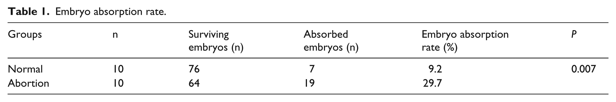
The mice were sacrificed by cervical spine dislocation on the 14th day. The placenta was taken from the mice to count the number of absorbed embryos and surviving embryos. As shown in Table 1 and Figure 1, the embryo absorption rate in spontaneous abortion group was 29.7%, which was significantly higher than that in normal pregnancy group (9.2%, P = 0.007).
Embryo absorption rate.

Representative pictures of uteri from normal pregnant mice and from abortion mice.
The levels of IL-6, IL-17A, IL-10, and TGF-β in serum and in decidua
The levels of IL-6, IL-17A, IL-10, and TGF-β in serum and in decidua were analyzed. The results are shown in Figure 2. The expressions of IL-6 and IL-17A were obviously higher in abortion group than those in normal group (P < 0.05), both in serum and in decidua. However, the levels of IL-10 and TGF-β were remarkably decreased in abortion mice than those in normal pregnancy mice, both in serum and in decidua (P < 0.05).

The expressions of IL-6, IL-17A, IL-10, and TGF-β (a) in serum and (b) in decidua. *Comparison with normal pregnancy group, P < 0.05.
Foxp3 and RORγt levels in decidua
In this study, the levels of Foxp3 and RORγt in decidua tissue were measured. As shown in Figure 3(a), the relative expression of Foxp3 messenger RNA (mRNA) in spontaneous abortion group was lower than that in normal pregnancy group and the difference was statistically significant (P < 0.05). However, the level of RORγt mRNA in spontaneous abortion group was significantly higher than that in normal pregnancy group (P < 0.05). Furthermore, the results of protein levels were consistent with the results of mRNA (Figure 3(b)). The expression of Foxp3 protein was remarkably declined and the expression of RORγt protein was notably increased in abortion rats (P < 0.05).

The relative levels of Foxp3 and RORγt (a) mRNA and (b) protein in decidua tissues of rats. *P < 0.05, comparison with normal group.
The proportion of Th17 and Treg cells in splenic lymphocyte suspension
We further wondered the changes of Th17 and Treg cells in abortion mice, so we analyzed the numbers of CD4+Foxp3+ and CD4+IL-17A+ in splenic lymphocyte suspension (Figure 4). We found that the percentage of Treg in abortion mice was significantly decreased compared with that in normal pregnancy mice (4.29% vs 8.28%, P < 0.05). Conversely, the higher levels of Th17 in abortion group were expressed than that in normal pregnancy group (4.38% vs 2.52%, P < 0.05).

The proportion of Treg and Th17 cells in splenic lymphocyte suspension detected by flow cytometry. *P < 0.05, compared with normal group.
Discussion
The spontaneous abortion may be caused by the unbalance immune mechanisms. During pregnancy, the maternal immune system has to tolerate the persistence of paternal alloantigens without affecting anti-infectious immune responsiveness. Here, we studied the changes of Th17/Treg cells in pregnancy mice and abortion mice.
Foxp3 is a specific transcription factor of Treg cells that plays an important role in function of Treg cells. 11 There was a report that the level of Foxp3 was significantly increased in abortion mice than that in normal pregnant and nonpregnant mice. 2 RORγt is a key transcription factor that consistently regulates the differentiation of Th17 cells. A research found that RORγt mRNA expression was significantly increased in the abortion sites compared with the implantation sites of abortion model mice. In addition, the levels of IL-17A mRNA were significantly higher in abortion sites than that in implantation sites. 1 These results are consistent with our study. These indicated the Treg/Th17 was involved in immune response in abortion and pregnancy.
TGF-β plays a pivotal role in maintaining the balance between Th17 and Treg, which can induce the expression of both Foxp3 and RORγt. 12 RORγt can induce the production of IL-17 in naive T cells; in turn, IL-17 promotes the proliferation and differentiation of a variety of cell types including Th17. 13 Our study showed that abortion resulted in the IL-10, TGF-β expression decreasing and IL-6, IL-17A increasing compared to normal pregnancy. The results suggested an immune response of Treg/Th17 occurred during pregnancy and abortion.
In summary, the levels of Treg cells were remarkably decreased and the levels of Th17 cells were significantly increased in abortion mice than those in normal pregnancy mice. The immune response of Th17/Treg cells may play a pivotal role in determination of pregnancy outcomes.
Footnotes
Acknowledgements
N.L. and Q.Q. contributed equally to this work.
Declaration of conflicting interests
The author(s) declared no potential conflicts of interest with respect to the research, authorship, and/or publication of this article.
Funding
The author(s) received no financial support for the research, authorship, and/or publication of this article.