
Abstract
Background
Diagnostic criteria for multiple sclerosis have been developed to guide the diagnostic process. In the latest revision of the McDonald criteria, the presence of oligoclonal bands may replace the need for dissemination in time. The aim of this study is to investigate if the less time-consuming analysis of immunoglobulin G index in cerebrospinal fluid can safely predict the findings of oligoclonal bands.
Methods
This is a retrospective study of patients with multiple sclerosis at three hospitals in South-East Norway where lumbar puncture is performed routinely. We included patients diagnosed with multiple sclerosis after 2005 with known oligoclonal band status and an immunoglobulin G index score.
Results
Of 1295 patients diagnosed during or after 2005, 93.8% were oligoclonal band positive at diagnosis. Of 842 multiple sclerosis patients with known immunoglobulin G index and oligoclonal band status, 93.3% were oligoclonal band positive and 76.7% had an elevated immunoglobulin G index. The positive predictive value of a high immunoglobulin G index when oligoclonal bands are positive was 99.4% (95% confidence interval 98.4–99.8%). The negative predictive value of a normal immunoglobulin G index when oligoclonal bands are negative was 26.5% (95% confidence interval 23.5–29.9%).
Conclusion
An immunoglobulin G index >0.7 has a positive predictive value >99% for oligoclonal bands. An elevated immunoglobulin G index adds diagnostic value versus oligoclonal bands and saves time in the diagnostic process.
Introduction
Multiple sclerosis (MS) is an inflammatory disease with secondary neurodegeneration that causes significant disability in patients over time. Disease onset is usually between 20 and 40 years of age and MS is one of the most common non-traumatic causes of disability in young adults. 1 Recent studies have shown increasing evidence of a better prognosis when disease-modifying drugs are initiated early in the disease course.2,3
The diagnostic criteria for MS are based on a combination of clinical, imaging and laboratory evidence for disease in the central nervous system (CNS). The impact of each of these elements has changed, although the need for evidence of dissemination in time (DIT) and dissemination in space (DIS) for a secure diagnosis has remained.4–7 Cerebrospinal fluid (CSF) analysis is not mandatory for the diagnosis of MS in patients with a clinical syndrome suggestive of the disease. However, in the 2017 revision of the McDonald diagnostic criteria, presence of ≥2 CSF-specific oligoclonal immunoglobulin G (IgG) bands (OCB) can be used in place of demonstrating DIT, 8 possibly leading to an earlier diagnosis. Several authors have erroneously assumed the newest McDonald criteria allow for OCB to prove DIT. In fact, the presence of OCB in patients with a typical clinical presentation, typical lesions fulfilling DIS and with alternative diagnoses reasonably ruled out will provide supporting evidence of the immune and inflammatory nature of the disease in patients, without having to wait for DIT to occur.9,10 Quantitative measurement of IgG in the CNS is less sensitive than isoelectric focusing (IEF), and due to lack of studies on the validity of an elevated IgG index, this was not included in the revised criteria and should be interpreted with caution.11,12
Normally, IgG is not produced intrathecally in any significant amount, and most of the IgG found in the CSF is derived peripherally and crosses the blood-brain barrier (BBB). The presence of IgG in the CSF is therefore not synonymous with CNS inflammation. However, in a range of neuroinflammatory conditions, most notably MS, oligoclonal IgG is produced in the CNS by a small number of B cell clones. Readily distinguishable IgG bands found in the CSF are picked up on the qualitative analysis IEF as separate bands, so-called OCB. 13 The presence of two or more OCB in the CSF without an identical match in the serum is considered pathological. Albumin, in contrast, is produced in the liver and the presence of albumin in the CSF is due to leakage through the BBB. The IgG index is the ratio of the quotients for IgG and albumin (IgGcsf/IgGs)/(Albcsf/Albs). It is a quantitative analysis of the relationship between CSF IgG and serum IgG, divided by the same relationship for albumin. As albumin is a smaller protein than IgG, it crosses the BBB more easily. In inflammatory CNS conditions, commonly MS, the IgG index is raised.
In most hospitals, the IgG index is ready within a day. The analysis of OCB, in contrast, is more time consuming. The aim of the current study was to ascertain whether the IgG index can replace or add diagnostic or clinical value versus OCB in the early diagnostic work up of patients with suspected MS.
Patients and methods
Patients
The BOT database is a local registry of all patients registered with a McDonald criteria confirmed MS diagnosis (International Statistical Classification of Diseases 10 G35) between 1919 and 2017 at Oslo University Hospital (OUS) and at the two regional hospital trusts of Vestre Viken Health Trust (VVHF) and Telemark Health Trust (STHF). These three hospitals serve a population of approximately 1 million people in South East Norway, approximately 20% of the total Norwegian population. The vast majority of the population and the patient cohort (>95%) are native Scandinavian.
Lumbar puncture has been a routine part of diagnostic work up in all three hospitals for the past 50 years. In the current study, we chose to only include patients diagnosed in 2005 and later because isoelectric focusing became the sole means of measuring OCB in all three hospitals in 2005. None of the patients received steroids before lumbar puncture.
CSF IgG index and OCB
OCB were detected using IEF followed by immunofixation with anti IgG, on the HYDRASYS system (Sebia, Lisses, France). Serum was collected at the same time as the lumbar puncture. IgG and albumin in serum and CSF were measured with the routine method at the different hospital trusts at the time. The presence of OCB was registered binary as positive (≥2) or negative (0–1). We recorded the IgG index both on a continuous scale and binary as normal or elevated. Because of some method variations the cut-off varied slightly between the three hospitals. In VVHF the cut-off for an elevated IgG index was 0.6, in STHF the cut-off was 0.63 and in OUS the cut-off was 0.7.
Data handling
We used SPSS software version 21 for data handling. The p values were measured using an independent sample t test for continuous variables and Pearson chi-square test for binary variables. For calculating positive and negative predictive value, sensitivity and specificity as well as confidence intervals (CI), we used the direct method and MedCalc Statistical Software (https://www.medcalc.org/). The study was approved by the regional ethics committee.
Results
We identified 2953 MS patients with a known OCB status diagnosed between 1941 and 2017, 87.9% of which were OCB positive. In total, 96.3% of the 1295 patients diagnosed in 2005 or after had a known OCB status, and 93.8% of these were OCB positive. MS patients without OCB or with a normal IgG index were on average more likely to have progressive disease at the time of diagnosis, reported longer time from onset to diagnosis and were older than those with OCB (Table 1).
Demographic and clinical findings of all 842 MS patients.

*significant.
IgG: immunoglobulin G; OCB: oligoclonal bands; IgG index: (cerebrospinal fluid (CSF)/serum IgG)/ (CSF/serum albumin); EDSS: Expanded Disability Status Scale; CIS: clinically isolated syndrome; RRMS: relapsing–remitting multiple sclerosis; SPMS: secondary progressive multiple sclerosis.
Overall, 842 MS patients had a known IgG index. This ranged from 0.10 to 5.96 (mean 1.08, SD 0.65). In total 23.3% had a normal IgG index, whereas 76.7% had an elevated index. We found that 99.8% of patients with an IgG index above 0.8 had OCBs in their CSF. All patients with IgG index above 0.86 had OCBs (Figure 1).

Scatter plot of immunoglobulin G (IgG) index results in oligoclonal band (OCB) negative and OCB positive multiple sclerosis (MS) patients.
All 842 patients with a known IgG index had a known OCB status (93.3% OCB positive, Table 2). The sensitivity of the IgG index predicting OCB outcome was 81.7% (95% CI 78.8–84.3%), and the specificity was 92.9% (95% CI 82.7–98.0%). The positive predictive value of an elevated IgG index was 99.4% (95% CI 98.4–99.8%), whereas the negative predictive value of a normal IgG index was 26.5% (95% CI 23.5–29.9%). Each of the three hospitals had similar individual positive predictive values (OUS 99.0%, VVHF 100%, STHF 99.4%) (Figure 2).
Cross table of OCB and IgG index findings. Percentage of elevated or normal IgG index with OCB or without OCB.

IgG: immunoglobulin G; OCB: oligoclonal bands; IgG index: (CSF/serum IgG)/ (CSF/serum albumin)
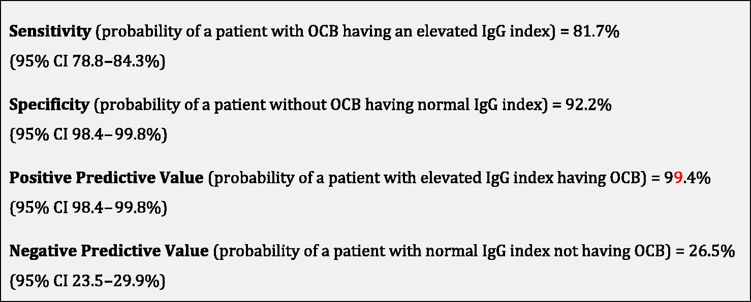
Sensitivity, specificity, positive predictive value and negative predictive value.CI: confidence interval; IgG: immunoglobulin G; OCB: oligoclonal bands.
Discussion
The current study demonstrates that an elevated IgG index has a positive predictive value above 99% predicting the presence of intrathecal OCBs. Thus, an elevated IgG index can be used as a proxy to OCB and DIT is not required.
In a Northern European population, around 95% of MS patients have OCB in the CSF.14,15 Different ethnic populations have significantly lower prevalence of OCB,16–18 although this difference may not be as obvious in immigrants to high-risk countries 19 and may in fact represent less-sensitive methodology and misdiagnosis. 20 In our population, which is mostly Northern-European, 93.8% had OCB.
In the current practice, it can take several days or weeks to get the OCB result and thus the MS diagnosis is potentially delayed. Meanwhile, calculating the IgG index takes less time and is cheaper and can be done within a day. In addition, it is rater independent. However, the IgG index and other quantitative IgG analysis are not equivalent to qualitative analysis using IEF due to lower sensitivity.12,21 Quantitative analysis has a diagnostic sensitivity of 60–70%15,22 and only 75% of patients will turn out to be OCB positive. 12 In our study, the probability of a patient with OCB having an elevated IgG index was 81.7%. Although the IgG index itself cannot predict the OCB outcome, our findings show an elevated IgG index has a very high predictive value of forecasting OCB. A normal IgG index, however, cannot be used to predict the presence or lack of OCB.
Like most hospitals, we use 0.6–0.7 as the cut-off for an elevated IgG index. We found that 99.8% of MS patients had positive OCB when the IgG index was above 0.8 and 100% of MS patients had ≥2 OCB when the IgG index was more than 0.86. Other studies have also proven strong correlations between a positive IgG index and the presence of intrathecal OCBs, with 96–100% of patients with an IgG index above 0.8 having positive OCB.15,23 A few smaller studies have found no correlation.24,25 However, most studies on OCB and IgG index have had small sample sizes or have been subject to testing bias, as patients with complicated disease history are more likely to undergo lumbar puncture. Moreover, many of the cited OCB and IgG index studies were done before the introduction of IEF. Our study includes a large and near-complete MS population, as lumbar puncture is performed routinely when diagnosing MS in Norway. In addition, our study only included patients diagnosed after the introduction of IEF in 2005.
CSF findings used in the routine diagnosis of MS serve two purposes: to confirm a diagnosis of MS early in the disease course, and support exclusion of differential diagnoses.26,27 A meta-analysis found that OCB has a specificity of 94% for MS. 13 However, when considering patients with MS or other neuroinflammatory conditions, the specificity fell to 61%. This underlines the importance of context. We emphasise that our findings cannot be extrapolated to all neurological patients with a high IgG index, but are reserved for those patients where other neurological conditions have been excluded and the clinician is merely waiting for a positive OCB or DIT to be able to diagnose MS. We found that MS patients without OCB or with a normal IgG index were on average more likely to have progressive disease at onset and were older than those with OCB or elevated IgG index. This is in line with findings from Siritho et al., 28 though not others.29,30 One likely explanation for the significant difference in disease phenotype between those with and those without OCB and elevated IgG index is misdiagnosis. 20 The lack of OCB has a very high negative predictive value. 31 Although the current ethos is to diagnose MS as early as possible, this can sometimes decrease the accuracy of the diagnosis. If one suspects other neuroinflammatory disorders or there is presence of red flags in the diagnostic work up, 32 both the presence of OCB and a positive IgG index score should be interpreted with caution.
A faster diagnosis of MS is important to initialize treatment early. 33 This study of data from routine lumbar punctures over many years in a large Norwegian MS population demonstrates that an IgG index >0.7 has a very high positive predictive value for the presence of OCB. A positive IgG index can therefore replace OCB and thus lead to an earlier diagnosis.
Footnotes
Acknowledgements
We would like to thank the patients in the BOT database. We are also grateful to our colleagues at the three neurological departments for performing the lumbar punctures, as well as colleagues at The Departments of Medical Biochemistry at Oslo University Hospital, Vestre Viken Hospital Trust and Telemark Hospital Trust for analysing the CSF and serum.
Declaration of conflicting interests
The author(s) declared the following potential conflicts of interest with respect to the research, authorship, and/or publication of this article: CSS has received unrestricted research grants from Sanofi and Novartis, advisory board and/or speaker honoraria from Sanofi, Merck and Biogen Idec. HØF has received unrestricted research grants from Biogen Idec and Novartis, advisory board and/or speaker honoraria from Sanofi, Merck and Biogen Idec. TL has no conflicts of interest. P.B-H has received speaker honoraria from Novartis, UCB, Teva, Merck and Biogen Idec. SMM has received an unrestricted travel grant from Biogen Idec and speaker honoraria from Biogen Idec and Novartis. EGC has received unrestricted research grants (Novartis and Sanofi), advisory boards and/or speaker honoraria (Almirall, Biogen Idec, Merck, Roche, Novartis, Sanofi and Teva).
Funding
The author(s) disclosed receipt of the following financial support for the research, authorship, and/or publication of this article: This study was funded by grants from Vestre Viken Hospital Trust, Telemark Hospital Trust and The Independent Order of Odd Fellows.