
Abstract
Background
The application of digital technology in jaw reconstruction is clinically significant as it enhances surgical precision and patient outcomes. This study aimed to conduct a comprehensive bibliometric analysis to explore global research trends, hotspots, and future directions in the application of digital technology for jaw reconstruction.
Methods
This study employs a bibliometric analysis design, utilizing a retrospective approach. The data were sourced from the Web of Science Core Collection (WoSCC) database, focusing on publications related to digital technology in jaw reconstruction from 1995 to 2024. The final sample includes 1069 articles authored by 4781 authors from 3081 institutions. Data analyses were conducted using VOSviewer, CiteSpace, and the R package “Bibliometrix.”
Results
The analysis revealed a total of 1069 articles, with publication trends showing a peak in 2022. China produced the most articles, while the United States led in citations. The most productive institutions were based in Italy, Germany, and China, and key contributing authors included Marchetti Claudio and Ciocca Leonardo. Journal of Cranio-Maxillofacial Surgery had the highest H-index among journals. Keyword co-occurrence revealed hotspots like “implants” and “computer-aided design,” while burst terms such as “virtual surgical planning” and “3D printing” signaled emerging frontiers.
Conclusion
This bibliometric analysis examined the global research landscape on digital technology in jaw reconstruction, identifying key developments such as rapid prototyping, virtual surgical planning, and 3D printing. Future research should focus on integrating these technologies into clinical workflows, reducing costs, improving training, and addressing regulatory challenges.
Introduction
Jaw reconstruction is a critical surgical procedure aimed at restoring the function and esthetics of the jaw bones, which may be compromised due to trauma, congenital defects, tumor resection, or degenerative diseases. 1 The pathogenesis of these conditions often involves damage to the jawbone structure, leading to functional and esthetic impairments that significantly affect patients’ quality of life. 2 These defects affect a significant portion of the global population, posing a considerable disease burden. For instance, the incidence of oral and maxillofacial tumors alone is estimated to be around 3% to 5% of all malignancies, with a considerable proportion requiring jaw reconstruction. Furthermore, trauma-related craniofacial injuries account for approximately 40 million cases annually.3,4 Despite advancements in surgical techniques, jaw reconstruction remains challenging, often necessitating multiple surgeries and prolonged recovery periods.
In recent years, the advent of digital technology has revolutionized the field of jaw reconstruction. These advancements have enabled surgeons to perform more precise and personalized surgeries, significantly improving patient outcomes. For instance, surgeons can use computer-aided design (CAD) software to create a precise 3D model of the patient's jawbone, taking into account their unique anatomical structure and any defects or deformities. Computer-aided manufacturing (CAM) machines, such as milling or laser cutting devices, can precisely carve the implant from a block of material based on the CAD model. 5 The 3D-printed implants can be tailored to match the patient's unique anatomical features, reducing the risk of complications and enhancing the esthetic results. 6 Additionally, using advanced software, surgeons can simulate the surgery on a digital model of the patient's jaw. This allows them to visualize the procedure, plan the placement of implants and bone grafts, and anticipate potential challenges. Intraoperative navigation systems use real-time imaging and tracking technology to provide surgeons with a 3D view of the patient's jaw during the surgery. 7 Recent reviews have summarized the development and applications of these digital technologies, including CAD/CAM, 3D printing, and virtual surgical planning, in maxillofacial reconstruction, highlighting their impact on surgical precision and workflow optimization. 8 However, despite these advancements, there remains a lack of bibliometric studies systematically mapping global research trends in this field. Issues such as high costs, technical complexity, and the need for specialized training remain barriers to widespread adoption.
Given the rapid development and increasing interest in digital technology for jaw reconstruction, a systematic analysis of the existing research is crucial. This is where bibliometric analysis comes into play. Bibliometric analysis is a quantitative method used to study publication patterns, research hotspots, and development trends in a specific field. By analyzing the citation networks, keyword co-occurrences, and other bibliometric indicators, researchers can identify the most influential papers, authors, and institutions, as well as emerging trends and future directions. 9 To date, while several studies have explored the application of digital technology in various medical fields,10,11 there is a lack of dedicated bibliometric analyses focusing on jaw reconstruction.
Therefore, the present study aims to bridge this gap by conducting a comprehensive bibliometric analysis of research on the application of digital technology in jaw reconstruction. Our analysis will not only summarize the current state of knowledge but also identify future directions for research.
Materials and methods
The literature search for digital technology in jaw reconstruction was conducted on the Web of Science Core Collection (WoSCC). The search formula was ((TS = (jaw OR maxillo-mandibular OR mandibular OR maxilla OR maxillofacial OR mandible)) AND TS = (reconstruction OR reconstructive surgery)) AND TS = (digital OR computer aided surgery OR computer aided design OR computer aided manufacturing OR CAS OR CAD OR CAM).12–15 To prevent discrepancies during database updates, the literature search was performed on 14 October 2024. For this study, the publication language was limited to English. Among the various document types, only articles were taken into account.
Statistical analysis
Bibliographic records were utilized to gather relevant data, and bibliometric indicators were computed using Microsoft Excel. An in-depth academic analysis was carried out with visualization tools including VOSviewer (V1.6.20), CiteSpace (V 6.3.R1), and R package “Bibliometrix” (V4.3.3).
VOSviewer created maps of co-occurrences among countries, institutions, authors, and keywords, revealing key research themes and collaborations. Node sizes represented frequency, with colors distinguishing research clusters or average publication years for keywords. Thicker lines denoted stronger co-occurrence relationships. 16
CiteSpace analyzed the burst keywords on digital technology in jaw reconstruction from 1995 to 2024, producing time-zone maps that traced historical advancements and emerging patterns. 17 When keywords served as nodes, the threshold (top N per slice) was set to 5, and the pruning method was set to “pathfinder” combined with network merging. Based on these parameter settings for each node, a visual analysis was performed. 18
R package “Bibliometrix” enabled advanced statistical analysis and computation of bibliometric indices like the H-index, G-index, and M-index for prominent authors and institutions. The H-index, the most widely adopted bibliometric metric, is defined as the number of papers an author has published that have each been cited at least h times. 19 The G-index measures the cumulative citation performance of a researcher's publications, 20 while the M-index reflects the average number of citations per year for articles contributing to the H-index. 21 Authors’ three indices were all retrieved from the WoSCC database.
To assess journal influence, Journal Citation Reports (JCR) metrics, including the 2023 impact factor (IF) and quartile ranking (Q1–Q4), were used. The IF reflects average citations per article, and quartile ranking categorizes journals into four impact groups, with Q1 being the highest.
Results
Overview of publications
A total of 1069 English-language articles regarding digital technology in jaw reconstruction between 1995 and 2024 were included in this study (Figure 1). Our findings demonstrated that 4781 authors from 3081 institutions participated in producing the publications (Figure 2(a)). From 2009 to 2022, the number of publications showed a fluctuating upward trend, reaching a peak in 2022 (102 articles) (Figure 2(b)).

Flow diagram of the bibliographic retrieval process.
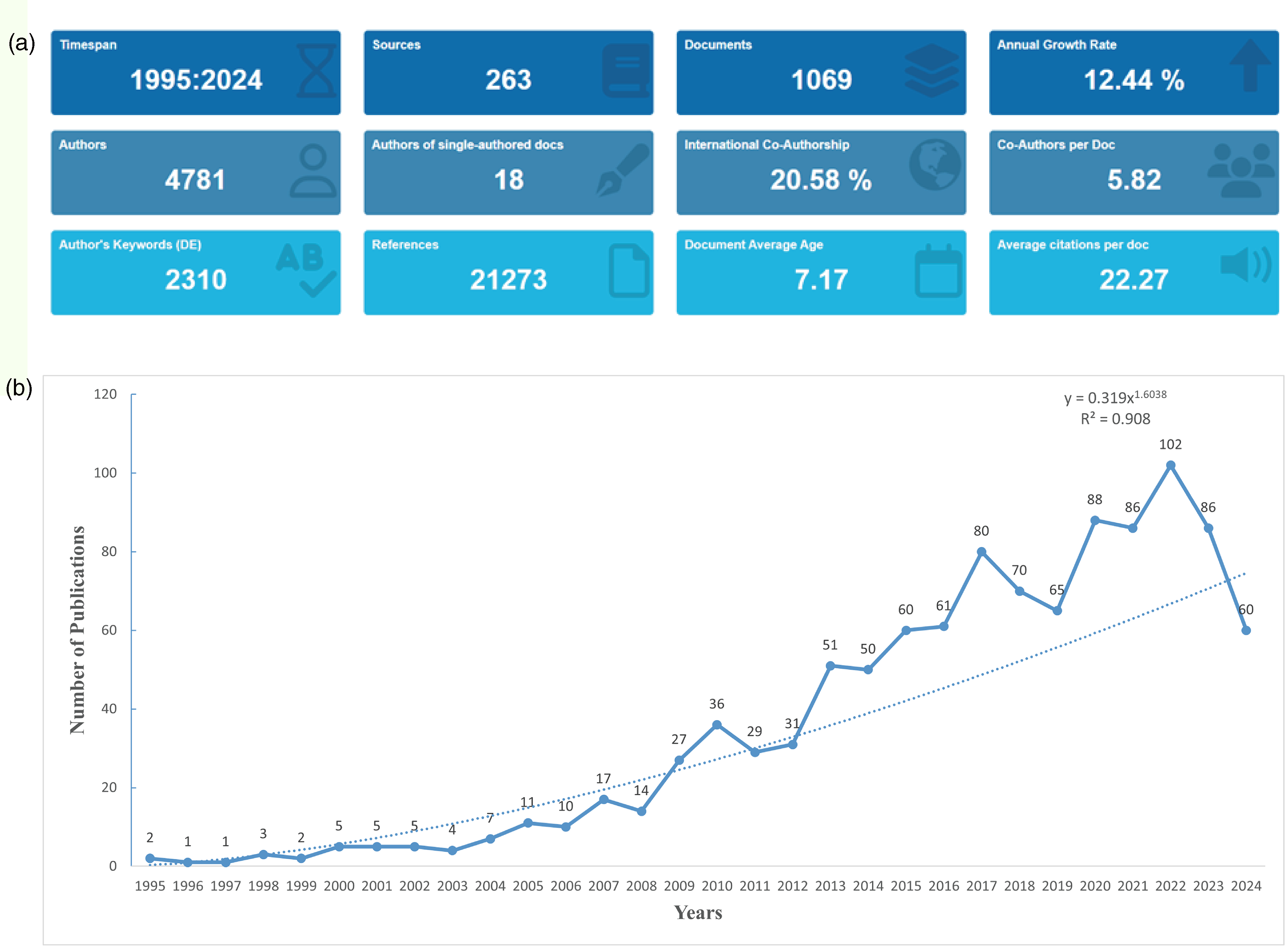
Analysis of the general information. (a) A summary of quantitative analysis of the publications. (b) Annual output of research from 1995 to 2024.
The top 50 most cited articles are listed in Table S1. The article titled “3D printing in dentistry” was published in the British Dental Journal (IF 2023 = 2.0) in 2015 and accumulated the most cited count of 645 citations. In addition, the article published in the Proceedings of the Lancet (IF 2023 = 98.4) in 2004, titled “Growth and transplantation of a custom vascularised bone graft in a man” has garnered a total of 526 citations. The article titled “Mandibular reconstruction using stereolithographic 3-dimensional printing modeling technology” published in the Oral surgery, oral medicine, oral pathology, oral radiology, and endodontics (IF 2023 = 42.1) in 2009 accumulated a total of 269 citations.
Distribution and collaborative networks of countries
China had the highest number of articles (n = 222, 20.77% of the total), but ranked third in total citations (TC = 3258). The United States had the second highest number of articles (n = 168, 15.72%) and led in terms of total citations (TC = 5418). Germany ranked third in the number of articles (n = 155, 14.50%) second in total citations (TC = 3670), and first in multiple country publications (MCP = 37) (Figure 3(a), Supplemental Table S2).

Analysis of countries. (a) Distribution of responding author's publications by country. (b) Visualization map depicting collaboration among different countries.
Among the 66 countries involved in international collaborations with a minimum of 1 article, the United States topped the list (strength = 104), actively collaborating with other countries such as Germany (strength = 70, ranking second), China (strength = 51, ranking third), the United Kingdom, and Switzerland (Figure 3(b)).
Distribution and collaborative networks of institutions
A total of 3081 institutions published research in this field. Among the top 10 institutions in terms of article count, University of Bologna in Italy led the pack with 75 articles, followed by the Berlin Institute of health in Germany (69 articles) and the Shanghai Jiao Tong University in China (62 articles) (Figure 4(a)).

Analysis of institutions. (a) Top 10 institutions ranked by article count. (b) Visualization map depicting collaboration among different institutions.
Among the 122 institutions involved in collaborations with a minimum of 4 articles, the Free University of Berlin had the highest number of collaborations with other institutions (strength = 41), followed by Humboldt-Universität zu Berlin (strength = 41) and Berlin Institute of Health (strength = 37), all in Germany (Figure 4(b)).
Authors and co-authors
A total of 4781 authors contributed to these publications. Marchetti Claudio led the high-impact authors list with an H-index of 14, G-index of 21, M-index of 1.08, the highest number of publications (TP = 21) and citations (TC = 233). Ciocca Leonardo followed with an H-index of 13, G-index of 14, M-index of 0.87, and 14 publications (ranking third). Tied for second place was Tarsitano Achille, who recorded an H-index of 13, G-index of 18, M-index of 1.18, and 18 publications (ranking second) (Supplemental Table S3).
Among the 151 authors involved in collaborations with a minimum of 4 articles, Marchetti Claudio had the highest number of collaborations with other authors (strength = 74), followed by Tarsitano Achille (strength = 66) and Ciocca Leonardo (strength = 43) (Figure 5).

The visualization map depicting collaboration among authors.
Contributions and collaborative networks of journals
The articles included have been published in 263 different journals. Journal of Cranio-Maxillofacial Surgery (IF 2023 = 2.1, Q2) led the high impact journals list with an H-index of 31, 85 publications (ranking third) and 1698 citations (ranking third). Journal of Oral and Maxillofacial Surgery (IF 2023 = 2.3, Q2) followed closed with an H-index of 29, most publications (TP = 101) and most citations (TC = 2355). Journal of Craniofacial Surgery (IF 2023 = 1, Q3) ranked third with an H-index of 20, and 93 publications (ranking second) (Supplemental Table S4).
The co-occurrence network quantified the frequency of concurrent citations among journals within articles, highlighting thematic convergence via strong links. Among 108 journals with at least 2 occurrences, the three key journals with the highest strength were Journal of Oral and Maxillofacial Surgery (strength = 589), Journal of Cranio-Maxillofacial Surgery (strength = 554), and Journal of Craniofacial Surgery (strength = 390) (Figure 6(a)).

Analysis of journals. (a) The co-occurrence networks of journals. (b) The coupling networks of journals.
The journal coupling network evaluated interconnectedness by analyzing shared references across articles, where strong links signify substantial overlap in these references and emphasize a shared intellectual foundation. Among 108 journals with at least 2 couplings, the three key journals with the highest strength were the same as above, including Journal of Oral and Maxillofacial Surgery (strength = 14,283), Journal of Cranio-Maxillofacial Surgery (strength = 13,323), and Journal of Craniofacial Surgery (strength = 12,033) (Figure 6(b)).
Keyword co-existence network and burst keywords
In total, 105 keywords with a minimum of 9 occurrences were identified for co-occurrence network analysis in Figure 7(a). Around 2015 (purple), the keywords focused on “system,” “model,” and “finite-element-analysis.” Between 2016 and 2017 (blue), keywords such as “defects,” “implants,” and “simulation” emerged as the dominant theme in this field. Around 2018 (green), “surgery,” “accuracy,” and “computer-aided-design” appeared. And around 2019 (yellow), “outcomes,” “rehabilitation,” and “plates” frequently emerged.

Analysis of keywords. (a) Visual analysis of keyword co-occurrence network. (b) Top 20 keywords with the strongest citation bursts.
The top 20 keywords with the strongest citation bursts between 1995 and 2024 are listed in Figure 7(b). In the early period (1998–2010), the keyword “CT” (strength = 4.75) began to surge in popularity in 1998 and had the longest surge duration among all keywords, spanning from 1998 to 2012. Subsequently, “maxillofacial surgery” (strength = 8.26), which emerged in 2008, demonstrated relatively high surge intensity. From 2011 to 2019, “rapid prototyping” (strength = 7.73), “experience” (strength = 9.49), and “simulation” (strength = 6.58) showed significant surges. Since 2020, the surges of “virtual surgical planning” (strength = 6.97), “3D printing” (strength = 4.31), “rehabilitation” (strength = 5.55), “classification” (strength = 5.35), and “management” (strength = 4.78) have continued until now.
Discussion
General information
This bibliometric analysis unveiled a comprehensive overview of the application of digital technology in jaw reconstruction, encompassing 1069 English articles published between 1995 and 2024. The inclusion of 4781 authors from 3081 institutions underscored the global collaborative effort in this field. The findings offered profound insights into evolution, collaboration patterns, and pivotal research themes within jaw reconstruction utilizing digital technology.
Regarding the annual publication trend, the surge from 2009 onwards can be attributed to advancements in digital technology, increasing awareness of jaw reconstruction needs, and growing research funding. When examining the national and institutional contributions, China and the United States stood out, reflecting their substantial investment in this field. This can be linked to their large population, high incidence of jaw-related issues, and substantial surgical demand. 22 The institutional landscape was dominated by Italian, German, and Chinese institutions, reflecting their significant contributions to jaw reconstruction research using digital technology. High-impact researchers like Marchetti Claudio, Ciocca Leonardo, and Tarsitano Achille have made substantial contributions, as evidenced by their high citation indices and extensive publication records. High-impact journals such as the Journal of Cranio-Maxillofacial Surgery, Journal of Oral and Maxillofacial Surgery, and Journal of Craniofacial Surgery focus on research themes that align with the evolution of jaw reconstruction techniques using digital technology.
The most cited literature represents the core technologies and viewpoints in this field. Dawood et al. described the types of 3D printing technologies available and their various applications in dentistry and in maxillofacial surgery. These applications include the production of drill guides for dental implants, the creation of physical models for prosthodontics, orthodontics, and surgery, the manufacturing of dental, craniofacial, and orthopedic implants, as well as the creation of coatings and frameworks for implant and dental restorations. 23 Warnke et al. uses 3D CT scanning, CAD, and a titanium mesh cage filled with bone mineral blocks and rhBMP-7 to create a custom bone transplant grown in the latissimus dorsi muscle, which was later used to repair a mandibular defect, concluding that this technique enables heterotopic bone induction with a lower operative burden and good 3D outcome. 24 Cohen et al. demonstrated that mandibular reconstruction, challenging due to its unique geometry, can be effectively achieved using 3D printing modeling technology with titanium bone plates and autogenous bone grafting, providing accuracy, speed, and cost-effectiveness. 25 The most cited articles underscore the need for continued research and development in this area to further refine techniques and expand the application of digital technology in jaw reconstruction, ultimately leading to better patient care and outcomes.
Research hotspots and trends
Keywords were identified to map out the evolution of research hotspots. The years around 2015 marked a foundational stage where researchers were focused on creating reliable and accurate models to simulate and analyze jaw structures. Digital models allow for precise measurements and planning, aiding in the creation of custom implants and surgical guides. For example, “finite-element-analysis” (FEA), a mathematical technique used to predict how a physical object will react to real-world forces, is crucial in jaw reconstruction to analyze the structural integrity of the jawbone and the performance of implants under various conditions. It helps with optimizing implant design and surgical planning.26,27
Between 2016 and 2017, a shift towards more practical applications became evident, with keywords like “defects,” “implants,” and “simulation” taking prominence. This transition indicated a growing interest in addressing specific clinical challenges, such as jaw defects, and exploring the potential of implants and simulation techniques in addressing these issues. 28 The emergence of “surgery,” “accuracy,” and “computer-aided-design” in 2018 further underscored the integration of digital technology into surgical planning and execution. By 2019, the focus had broadened to include broader anatomical considerations and outcome evaluations, suggesting a maturing field that is not only concerned with technical advancements but also with patient rehabilitation outcomes and the broader implications of jaw reconstruction procedures. 29
The analysis of burst keywords provided a deeper insight into the pivotal moments and emerging trends in the field. The early surge of “CT” (Computed Tomography) in 1998 marked a significant technological leap. In jaw reconstruction, CT scans provide precise 3D images of the jaw and surrounding structures, which are crucial for planning and executing complex surgeries. This technology allows for accurate assessment of jaw defects and the development of personalized treatment plans. 30
From 2011 to 2019, the surge of keywords signaled a period of rapid technological innovation and clinical application. “Rapid prototyping” (RP) is a manufacturing technique used to create 3D physical models from digital data quickly and accurately. In jaw reconstruction, rapid prototyping technologies such as stereolithography and fused deposition modeling can be used to create physical models of the jaw for preoperative planning, surgical simulation, and the fabrication of custom implants. 31 Additionally, surgical “simulation” using digital technologies allows for the practice of jaw reconstruction procedures in a virtual environment, enhancing surgeon skill and reducing the risk of complications during actual surgeries. 32
Since 2020, the continued surge of “virtual surgical planning,” “3D printing,” and “management” highlighted the evolution of jaw reconstruction research toward more sophisticated and integrated approaches. Virtual surgical planning (VSP) is a critical component of digital jaw reconstruction, enabling surgeons to customize their surgical approach, predict outcomes, and communicate effectively with patients and other healthcare professionals. 33 “3D printing” builds 3D objects by depositing layers of material under computer control. 34 Matthew et al. indicated that VSP and 3D printed cutting guides (3D/VSP) significantly reduced the rate of radiographic nonunion and flap-related complications compared to conventional methods in fibula free flap (FFF) reconstruction for mandibular defects. 35 Donald et al. examined the use of CSP, 3D-printed osteotomy guides, and custom-milled mandibular reconstruction plates in mandibular osteoradionecrosis reconstructions, finding accurate results, acceptable complications, and improved functional outcomes. 36 In summary, VSP, in conjunction with 3D printing, has revolutionized preoperative planning and implant creation.
The evolution of research hotspots indicates that early research focused on developing and refining computational models and imaging technologies, while recent work has emphasized the integration of these technologies into surgical planning and execution. However, despite these advancements, several research bottlenecks remain. These challenges mainly relate to cost, software standardization, and surgeon training, which restrict the widespread clinical adoption of digital workflows. These findings are consistent with previous bibliometric and clinical studies that have identified similar developmental patterns in maxillofacial digital reconstruction. Kim et al. reported that the integration of 3D printing and VSP has revolutionized surgical precision but remains limited by software complexity and training barriers. 37 Similarly, Zoabi et al. demonstrated that while additive manufacturing and point-of-care 3D printing significantly improve maxillofacial surgical outcomes, their clinical adoption still faces cost and standardization challenges. 38 Furthermore, Tian et al. conducted a bibliometric study in plastic and reconstructive surgery, confirming that “3D printing,” “virtual surgical planning,” and “reconstructive surgery” remain leading research hotspots. 39 These parallel findings reinforce that digital technologies are globally transforming reconstructive surgery but still require multidisciplinary integration and standardized clinical pathways. Looking ahead, the future direction of research in this field is likely to be driven by continued technological innovations and clinical demands. Efforts to overcome existing bottlenecks, such as reducing costs and improving training, will be critical in facilitating the wider adoption of digital technology in jaw reconstruction. Additionally, there is a need for more long-term studies to evaluate the durability and outcomes of 3D-printed implants and other emerging technologies.
Collectively, the findings of this bibliometric study have several implications. For researchers, the identification of emerging keywords and co-occurrence clusters provides a roadmap for future investigations, particularly in artificial intelligence-assisted planning and bioprinting. For clinicians, understanding global trends in digital jaw reconstruction can facilitate the adoption of evidence-based, patient-specific surgical workflows. Moreover, policymakers and institutions can use these insights to optimize funding priorities and promote interdisciplinary collaboration between surgeons, engineers, and data scientists.
Limitations
This study has limitations. Firstly, the study was limited to English-language articles, potentially excluding relevant research published in other languages, which may lead to an incomplete picture of the global research landscape. Secondly, the analysis relied on published articles, which may not capture all research conducted in this field. This could lead to an overestimation of the impact of certain research areas. Furthermore, although the study identified temporal trends, the analysis may not capture short-term fluctuations or emerging trends with sufficient granularity.
Conclusion
This bibliometric analysis comprehensively mapped the evolution, current status, and future trajectory of digital technology in jaw reconstruction from 1995 to 2024. Our findings identified rapid prototyping, virtual surgical planning, and 3D printing as the main technological drivers of progress in this domain. However, integration of these tools into daily clinical workflows remains limited by cost, training, and standardization barriers. Future research should emphasize multidisciplinary collaboration among surgeons, engineers, and data scientists to improve accessibility and clinical translation. Moreover, longitudinal studies evaluating the long-term performance, cost-effectiveness, and patient outcomes of digital-assisted reconstructions are crucial. Ongoing bibliometric surveillance will continue to illuminate emerging directions and guide resource allocation within this evolving field.
Supplemental Material
sj-docx-1-dhj-10.1177_20552076261431436 - Supplemental material for Global trends in the application of digital technology in jaw reconstruction: A bibliometric and visualization analysis
Supplemental material, sj-docx-1-dhj-10.1177_20552076261431436 for Global trends in the application of digital technology in jaw reconstruction: A bibliometric and visualization analysis by Xu Xiang, Shumin Ma, Ping Shi, Li Yang and Jian Kang in DIGITAL HEALTH
Footnotes
Ethics approval and consent to participate
Not applicable.
Author contributions
Xu Xiang and Shumin Ma: Conception and design.
Jian Kang: Administrative support.
Shumin Ma, Shi Ping, Li Yang: Data analysis and interpretation.
All authors: Manuscript writing; Final approval of manuscript.
Funding
The authors disclosed receipt of the following financial support for the research, authorship, and/or publication of this article: Tianjin Key Medical Discipline Construction Project (grant no.TJYXZDXK-3-033C).
Declaration of conflicting interests
The authors declared no potential conflicts of interest with respect to the research, authorship, and/or publication of this article.
Availability of data and materials
All data generated or analyzed during this study are included in this published article.
Guarantor
Corresponding author.
Supplemental material
Supplemental material for this article is available online.
References
Supplementary Material
Please find the following supplemental material available below.
For Open Access articles published under a Creative Commons License, all supplemental material carries the same license as the article it is associated with.
For non-Open Access articles published, all supplemental material carries a non-exclusive license, and permission requests for re-use of supplemental material or any part of supplemental material shall be sent directly to the copyright owner as specified in the copyright notice associated with the article.