
Abstract
Dermatological conditions in pregnancy pose unique challenges due to concerns for maternal and fetal health. We present a case of a 32-year-old primigravida who, at 36 weeks of gestation, exhibited melanotic papules and neoplasms on her neck, chest, and breasts. Seeking evaluation for potential effects on her unborn child and breastfeeding, she presented to our dermatological outpatient facility. Physical examination revealed varied pigmented papules and verrucous proliferations. Laboratory tests and imaging were unremarkable, with histological analysis confirming fibromas and pityriasis versicolor. The patient declined treatment during pregnancy, and postpartum, spontaneous regression of lesions occurred, with complete resolution within 1 year. The child exhibited normal development, with no recurrence observed at the 2-year follow-up. This case underscores the importance of multidisciplinary care and long-term monitoring in managing dermatological manifestations during pregnancy.
Introduction
Soft fibromas, or skin tags, also known as fibroepithelial polyps, are benign fleshy tumors occasionally encountered during pregnancy. This case report highlights the unique presentation of multiple soft fibromas complicated by pityriasis versicolor in a pregnant woman and underscores the need for dermatological assessments in managing skin conditions during pregnancy.
Case report
A 32-year-old primigravida presented at 36 weeks of gestation with melanotic papules and neoplasms on her neck, chest, and breasts, which had developed gradually over 4 months. She expressed concern regarding potential implications for her unborn child and breastfeeding and sought evaluation at our dermatological outpatient facility on April 20, 2022.
Clinical findings
Physical examination revealed flesh-toned, umber, and ebon papules, some pedunculated, distributed across the neck, chest, breasts, and abdomen, along with verrucous proliferations on the nipples and areolae (Figure 1(a), (c), and (d)). Also visible was a patchy brown rash on the chest (Figure 1(b)). Laboratory investigations, including complete blood count, glycemic and lipid profiles, and blood chemistry, were within normal limits. Mammary ultrasonography showed no anomalies. Direct immunofluorescence microscopy of chest lesions revealed fungal hyphae and spores. Dermoscopic analysis of the right areola demonstrated translucent, ochreous striations and a snowflake pattern of vasculature (Figure 2(a)). Histological examination of the neck lesion specimen revealed papillary epithelial proliferation, hyperkeratosis, dyskeratosis, fibroblastic overgrowth, inflammatory infiltrates, and vascular changes, consistent with fibromas (Figure 2(b) and (c)).
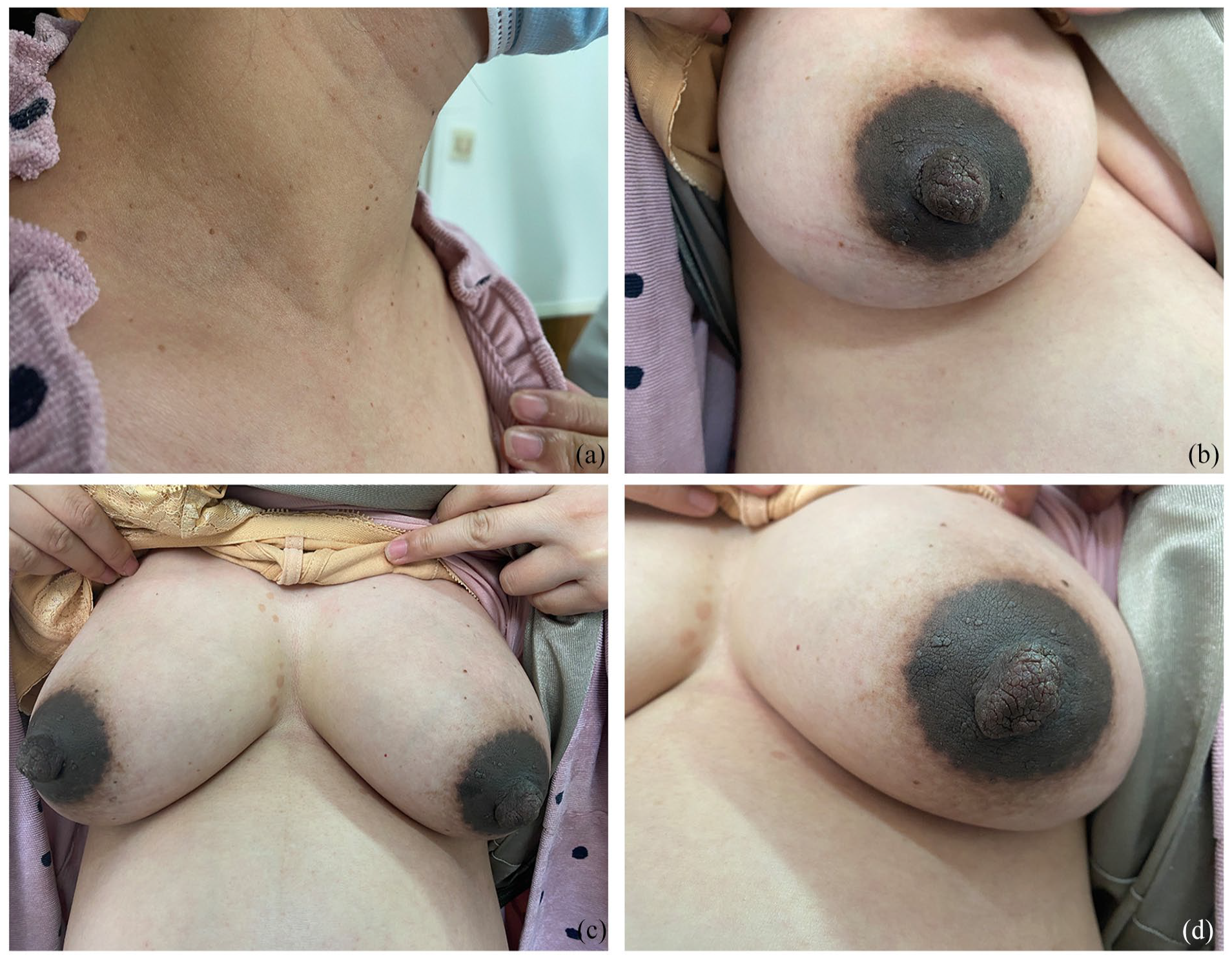
(a) The lesions on the neck. (b) The patchy brown rash on the chest. (c) The verrucous proliferations on the right nipples and areolae. (d) The verrucous proliferations on the left nipples and areolae.

(a) Dermoscopic observations indicate irregular epidermal projections and focal dotted vessels. (b) (H&E, ×10), (c) (H&E, ×20) Papillary epithelial proliferation, hyperkeratosis, dyskeratosis, fibroblastic overgrowth, inflammatory infiltrates, and vascular changes, consistent with fibromas.
Treatment and follow-up
The patient declined treatment during pregnancy. She had an uneventful vaginal delivery, delivering a healthy male neonate. At 6-month reevaluation, chest pigmentation had regressed, and neck lesions had largely resolved spontaneously. By 1 year postpartum, neck lesions had completely vanished, and chest lesions had significantly diminished. The child exhibited normal growth and development. Follow-up at 2 years showed no recurrence of dermatological findings.
Discussion
Soft fibromas are common benign skin lesions characterized by soft, flesh-colored growths. They mostly occur in areas of friction or skin folds and are often asymptomatic but can cause discomfort or cosmetic concerns, especially when multiple or located in prominent areas. The prevalence of soft fibromas approximates 23.5%. 1 Differential diagnoses for these lesions include Human Papillomavirus (HPV) and neurofibromas. Treatment typically involves cryotherapy or laser therapy for smaller lesions, while excision is reserved for larger fibromas.
The exact etiology of soft fibromas remains unclear, but hormonal factors, genetic predisposition, and frictional irritation have been implicated in their development. 2 These cutaneous markers may portend metabolic derangements such as obesity, diabetes mellitus, and metabolic syndrome. 3 Moreover, hormonal disequilibrium, manifesting in elevated estrogen and progesterone levels during gestation or through the use of hormonal contraceptives, may also precipitate fibroepithelial polyp development.4,5 In this case, the patient exhibited soft fibroma lesions on her neck, bilateral nipples, areolar areas, and abdomen. The lesions coincided with mid-pregnancy at 21 weeks and gained prominence by the 28th week. After delivery, the lesions regressed over a year, suggesting a potential association with metabolic and hormonal shifts during pregnancy or the postpartum shift toward a “fetal-centric” state.
Pregnancy brings in dramatic immunological, metabolic, and endocrinal adaptations to foster normal fetal development, particularly in the terminal stages of gestation. 6 Such changes encompass reduced insulin sensitivity, lipid metabolism alterations, and augmented free fatty acid levels.7,8 Throughout gestation, there is a remarkable upsurge in placental lactogen concentrations, escalating to levels that are 20-fold above those prior to conception, followed by a swift decline post-delivery. It orchestrates the management of insulin resistance during pregnancy, facilitates the adaptive response of maternal pancreatic β-cells, and plays a regulatory role in fetal growth. 9 In addition, placental lactogen incites maternal lipolysis and bolsters energy metabolism, augments the transfer of glucose to the developing fetus, underpins fetal development, and stimulates the proliferation of epithelial cells within the placenta and mammary ducts, highlighting its integral role as a hormone of gestational significance. 10
Concurrently, the patient developed pityriasis versicolor, a superficial mycosis of the stratum corneum, caused by Malassezia furfur. This phenomenon may be attributable to the immunosuppressive impact of soaring serum estrogen levels during gestation, coupled with the progressive establishment of a hormonal environment conducive to high local concentrations of free fatty acids and glycerol, thus fostering fungal proliferation. 11 Studies suggest that the exacerbation of soft fibromas during pregnancy, accompanied by the onset of pityriasis versicolor with symptoms resolving spontaneously postpartum, implicates the influence of hormonal changes during pregnancy as a potential risk factor for the development of skin fibromas. Case studies are limited in this area, but the hormonal response observed in fibromas suggests the involvement of local epidermal papillary hyperplasia and connective tissue growth, implicating various signaling pathways and mechanisms.
Conclusion
This case underscores the challenges of managing dermatological conditions during pregnancy and underscores the crucial role of multidisciplinary care involving dermatologists and obstetricians to enhance maternal and fetal outcomes. Some cases show spontaneous regression of lesions after childbirth, highlighting the necessity for personalized management approaches. Long-term follow-up is vital to monitor recurrence.
Footnotes
Declaration of conflicting interests
The author(s) declared no potential conflicts of interest with respect to the research, authorship, and/or publication of this article.
Funding
The author(s) received no financial support for the research, authorship, and/or publication of this article.