
Abstract
Snakebite envenomation is a life-threatening injury and a neglected public health issue in Africa. We report the case of a child that presented 6 days following a forearm snakebite with compartment syndrome and necrotizing fasciitis of the upper limb extending to the neck and chest who developed mediastinitis. She underwent multiple surgical debridements and the mediastinitis was managed non-surgically with antibiotics and postural drainage leading to recovery. The wounds were later grafted and the child discharged. Extension of necrotizing fasciitis from the limbs to the chest wall with development of mediastinitis is extremely rare with one previous case reported in a woman with poorly controlled diabetes mellitus on immunosuppressive therapy. We report this case due to its unusual presentation with no previous reports found on the development of mediastinitis following snakebite. In conclusion, physicians should have a high index of suspicion for mediastinitis in patients with necrotizing fasciitis extending to the neck and chest following snakebite.
Keywords
Introduction
Snakebite is a life-threatening injury described by the World Health Organization as a neglected public health issue in many tropical and subtropical countries. An estimated 5.4 million snakebites occur annually, resulting in more than 95,000 deaths and leaving a further 300,000 survivors with permanent disability or disfigurement. 1
In Zambia, snakebites often occur in rural areas where antivenom is not readily available. Children are at high risk, with venom having more severe effects that develop faster than in adults due to their smaller body mass. 2 Unsafe first-aid practices along with widespread use of traditional medicines and late presentation to the hospital further exacerbate the effects of snakebites. 3
Necrotizing fasciitis is a serious illness that commonly affects the lower limbs, abdominal wall and the perineum and can develop following snakebites.4,5 Progression of chest wall necrotizing fasciitis to mediastinitis is extremely rare. 6 Mediastinitis has a high mortality rate worldwide, with a poor prognosis particularly in the presence of comorbidity. 7 Early diagnosis with computed tomography (CT) scan of neck and thorax, immediate surgical debridement and control of sepsis are the cornerstone of management.
We report the case of a young girl who developed compartment syndrome, extensive necrotizing fasciitis and mediastinitis following a snakebite. She presented to our facility 6 days after the snakebite having travelled over 80 km from her village in a rural part of Zambia.
Case presentation
An 11-year-old girl was bitten by an unidentified snake on the volar aspect of her left forearm while playing at the farm. A tourniquet was tied on her arm for an unknown duration and traditional medicine applied to the wound. Initially, the forearm pain was sharp but gradually evolved to a severe throbbing pain spreading to the chest wall with associated swelling of the affected regions. A day later, the child was taken to her local hospital where fever and rapidly advancing dark-brown discolouration of the forearm was noted. She was then transferred to our facility due to toxaemia, arriving 6 days following the bite. There was no history suggestive of systemic envenomation and she had no known chronic illness.
On admission, she was ill-looking with a temperature of 38.4°C, tachycardia and tachypnoea. She was pale with haemoglobin level at 8.2 g/dL, white blood cell count at 14.6 × 10³, neutrophilia and platelet count at 53 × 103. Local examination revealed patchy dark-brown skin discoloration of the upper limb and anterior chest with multiple foul-smelling skin blisters. The skin of the forearm and hand was tense and tender with visible bite marks. The tenderness was exacerbated by passive movement of the fingers; however, the hand remained pink and warm with a palpable pulse.
A diagnosis of necrotizing fasciitis with acute compartment syndrome was made, and the patient was taken to theatre for surgical fasciotomy and debridement. Intraoperative findings included extensive soft tissue necrosis with overlying dermal gangrene and multiple pockets of pus in the arm, hand and anterior chest wall. Forearm fasciotomy was performed, and muscles bulged out revealing pale non-contractile muscles with poor bleeders. The skin blisters were de-roofed, pus drained and wounds washed. Minimal debridement was done to reduce operating time as the patient’s condition was critical. The muscles were left intact and the wounds dressed. Postoperatively, high-flow oxygen, whole blood, analgesics and high-dose antibiotics were commenced along with a high-protein diet and micronutrient supplements. The parents were counselled on the possibility of amputation if the limb showed no recovery on the second-look debridement scheduled at 48 h. Following the operation, fevers settled and the child recorded a good urine output.
During second-look debridement, the muscles were found to have recovered. Minimal pus was found, though the dermal gangrene had extended to include most of the upper limb and the chest up to the level of the xiphoid process (Figure 1). The underlying necrosis of the subcutaneous tissue extended beneath the viable skin of the anterior chest wall to the mid-axillary lines bilaterally. Multiple drains were inserted into the fascial plane to drain the anticipated fluid and pus under the skin pockets. The forearm tendons exposed following fasciotomy were covered by approximating the skin.

Muscle recovery and extensive dermal gangrene 48 h after fasciotomy.
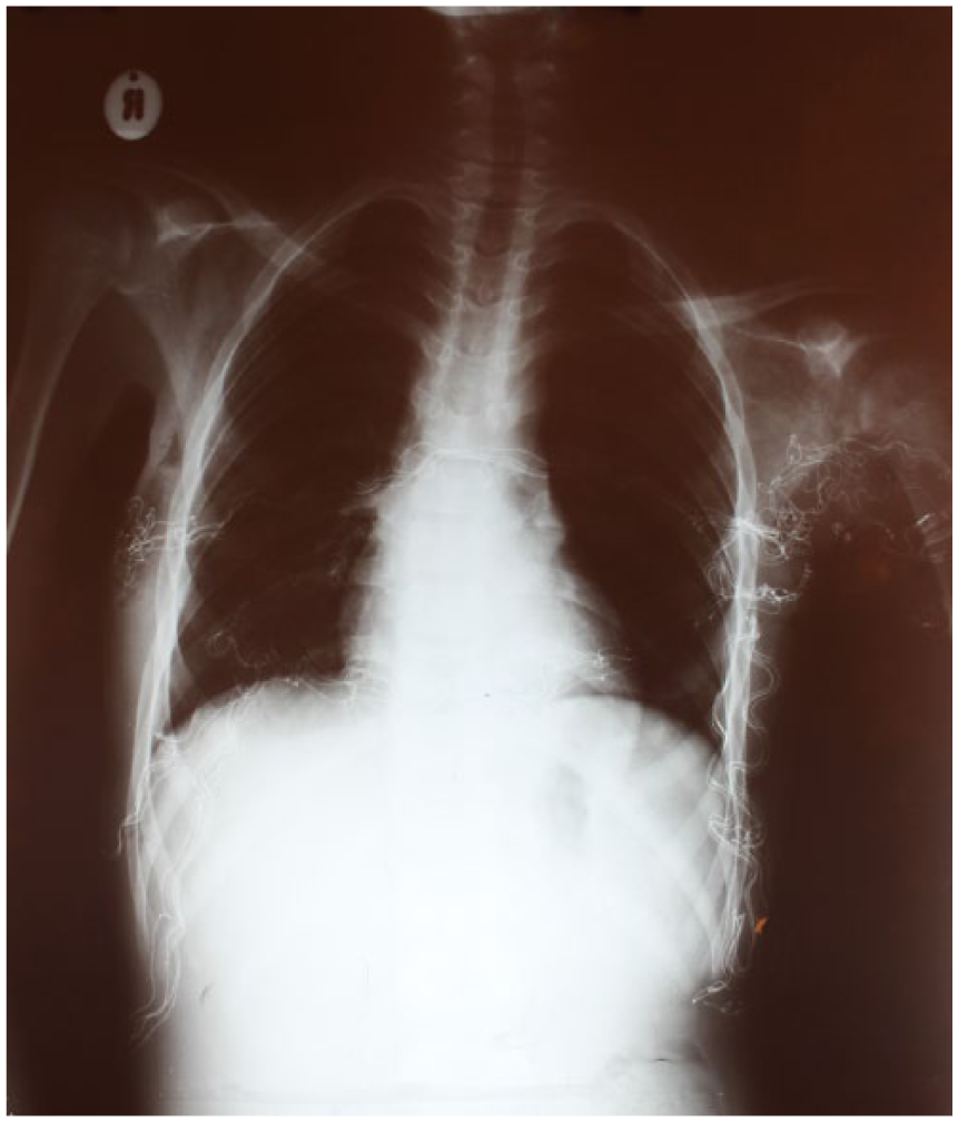
Staged debridement was repeated every 48 h with the patient making steady recovery. However, 6 days post admission she developed an intermittent dry cough associated with central chest pain. She was tachycardic with bilateral pitting pedal oedema. Chest x-ray showed widening of the mediastinum with clear lung fields (Figure 2). Pus was noted at the jugular notch draining from the mediastinum and a diagnosis of mediastinitis was made. A drain was placed beneath the jugular notch, diuretics started and the patient nursed in Trendelenburg position to facilitate pus drainage. A week later, the chest pain and cough decreased, and the mediastinum pus drainage ceased with repeat x-rays showing resolution of the mediastinal widening (Figure 3).

Posterior–anterior chest x-ray showing mediastinal widening.
Repeat posterior–anterior chest x-ray showing resolution of mediastinal widening.
Wound reconstruction was performed 22 days post admission (Figure 4). A split-thickness skin graft harvested from the lower limbs was meshed by hand and secured onto the wound. A sheet (not meshed) thick-split skin graft was used to cover the flexural area of the elbow joint. In total, 20% of total body surface area (TBSA) was grafted with a local flap mobilized and used to complete coverage of the left axilla. The skin at the base of the neck was sutured to achieve secondary closure and a plaster splint applied with the elbow semi-flexed at 120° and the hand in functional position.

The wound immediately prior to reconstruction.
Graft wounds were exposed on day 5 with good take of the graft except 0.5% TBSA on the back that sheared off earlier during movement of the child on the ward and dehiscence of the lower neck sutured wound (Figure 5). Donor site wounds were exposed on day 15 and healed well. Hand function was compromised with reduced power and movement of the ring and little finger while returning the ability to hold a pencil using her thumb and index finger. Physiotherapy was commenced and the child was discharged.

Wound healing 5 days post reconstruction.
Discussion
Snakebite envenoming results in 80,000–140,000 deaths globally every year, with the majority of deaths occurring in India and sub-Saharan Africa. 8 Many survivors develop long-term disability and suffer psychological sequel. 8
In our case, the snake was not identified; however, the clinical presentation is highly suggestive of cytotoxic venom damage. This contains various mixtures of myotoxic phospholipase (PPL) and metalloproteinases (MTP). 8 PPL-based toxins act by causing damage to phospholipid cell membranes of muscle leading to myonecrosis, while MTP toxins hydrolyse the basement membrane of the capillary endothelia by breaking down type IV collagen and damage the endothelial cell-to-cell adhesion.8–10 The result is compromised microvascular structural integrity, increased permeability, fluid extravasation and development of oedema. MTP toxins additionally degrade the dermal–epidermal interface causing skin damage and blister formation as observed in our case. 11 Cytotoxic venoms also cause damage to other local soft tissues including lymphatics, blood vessels and nerves. The net result is the accumulation of necrotic tissue that triggers an intense local inflammatory response. The introduction of bacteria to the wound either from the snakebite or from applied traditional herbs can rapidly lead to the development of infections such as necrotizing fasciitis.
While the development of necrotizing fasciitis following snakebite on limbs has been reported previously, extension of the infection from the limbs to the chest wall is uncommon. 5 Further development of mediastinitis in chest wall necrotizing fasciitis is extremely rare with one previous case reported in a 54-year-old woman with poorly controlled diabetes mellitus on methotrexate immunosuppressive therapy for psoriasis. 6 The diagnosis was confirmed on CT scan and the patient managed surgically with thoracotomy and debridement, which stabilized her condition. In our case, the diagnosis was made clinically upon noting pus draining from the retrosternal space in the presence of tachycardia, cough and chest pain, and supported by the chest x-ray finding of a widened mediastinum as a CT scan was unavailable at our hospital. Due to the child’s critical condition, the mediastinitis was managed non-surgically with antibiotics and postural pus drainage leading to recovery.
A split-thickness skin graft was selected for surgical reconstruction to achieve rapid life-saving wound closure in view of the child’s condition. A local transposition fasciocutaneous flap was used to cover the axilla, preserving shoulder join movement. However, due to the delayed treatment of compartment syndrome, the patient is likely to develop hand contractures and further reconstruction and rehabilitation therapy may be required.
Conclusion
Clinicians should have a high index of suspicion for mediastinitis in patients with necrotizing fasciitis extending to the neck and chest following snakebite. We report this case due to its unusual presentation with no previous reports found on the development of mediastinitis following snakebite. This case also demonstrates the urgent need for ready access of antivenom in sub-Saharan Africa and the need to educate the public on correct first-aid practices.
Footnotes
Acknowledgements
Special thanks to the Arthur Davison Children’s Hospital surgery ward staff and to Prof. Kasonde Bowa for the general support provided.
Declaration of conflicting interests
The author(s) declared no potential conflicts of interest with respect to the research, authorship and/or publication of this article.
Ethical approval
Ethical approval to report this case was obtained from Tropical Disease Research Centre (IRB Registration No. 00002911, FWA Approval No. 00003729).
Funding
The author(s) received no financial support for the research, authorship and/or publication of this article.
Informed consent
Full informed consent was obtained in writing from the parents of the child to write and publish the case report including images of the child.