
Abstract
The role of incompetent perforating veins in the aetiology of varicose veins is not well understood. Anecdotally, competitive cyclists appeared to be more prone to varicose veins than the general population. We present a case of a 63-year-old amateur competitive cyclist who acutely developed a painful varicosity of her left calf while straining during a hill climb in 106-mile cycle race. Duplex ultrasonography has shown an underlying incompetent perforating vein, feeding the varicosity directly through the underlying muscle. With no other significant venous reflux in either leg, we believe this case shows a clear causative association between the stresses put across the lower leg during competitive cycling and developing a varicose vein via an incompetent perforating vein. We believe this should lead to further investigations as to any link between cycling, perforator vein incompetence and the development of varicose veins.
Introduction
Perforator veins (PV) connect the superficial veins of the leg with the deep veins, ‘perforating’ the deep fascia which separates the deep (muscle) compartment from the superficial compartment of the leg. There are approximately 150 PV in each leg. 1 Their role in venous drainage is uncertain, and their role in association with varicose veins is controversial. 2 However, it is generally accepted that in normal function, PV have a net inward flow, taking blood from the superficial veins to the deep veins. 3
Although it has also been shown that 21% of people with no clinical varicose veins can be shown to have at least one incompetent perforating vein (IPV), 3 it is also clear that there is an association between lower limb venous disease and IPVs. There is a strong association between the presence and number of IPV in patients with recurrent varicose veins compared to those with primary varicose veins, 4 and also the number of IPV and worsening clinical etiology anatomy pathophysiology (CEAP) classificiation. 5
Anecdotally, we have noticed that our patients, who present with varicose veins and who cycle competitively but as an amateur, often appear to have quite severe varicose veins and, on scanning with venous duplex ultrasonography, appear to have an increased number of lower limb IPV contributing to their varicose vein pattern.
Here, we report a case of a patient who presented to our clinic with only one symptomatic varicosity. It appeared suddenly 6 weeks ago, accompanied by a sharp pain, while cycling uphill during a 106-mile ride. This large and tender varicosity was shown to be caused by an IPV which was visualised using three-dimensional (3D) Doppler ultrasound. Duplex scanning confirmed this was an isolated phenomenon and there was no other venous reflux or obstruction in either leg.
Case study
A 63-year-old female presented with a prominent symptomatic varicose vein on the medial aspect of her left lower leg. It caused her pain especially during vigorous exercise. She is very fit and exercises regularly. The vein had appeared suddenly 6 weeks previously.
During a 106-mile cycle ride, on a steep hill, she was standing on the pedals going uphill, when she felt a sharp pain in combination with a ‘snapping’ sensation in her left lower leg, medially. She stopped immediately, thinking she had injured herself, and she saw a large, prominent varicose vein which had not been visible before, exactly where the pain had been (Figure 1). Initially, the surrounding area was tender and red, although she reported that this settled down over the next few days. There was no local tenderness in the surrounding soft tissue, nor pain on movement which might suggest a pulled muscle. Clinical examination confirmed this large single varicosity with a varicose vein emerging from it inferiorly. There were no other varicose veins in either leg.
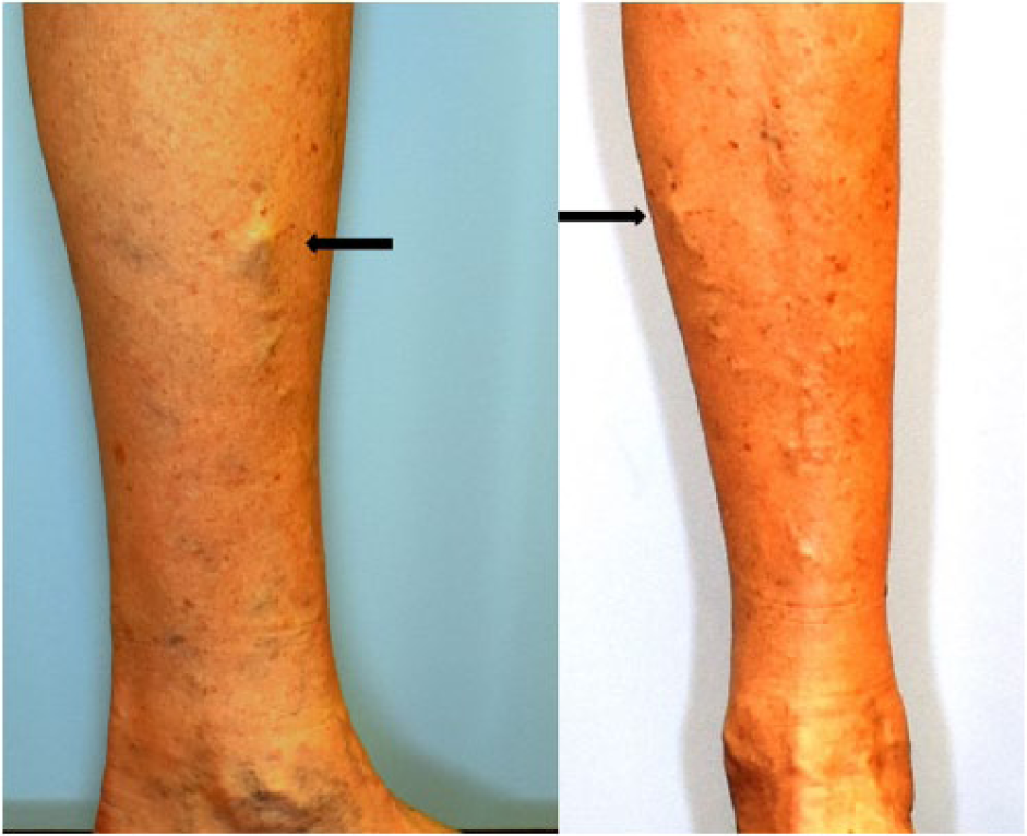
Photograph of left lower leg medial aspect (left) and anterior aspect (right). Black arrow marks the varicosity that emerged acutely during strenuous cycling. Varicose veins can be seen descending from the varicosity.
Duplex ultrasonography revealed an IPV measuring 4.9 mm diameter at the fascia, directly below the prominent varix, which was showing gross reflux (>500 ms; Figure 2). There was no other reflux found in any of the leg veins on either side. There was no haematoma, nor sign of any disruption to the muscle or soft tissue in this area on ultrasound.

Duplex ultrasound image showing large incompetent perforating vein directly underlying varicosity in Figure 1.
Discussion
Cycling is a very popular sport, pastime, and method of transport. Reports of any vascular complications due to cycling are rare and are predominately arterial – the most often quoted is stenosis or thrombosis of the external iliac artery secondary to endofibrosis. 6 A venous version of this has been described, where a 70-year-old cyclist presented with an iliofemoral deep vein thrombosis (DVT) secondary to an external iliac vein stenosis 7 and a second case where the external iliac vein thrombosed in a 57-year old with a previous history of external iliac artery thrombosis. 8 However, although anecdotally many people talk about cyclists having varicose veins, it is hard to find any scientific literature proving any link between the two.
As the usual clothing for competitive cyclists is wearing shorts, their legs are usually on show. In addition, as an endurance sport, competitive cyclists tend to have little sub-cutaneous fat, making any superficial leg veins more likely to appear prominent. Hence, without a formal study with appropriate controls, it is hard to know whether there is a genuine link between varicose veins and cycling.
This case strongly suggests that in this patient, her large and tender varicose vein was caused by strenuous cycling. She had no other varicose vein or venous reflux in either leg, and there is a clear history of straining when cycling uphill, a sudden sharp pain in the left leg, the immediate appearance of the bulging varicosity which had not been there previously and surrounding inflammation that settled over the next few days.
This combination of factors suggests one of two possible causes.
The more obvious process is that the sudden and intense contraction of the muscle caused a competent PV to rupture its valve or valve mechanism, causing it to become an IPV, dilating the vein superficial to it and causing the pain and inflammation.
However, it is also possible that the IPV was already present and incompetent, but without any clinical symptoms or signs, and the sudden pain and appearance of the varicosity were due to acute dilatation of the surface vein, directly over the IPV, due to a jet of blood being forcibly ejected during muscular contraction.
In either scenario, it is clear that this symptomatic varicose vein appeared as a result of strenuous pedaling and is associated with an IPV.
Although this one case cannot be used as proof that there is a link between varicose veins, IPV and competitive cycling, this coupled with the anecdotal reports of many competitive cyclists suggests that this might be a fertile area for further investigation.
Footnotes
Declaration of conflicting interests
The author(s) declared no potential conflicts of interest with respect to the research, authorship and/or publication of this article.
Funding
The author(s) received no financial support for the research, authorship and/or publication of this article.
Ethical approval
Our institution does not require ethical approval for reporting individual cases or case series.
Informed consent
Written informed consent was obtained from the patient for their anonymized information to be published in this article.