
Abstract
Asthma is a heterogeneous disease, usually characterized by chronic airway inflammation. Various clinical conditions can mimic asthma, such as foreign body aspiration, subglottic stenosis, congestive heart failure, diffuse panbronchiolitis, aortic arch anomalies, reactive airway dysfunction syndrome, chronic obstructive pulmonary disease, retrosternal goiter, vocal cord tumors, other airway tumors, and vocal cord dysfunction. Upper airway obstruction can be a life-threatening emergency. Here, we present the case of a 58-year-old female with recurrent hospital visits for wheezing and exacerbations of asthma, who was later found to have a vocal cord nodule confirmed to be squamous cell carcinoma, which was mimicking like asthma.
Introduction
Asthma is a heterogeneous disease, usually characterized by chronic airway inflammation. It can be present with wheezing, chest tightness, cough, and shortness of breath, all of which may vary in intensity and over time. 1 The diagnosis of asthma requires an appropriate history, consisting of respiratory symptoms, diurnal variation of symptoms, and exposure to triggers, as well as demonstration of variable expiratory airflow obstruction. 2
Wheezing is a common manifestation of respiratory illness in adults. Wheezing is not always indicative of asthma, and not all asthma produces wheezing. 3 Causes of wheezing can be divided into three categories: extrathoracic upper airway obstruction, intrathoracic upper airway obstruction, and lower airway obstruction.
Upper airway obstruction can be a life-threatening emergency. The most common causes of upper airway obstruction are paroxysmal vocal cord motion, retropharyngeal abscess, benign airway tumors, anaphylaxis, bilateral vocal cord paralysis, foreign body aspiration, tracheal stenosis, intrathoracic goiter, tracheomalacia, and a right-sided aortic arch.4,5
The vocal cord abnormality that most commonly mimics asthma is vocal cord dysfunction. However, the differential diagnosis includes other disorders that should also be kept in mind, such as vocal cord polyps, vocal cord granulomas, unilateral or bilateral vocal cord paralysis, and vocal cord tumors. 6
Vocal cord tumors can produce asthma-like symptoms at the time of presentation. These tumors are relatively common. Approximately 13,430 new cases of laryngeal cancer are identified each year in United States. The number of new cases of larynx cancer is 3.2 per 100,000 men and women per year.
Case report
A 58-year-old Hispanic woman arrived at our emergency department with a chief complaint of wheezing and dry cough for 2 days. She had no fever, sore throat, chest pain, hemoptysis, night sweats, odynophagia, no hoarseness of voice, abdominal pain, hematemesis, or melena. Her past medical history was significant for hypertension, diabetes mellitus type II, hyperlipidemia, and gastroesophageal reflux disease. She was recently discharged from the emergency department after management of shortness of breath and wheeze 1 week prior to the current visit. Our patient had four emergency room visits in past 6 months before this hospitalization. Patient also visited her primary care physician twice for similar complaints. All her symptoms started 6 months ago. Prior to this emergency room visit, she was seen at a pulmonary clinic for predominant symptoms of wheezing.
The patient’s surgical history was significant for a cesarean section 20 years ago. Her mother died of breast carcinoma. She had 15-pack-year history of smoking, and she consumed alcohol socially. She denied illicit drug use and reported no allergies. She was taking metformin, lisinopril, chlorthalidone, metoprolol, atorvastatin, aspirin, omeprazole, albuterol inhaler as needed. She lived with her family and was a home-maker with no known occupational exposure to toxic substances.
Physical examination on presentation to the emergency department revealed an emaciated woman with these findings: temperature, 97.6°F; pulse, 102 beats/min; respiratory rate, 18 breaths/min; blood pressure, 112/69 mm Hg; pulse oxygen saturation, 98% on ambient air; and body mass index, 15 kg/m2. Her oral mucosa was moist, and skin turgor was normal. She had no palpable lymphadenopathy. She had equal air entry bilaterally with inspiratory wheezes on lung auscultation. She had a sinus tachycardia but normal heart sounds. She had no organomegaly on abdominal examination, and her neurological examination was unremarkable.
Initial laboratory results which included hematology profile, serum chemistry, liver function blood tests, and serum calcium levels were within normal limits. Chest radiography showed no pulmonary infiltrates. She was subsequently discharged from hospital after her wheeze subsided. Spirometry done in pulmonary clinic revealed flattening of the inspiratory flow loop (Figure 1).

Spirometry showing flattening of the inspiratory flow–volume loop.
Computed tomography (CT) of the neck performed showed a 1.4 × 1.4 cm2 right vocal cord mass causing airway narrowing (Figure 2).
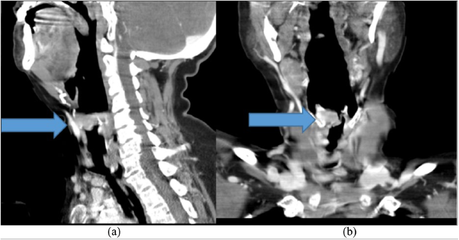
CT of the neck showing a 1.4 × 1.4 cm2 exophytic right vocal cord nodules: (a) sagittal view and (b) coronal view.
Members of the otolaryngology consultation service performed fiberoptic laryngoscopy, which showed a large exophytic mass arising from the right true vocal cord but normal vocal cord mobility (Figure 3).
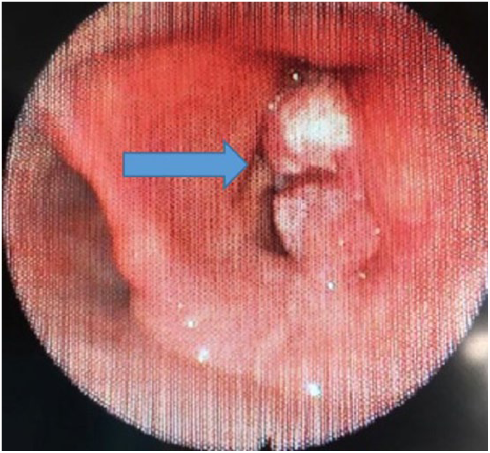
Fiberoptic laryngoscopy showing a large exophytic mass attached to the right true vocal cord.
Subsequently, she underwent excisional biopsy of the vocal cord nodules and underwent emergency tracheostomy to secure the airway. Histopathology of the mass showed moderately differentiated invasive squamous cell carcinoma T3N0M0. The patient’s postoperative hospital course was unremarkable, and she was discharged home with plans for outpatient chemoradiation therapy.
Discussion
Asthma is a common respiratory illness, which is managed with inhaled and oral agents such as bronchodilators, corticosteroids, and leukotriene receptor antagonists. Patients with chronic symptoms suggestive of asthma, normal spirometry, poor response to asthma medications, and frequent medical visits should be evaluated for the presence of other conditions that mimic asthma.
True vocal cord carcinoma is the most common of all laryngeal cancers. Most of these tumors occur on the anterior two-thirds of the cord, although a small percentage is located on the anterior commissure. They rarely occur on the posterior commissure. The growth characteristics of true vocal cord carcinomas are determined by the unique anatomy of the vocal cords. The sparsity of lymphatic drainage in all areas of the vocal cord except the posterior commissure makes metastasis of early lesions extremely unlikely. 7 The first reported case of head and neck squamous cell carcinoma was in 1868. 8
Various conditions can mimic asthma; common mimics include foreign body aspiration, vocal cord dysfunction, vocal cord abnormalities, exercise-induced bronchospasm, and gastroesophageal reflux disease. Vocal fold paresis from prolonged intubation, idiopathic vocal fold paralysis, recurrent laryngeal nerve damage during surgery, and vocal fold paralysis caused by head and neck malignancy can all cause abnormal vocal fold movement and thereby produce symptoms of vocal cord dysfunction that mimic asthma. 9 The other uncommon conditions which may mimic asthma-like symptoms are subglottic stenosis, congestive heart failure, diffuse panbronchiolitis, reactive airway dysfunction syndrome, chronic obstructive pulmonary disease.
Psychogenic respiratory distress mimicking asthma can be a conversion reaction that causes paradoxical vocal cord dysfunction. 10 Non-asphyxiating foreign body aspiration usually presents as a cough, but it may also cause wheezing, shortness of breath, and hemoptysis. The wheezing in these patients mimics asthma. 11 Laryngotracheal stenosis is a rare condition, which can present as a complication of granulomatosis with polyangiitis; it can also produce wheezing that mimics asthma. 12 As reported by Özgül et al., 13 an ectopic parathyroid adenoma at the distal part of the trachea can also produce asthma-like wheezing. Endobronchial lipomas, which are rare benign tumors of the lung, can likewise present with asthma-like wheezing. 14 In addition, endobronchial tuberculosis presenting as a polypoid, necrotic endobronchial lesion has been reported to produce symptoms mimicking asthma. 15
In the herein described case, a true vocal cord nodules, which was found to be squamous cell cancer on biopsy, produced symptoms mimicking asthma that led to recurrent office and emergency room visits. Among carcinomas of the head and neck, 45% are laryngeal cancer. These cancers are most often found in males over 50 years of age. Common risk factors for laryngeal and hypopharyngeal cancers are male sex; tobacco use and alcohol consumption; human papilloma virus infection; genetic predilection; and exposure to wood dust, paint fumes, and certain chemicals used in metal-working, petroleum, plastics, and asbestos textile industries.16,17 A study by Zhao et al. 18 suggests that an association exists between overexpression of histone deacetylase 1 and the clinical characteristics of laryngeal squamous cell carcinoma.
Symptoms of vocal cord cancer vary, depending on the structures involved by the malignancy and the accompanying inflammatory reaction. The symptoms may include wheezing, stridor, dyspnea, dysphagia, dysphonia, pain, halitosis, blood-tinged sputum, and a neck mass. Investigations that can be performed include direct laryngoscopy, CT of the neck and chest, plain chest radiography, positron emission tomography, and pulmonary function tests (PFT) (Figure 4). Inspection of the larynx is best accomplished using a flexible laryngoscope, and direct laryngoscopy also provides an opportunity to obtain biopsies of the tumor.

An algorithm for diagnosis of inspiratory wheeze.
PFT, starting with spirometry before and after the administration of inhaled bronchodilator, can be performed for patients who have wheezing. The flow–volume loop helps determine whether airflow obstruction is caused by an intrathoracic or extrathoracic obstruction and whether the obstruction is variable or fixed. Variable extrathoracic obstruction caused by vocal cord paralysis is typically apparent only during maximum inspiration. The maximal expiratory flow–volume curve and expiratory spirometry are usually normal in this setting because the extrathoracic airway will be pushed open during expiration. In addition to visual inspection of the graph, the ratio of inspiratory to expiratory flow rates can help distinguish a variable extrathoracic lesion from an intrathoracic upper airway lesion.19,20 In our case, patient had spirometry that showed inspiratory loop flattening suggesting extrathoracic obstruction.
CT with contrast of the chest and neck can identify various structures that cause extrinsic compression of the trachea, such as a mediastinal mass, lymphadenopathy, an aneurysm, airway stenosis, a central airway neoplasm, congenital airway disorders, tracheobronchomalacia, and vascular rings. These conditions may all cause wheezing. 21
The National Comprehensive Cancer Network 22 Practice Guidelines in Oncology currently recommend concurrent radiation therapy and cisplatin chemotherapy to achieve laryngeal preservation in cases of locally advanced laryngeal cancer. There are various surgical options, including transoral laser microsurgery, open partial laryngectomy, and total laryngectomy. 23 Our case emphasizes on the fact that different etiologies can mimic asthma and recognizing vocal cord nodules causing obstruction of upper airway as an emergency.
Conclusion
Various conditions may mimic asthma, and patients with poor response to asthma medications and frequent office or hospital visits should be further evaluated for the presence of an asthma mimic. Awareness of vocal cord nodules and its appropriate evaluation is critical in patients presenting with hoarseness, stridor, and/or wheezing. Timely PFT and direct laryngoscopy, as performed for the patient described in this report, can aid in the identification of vocal cord nodules and lead to a definitive diagnosis.
Footnotes
Acknowledgements
M.Kh. and M.Ka. searched the literature and wrote the manuscript. M.Kh. conceived and edited the manuscript. M.Kh. supervised the patient treatment, critically revised and edited the manuscript. T.S. and A.A. were involved in patient care along with M.Kh. All authors have made significant contributions to the manuscript and have reviewed it before submission. All authors have confirmed that the manuscript is not under consideration for review at any other Journal. All authors have read and approved the final manuscript.
Declaration of conflicting interests
The author(s) declared no potential conflicts of interest with respect to the research, authorship, and/or publication of this article.
Ethical approval
Our hospital does not require IRB approval for case reports. Our institution does not require ethical approval for reporting individual cases or case series.
Funding
The author(s) received no financial support for the research, authorship, and/or publication of this article.
Informed consent
Written informed consent was obtained from patient and is with corresponding author.