
Abstract
Purpose:
To assess the incidence of pulp stones in molar and premolar teeth of Southern Saudi Arabian adult sub-population.
Materials and Methods:
Six hundred patient files were randomly selected from the database (records) of the College of Dentistry outpatient department screened by Oral Medicine and Radiology division. Orthopantomogram and bitewing radiographs using radiovisiographs of first and second maxillary and mandibular molars and premolars were interpreted by three examiners. Pulp stones were identified as definite radiopaque masses and scored as present or absent. Data were subjected to statistical analysis using SPSS version 19.
Results:
Out of total 600 patients, pulp stones were found in 88 (14.7%) patients. Females showed statistically significant higher presence of pulp stones in mandibular molars than males. The patients belonging to middle age group (31–40 years) showed statistically significant higher presence of pulp stones than the other two groups in all first and second molar teeth in both arches. The presence of pulp stones was observed to be higher in maxillary arch than mandibular arch with left side having more frequency than the right side of the arch.
Conclusion:
The prevalence of pulp stones in the study population was found to be 14.7%, and it was higher in molars than premolars. It is quite significant to know the frequency distribution of pulp stones especially for an endodontist or a dentist to plan the root canal treatment of such teeth accordingly.
Introduction
Pulp stones result from the calcification process that may occur in diffuse or discrete forms. They may be free in the pulp tissue or may be attached or embedded into dentine.1,2 Occlusion of the canals due to pulp stones especially at the apical root curvature results in difficulties during Root canal treatment. 3 Pulp stones are being observed in healthy, diseased, or even in unerupted teeth 4 in different sizes varying from microscopic to large masses, and the number ranges from 1 to 12 or more in a single tooth. 2 Irregular pulp stones composed of dentine and lined by odontoblasts are the true pulp stones 5 whereas false pulp stones are formed by mineralization of degenerating cells. 2
The exact etiology of calcification of pulp is unknown; however, many factors have been associated with its formation. Irritants existing since long periods such as dental caries, deep tooth-restorations or chronic inflammation, 6 factors like age, disturbance in pulpal blood-circulation, tooth-movements during orthodontic treatment, 7 idiopathic reasons, epithelium and pulp tissue interaction, 8 and genetic predisposition 9 are being linked to tooth pulp calcification. The prevalence of pulp stones shows wide range of variation from 8% to 90%. 10 Even higher values of the frequency of pulp stones has been observed through histological evaluations. 11 In a study conducted by Al-Nazhan and Al-Shamrani, 12 the prevalence of pulp stones was found to be 10.2% in a Saudi Arabian population. Tamse et al. 11 reported a higher prevalence of pulp stones in molar teeth compared to premolars, in females more than males while Patil et al. 13 found the higher frequency in males of North Saudi Arabian population.
The presence of pulp stones may pose some procedural difficulties to the dental specialist while performing the endodontic treatments, hence knowing about its frequency distribution would be beneficial in handling such cases while planning and performing the treatment. Keeping the above points in view, this study was undertaken to assess the incidence of pulp stones in molar and premolar teeth of Southern Saudi Arabian sub-population.
Materials and methods
A retrospective observational study was carried out at the College of Dentistry, King Khalid University, Abha, Saudi Arabia, from the records of patients obtained from the database. A total of 600 patient files were selected for the study screened by the department of Oral Diagnosis and Radiology having Orthopantomogram (OPG) and Bitewings in the period from March 2019 to February 2020 (King Khalid University College of Dentistry records). Prior to the conduct of study, ethical approval was taken from the Scientific Research Committee, King Khalid University (SRC/ETH/2018-19/077).
A total of 9600 teeth were examined from 600 patient radiographs. All those patients having full set of permanent dentition (third molars present/absent), aged 20–50 years were included for the study. Patients who were less than 20 years of age and whose radiographs were of poor quality were excluded from the study. The OPG bitewing radiographs using radiovisiographs (RVGs) of first and second maxillary and mandibular molars and premolars were interpreted by three examiners. Inter- and intra-examiner reliability was assessed during the pilot study that was carried out prior to the main study so as to reduce the possibility of observer’s bias. The reading of radiographs was repeated after 14 days, and then the intra- and inter-examiner reliability was calculated by Cohen’s Kappa and was found to be satisfactory. Pulp stones were identified as definite radio-opaque masses inside the pulp chambers of the first and second premolars, and first and second molars. They were scored as present or absent.
Sample size estimation
The sample size n is shown as follows

where Zα/2 is the critical value of the normal distribution at α/2 (e.g. for a confidence level of 95%, α is 0.05 and the critical value is 1.96), and Z β is the critical value of the normal distribution at β (e.g. for a power of 80%, β is 0.2 and the critical value is 0.84). p1 and p2 are the expected sample proportions of the two groups.
By the literature survey or pilot study, the expected proportion of the parameters in two groups was 8.6% and 13.7%. So, taking 5% level of significance, 95% confidence interval and 80% power, with the help of given formula the sample size was estimated to be 594, which was rounded-off to 600 in this study.
Statistical analysis
All analyses were performed by SPSS statistics program for windows version (SPSS version 19.0, SPSS Inc., Chicago, IL, USA). Chi-square analyses were used to compare the frequency of pulp stones between sexes, tooth types, dental arches, and sides (right and left). Significance was tested at p < 0.05.
Results
The total 600 patient files comprised of 300 male and 300 female patients. Frequency distribution of subjects based on demography is shown in Table 1. The overall prevalence of pulp stones was found to be 14.7%, and it was observed more in females (16%) than in males (13.3%) (Table 2). Pulp stones are shown in Figure 1 (OPG) and Figure 2 (bitewing radiograph).
Frequency distribution table showing descriptive statistics for demographic data of the sample.

Frequency distribution of pulp stones according to gender and age groups.

NSstatistically non_significant.
statistically significant.

OPG showing pulp stones in molar teeth.

Bitewing radiograph showing pulp stones.
Pulp stones were observed in 437 teeth (4.6%) of the whole 9600 teeth examined for 600 patients. Molars exhibited more pulp stones compared to premolars with a statistically significant difference. The presence of tooth-pulp stones in maxillary arch was more than in mandibular arch, on the left side more than the right side (Table 3). Higher frequency of pulp stones was seen in maxillary left first molar teeth of females compared to males with a statistically non-significant difference (p = 0.127) (Table 4). The middle age group (31–40 years) showed significantly higher presence of pulp stones than the other two groups in all first and second molar teeth in both arches (Table 5).
Frequency distribution of pulp stones according to teeth groups (molars and premolars), side (right and left), and arch (maxillary and mandibular arch).

statistically highly significant.
Frequency distribution of pulp stones in maxillary left first molar teeth based on gender and age.
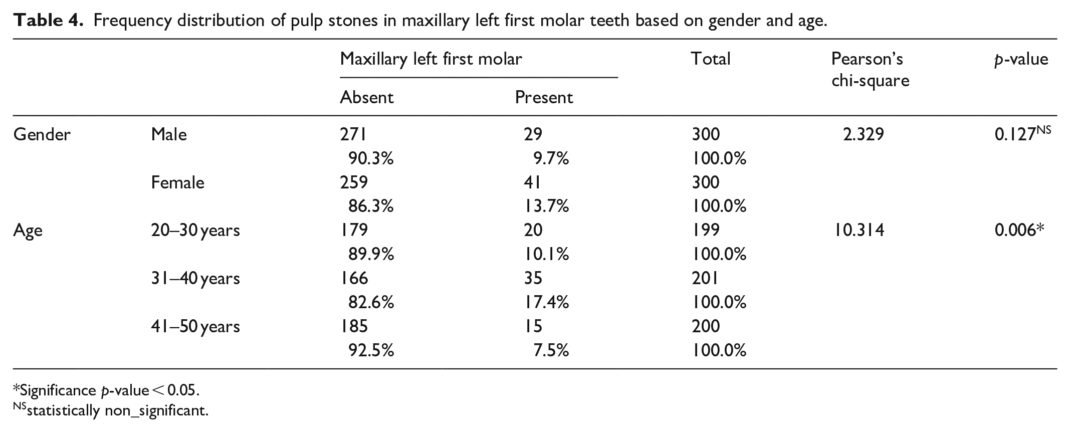
Significance p-value < 0.05.
NSstatistically non_significant.
Descriptive and inferential statistics for incidence of pulp stone between among different age groups.
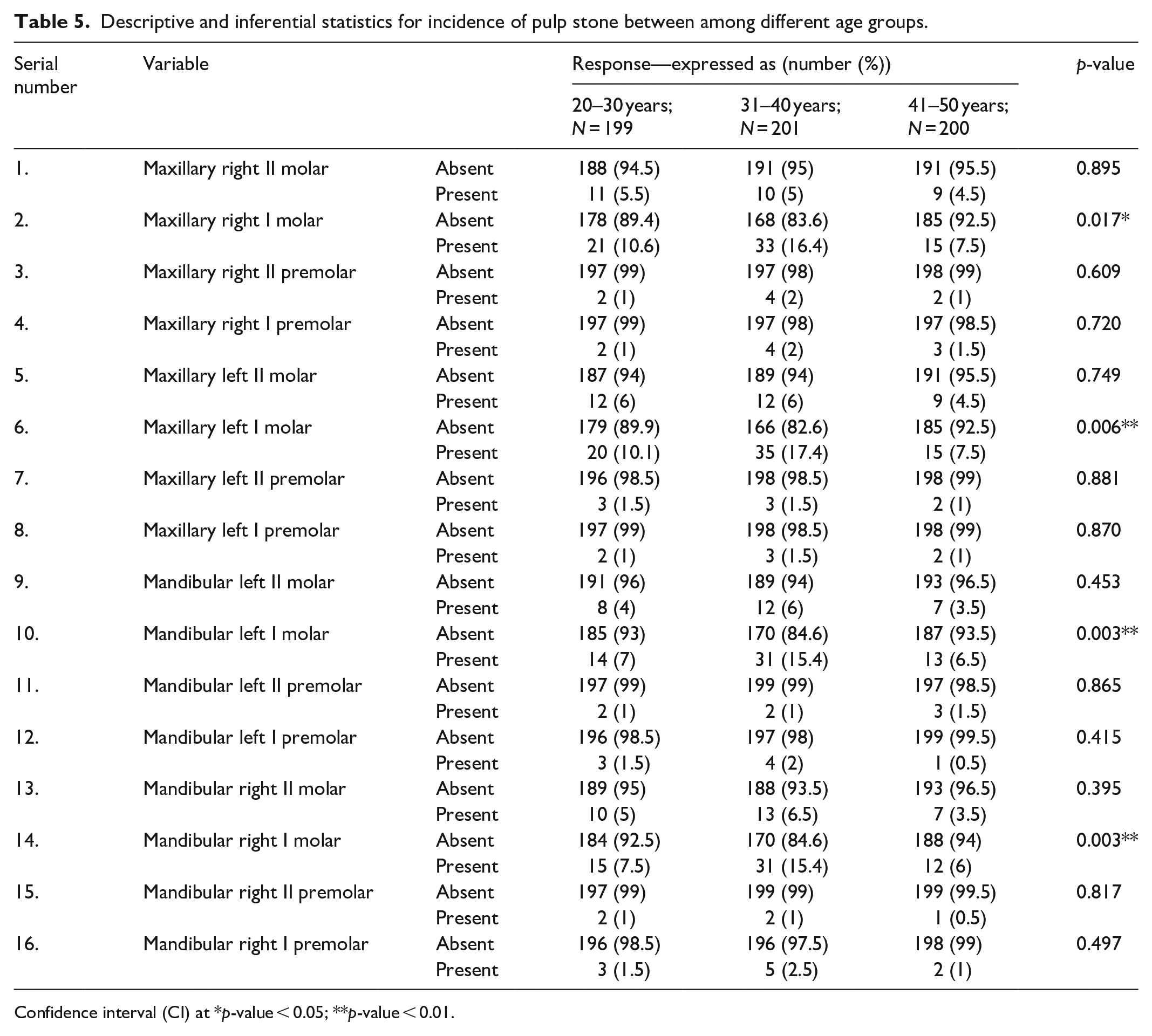
Confidence interval (CI) at *p-value < 0.05; **p-value < 0.01.
Discussion
The results of the study highlighted the distribution of pulp stones among Saudi Arabian patients across different age groups and demographics. In this study, the prevalence of pulp stones was found to be 14.7%. When compared to the literature review, Turkal et al. 14 observed that the prevalence of pulp stones among the study participants was 12.7%. The occurrence of pulp stones was found to be higher in the maxilla than the mandible. As per the conclusion drawn by a study by Sandeepa et al., 15 the prevalence of pulp stones in the participants was 7.5%, with more presence in first molars. Al-Nazhan and Al-Shamrani 12 conducted a study to determine the prevalence of pulp stones in a group of patients using radiographs wherein the researchers found that pulp stones were identified in 10.2% of the examined teeth. Considering the incidence of pulp stones, Patil et al. 13 observed that the first molars have higher presence of pulp stones in Saudi Arabian population; however, there was no statistical significant difference in the maxillary and mandibular arches. Sadoon et al. 16 observed that the presence of pulp stones was higher in the molars and non-intact teeth of the participants. According to Jayam et al., 17 the incidence of pulp stones in maxillary first molars was higher than the mandibular first molars. As per the results of a study by Sisman et al., 18 the presence of pulp stones found in participants was 9.07%, where they were highly present in molars than premolars. All these study findings were in alignment to the results obtained in this study that might be contributed to the fact that first molars erupt earlier to the premolar teeth.
This study has also evaluated the association of gender with the prevalence of pulp stones. It was inferred from the current analysis that a higher number of pulp stones exist in the mandibular molar teeth in females, as compared to those in males with a statistically significant difference. In comparison to the literature, there were a few studies that found no statistical significant difference for gender, while several others found a concrete difference. Studies by Alsweed et al., 19 Patil et al., 20 and Panwar et al. 21 drew no association between gender and the prevalence of pulp stones.
There were also a few studies that found male population having higher pulp stones, as opposed to this study. Patil et al. 13 found that pulp stones were more common in males compared to females. A significant difference in pulp stone development between male and female patients was observed by Mathew et al., 22 where more pulp stones were found among male participants than in female participants. These revelations were in contrast with our research findings. However, there are other researchers who have determined a stark difference in the occurrence of higher pulp stones in females than males. Turkal et al. 14 observed that the occurrence of pulp stones is higher in females than males. Yousuf and Antony 23 determined that the incidence and distribution of pulp stone varies as per the population. Female population was revealed to have a higher presence of pulp stones than the male population. Ravanshad et al. 24 observed that pulp stones are more common in females than in males. According to Jayam et al., 17 females had higher prevalence of pulp stones than the male counterparts. These research findings were in accordance with this study results which might be due to the hormonal changes occurring in females.
When considering the age factor, it was observed from the analysis that the patients of middle age group (31–40 years) had higher prevalence of pulp stones in their first and second molars in both arches. For maxillary left first molars, a majority of 17.4% of the patients who were of 31–40 years of age had higher number of pulp stones. A similar finding was drawn for maxillary right first molars, mandibular left first molar, and mandibular right first molar, where middle-aged patients had higher incidence of pulp stones as compared to those between 20 and 30 years and 41 and 50 years of age groups. When compared to the literature review, Sadoon et al. 16 and Al-Nazhan and Al-Shamrani 12 found a concrete relationship of age with pulp stones.
While a few studies such as Patil et al. 13 found that pulp stones are mostly observed in people above 50 years of age and Mathew et al. 22 observed that a majority of the population aged 46–60 years were highly prone to developing pulp stones, there were opposing research works as well such as the one reported by Alsweed et al. 19 according to whom, the prevalence of pulp stones amounted to be 4.6%, where the incidence of pulp stones was higher in younger patients as compared to the older ones.
It was also established in this study that the presence of tooth-pulp stones in the maxillary arch was more than those present in the mandibular arch. They were also more on left side as compared to the right-side teeth. On comparing with the previous literature, several studies observed similar results, where Sandeepa et al. 15 and Panwar et al. 21 found that pulp stones were most commonly found in the maxillary first molars than mandibular first molars. Kalaji et al. 25 also observed in their study that the occurrence of pulp stones is evidently higher in the maxilla than the mandible for each tooth type. According to Yousuf and Antony, 23 maxillary arch is the most affected arch, and also the pulp stones were higher in the maxillary first molar teeth. A statistically significant difference was observed between the incidence of pulp stones in maxillary and mandibular arches by Ravanshad et al., 24 who reported that the frequency was 26% in maxillary molar teeth while it was 18.7% in mandibular ones. However, a few studies found no such association, where Sadoon et al. 16 and Patil et al. 20 stated that there are no significant differences between the presence of pulp stones in maxilla and mandible arch.
In this study, the panoramic radiographs were utilized for studying pulp stones as the panoramic radiography is integral part of dental examinations in various hospitals or clinical settings. 26 Minimal ionizing radiation is required and all teeth can be examined at the same time. 27 However, the two-dimensional technique employed for panoramic radiography may sometimes under-estimate the actual frequency of pulp stones. To overcome the under-reporting, three-dimensional techniques such as CBCT (cone beam computed tomography) is the recommended technique as it provides the minute details with separate examination of all teeth. 28
As far as clinical significance pulp stones of knowing the frequency of pulp stones is concerned, it is quite significant for an endodontist or a dentist to plan the treatment especially root canal treatment accordingly as their presence may pose procedural difficulties. Ultrasonic instruments can be used for removal of pulp stones. In teeth with narrow canals, sodium hypochlorite or EDTA can be used as a dissolving agent in addition to the ultra-sonic instrumentation. Proper instrumentation, access opening, and magnification are necessary to overcome the hindrance posed by pulp stones while performing root canal treatment.
Limitations
CBCT studies would yield better and accurate results for detection of pulp stones in teeth. The number and size of pulp stones could not be evaluated. The parafunctional habits of patients were not recorded due to time constraints.
Conclusion
Prevalence of pulp stones among the study population was found to be 14.7% with more predilections in females than in males. Pulp stones were found more in maxillary teeth with higher prevalence in molar teeth than premolars. Middle-aged patients were found to have more pulp stones than the other age groups.
Footnotes
Declaration of conflicting interests
The author(s) declared no potential conflicts of interest with respect to the research, authorship, and/or publication of this article.
Ethical approval
Ethical approval for this study was obtained from the King Khalid University College of Dentistry Research Ethics Review Committee prior to the study (SRC/ETH/2018-19/077).
Funding
The author(s) received no financial support for the research, authorship, and/or publication of this article.
Informed consent
Verbal informed consent was obtained from legally authorized representatives before the study. Because the study was done on previous records (radiographs) taken from college database, so only verbal consent is required in that case as per our institutional rules. We can take either written or verbal consent when the nature of the study is a retrospective one (from the previous records). If the study would have been conducted in-vivo, directly on the patients, then written consent is compulsory. The method was approved by IRB.