
Abstract
Objectives:
This cross-sectional study aimed to individually and cumulatively compare sensitivity and specificity of the (1) ankle brachial index and (2) pulse volume waveform analysis recorded by the same automated device, with the presence or absence of peripheral arterial disease being verified by ultrasound duplex scan.
Methods:
Patients (n=205) referred for lower limb arterial assessment underwent ankle brachial index measurement and pulse volume waveform recording using volume plethysmography, followed by ultrasound duplex scan. The presence of peripheral arterial disease was recorded if ankle brachial index <0.9; pulse volume waveform was graded as 2, 3 or 4; or if haemodynamically significant stenosis >50% was evident with ultrasound duplex scan. Outcome measure was agreement between the measured ankle brachial index and interpretation of pulse volume waveform for peripheral arterial disease diagnosis, using ultrasound duplex scan as the reference standard.
Results:
Sensitivity of ankle brachial index was 79%, specificity 91% and overall accuracy 88%. Pulse volume waveform sensitivity was 97%, specificity 81% and overall accuracy 85%. The combined sensitivity of ankle brachial index and pulse volume waveform was 100%, specificity 76% and overall accuracy 85%.
Conclusion:
Combining these two diagnostic modalities within one device provided a highly accurate method of ruling out peripheral arterial disease, which could be utilised in primary care to safely reduce unnecessary secondary care referrals.
Keywords
Introduction
There is, at present, a clear and recognised need to optimise the diagnosis of peripheral arterial disease (PAD), particularly in non-specialist settings such as primary care, and this arises from several key facts. First, PAD is a highly prevalent condition; in 2010, it was estimated that globally, it affected more than 202 million people and furthermore, this prevalence is predicted to further escalate. 1 The disease itself, although frequently asymptomatic, can cause considerable patient suffering with symptoms such as lower limb pain, ulceration and gangrene which, in worse-case scenarios, can necessitate limb amputation. A further and perhaps the most eminent consequence of PAD arises from the fact that it is a manifestation of systemic atherosclerosis and therefore is a powerful predictor of coronary heart disease and cerebrovascular disease. 2 Multiple longitudinal studies have demonstrated that PAD (both asymptomatic and symptomatic) has been associated with a three to sixfold increased risk of death from cardiovascular causes. 3
PAD, however, is frequently asymptomatic, particularly in those less mobile and therefore is under-diagnosed; 4 hence it has been termed ‘a silent but lethal epidemic’. 5 This has resulted in calls for the instigation of primary care PAD screening which would identify those at increased risk and potentially allow alteration of the disease trajectory via secondary risk factor modification. 6
The ankle brachial index (ABI) has been the foundation of non-invasive PAD diagnosis for several decades, hence making it seemingly pivotal to any primary care PAD screening strategy. However, studies have demonstrated that the ABI has not been readily adopted by primary care clinicians and that it is, in fact, infrequently and often incorrectly utilised in non-specialist healthcare settings.7,8 Lack of knowledge and skills to undertake the procedure utilising a hand-held Doppler ultrasound probe and manual sphygmomanometer has been identified as a factor associated with this low use. 9 In addition, the time-consuming nature of this method and the need to rest subjects for at least 10 min prior to the procedure also significantly limit its use in busy healthcare settings.7,8 In recent years, several manufacturers have developed automated ABI devices which aim to address such issues by negating the need for both operator skill and a rest period. Research investigating whether such devices have sufficient diagnostic accuracy to replace the traditional Doppler method has proven inconclusive. 10
A further, well-recognised limitation of the ABI is that it can become artefactually elevated and non-diagnostic in certain patient groups such as diabetics, the elderly and those with renal disease. This therefore underlines the need for a secondary mode of assessment for the diagnosis of PAD. Pulse volume waveform (PVW) interpretation constitutes a further non-invasive, diagnostic procedure that can be utilised to evaluate blood flow in the extremities. Its use is recommended by both the European Society of Cardiology and the American College of Cardiology/American Heart Association as a second-level assessment tool for patients with suspected PAD.2,11 It has been used in vascular laboratories for PAD assessment for several decades; however, recent technological advances have resulted in this modality becoming more amenable for use in other settings such as community and primary care. Interpretation of PVWs can be undertaken by visually comparing them to a four-level grading system (Figure 1). 13 There is, however, limited evidence regarding the feasibility and practicality of incorporating this technology into routine, non-specialist practice.

Pulse volume waveform interpretation (according to four-level grading system). 13
The aims of this study were twofold: first, to evaluate the accuracy of the automated ABI measurement and PVW analysis for the diagnosis of PAD using duplex ultrasound scanning as the reference standard and second, to consider the utility of a device which incorporates both automated ABI and PVW for use in the primary care setting.
Materials and method
This cross-sectional study recruited 205 consecutive patients who had been referred for lower limb arterial investigations to one of two medical physics/vascular outpatients departments within two UK teaching hospitals. Inclusion criteria included those referred for lower limb arterial investigations who were ⩾18 years of age and able to provide informed consent. Patients who had lymphoedema, thrombophlebitis or cellulitis were excluded from participation, as were those who were suspected as having a deep vein thrombosis (DVT) (current or in the preceding 6 months), those who had undergone bilateral mastectomy with lymph node removal, those with bilateral upper or lower limb amputation and those who were unable to lie supine. The study was approved by the Research Ethics Committee 2 (Cardiff, Wales, REC No: 13/WA/0072) and written informed consent was gained from each participant.
Prior to the arterial assessment procedures, participants were asked to complete a brief questionnaire which captured basic demographic data (gender, age, smoking status), past medical history, family history of cardiovascular disease and reason for referral. Next, while supine, participants underwent ABI measurement using an automated device (Dopplex® ABIlity, DA100PB; Huntleigh Healthcare, Cardiff, UK), which utilises volume plethysmography to measure and calculate the ABI and provides a paper printout of the PVW for each leg (Figure 2); further detail of the device is provided in a previously published paper. 12 The device was used in accordance with the manufacturer’s guidelines and was operated by a podiatrist (J.E.A.L.) or vascular nurse practitioner (E.T.). J.E.A.L. subsequently graded the obtained PVWs according to Rumwell and McPharlin’s grading system (Figure 1). 13
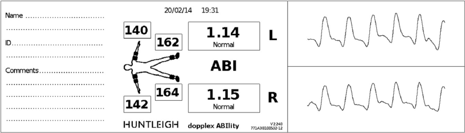
Example of a results printout from the automated device.
Duplex ultrasound scans of the lower limb arteries were then performed by a highly experienced medical physicist (P.W.), who was blinded to the ABI and PVW results (equipment utilised: Toshiba Aplio 500 with linear PLT-704SBT and curvi-linear PVT-375BT probes). The participant again lay supine on the scanning couch with the lower limbs exposed. The distal common femoral artery (CFA) was imaged and the Doppler waveform (DW) was assessed visually for any loss of triphasic flow due to significant iliac disease. If the DW showed indications of this, then the iliac arteries were assessed for the presence of atherosclerotic disease. The scan continued distally from the CFA assessing the superficial femoral artery (SFA) and popliteal arteries in the longitudinal plane. The extent and severity of any arterial disease were assessed using triplex mode by measuring the peak systolic velocity (PSV) from the DW just proximal to and through the stenosis (Figure 3). Disease severity was classified using standard criteria outlined in Table 1.

Example of an ultrasound Duplex scan image.
Grading of stenoses according to PSV ratio of velocities. 14

PSV: peak systolic velocity; PAD: peripheral arterial disease.
For the purpose of this study, the results of each test for each limb were graded as ‘PAD present’ if ABI ⩽0.9; PVW = grade 2, 3 or 4 and duplex scan demonstrating ⩾50% stenosis.
Statistical analysis was undertaken using IBM SPSS software (version 21; New York, USA). The sensitivity, specificity, positive predictive value and negative predictive value of the ABI and PVW were calculated, against the duplex ultrasound scan results as the reference standard. A receiver operating characteristic (ROC) curve was utilised to further assess the accuracy of the ABI and to determine the optimal ABI cut-off point for the diagnosis of PAD. Agreement between the three tests was assessed using Cohen’s kappa. 15 Significance was set at p < 0.05.
Results
The flow charts of the study are shown in Figure 4(a) and (b) and participant demographics are presented in Table 2. Of the 189 participants who completed a full set of study measurements, the mean age was 67 ± 12 years and 67% of the sample were male. In total, 36% of the participants were found to have PAD, in either one or both legs, as defined by the reference standard duplex ultrasound scan. PAD was found to be positively associated with male gender (p = 0.003), diabetes (p = 0.02), smoking history (p = 0.04), referral for leg pain (p = 0.01) and previously diagnosed PAD (p < 0.001) or vascular surgery (p < 0.001).
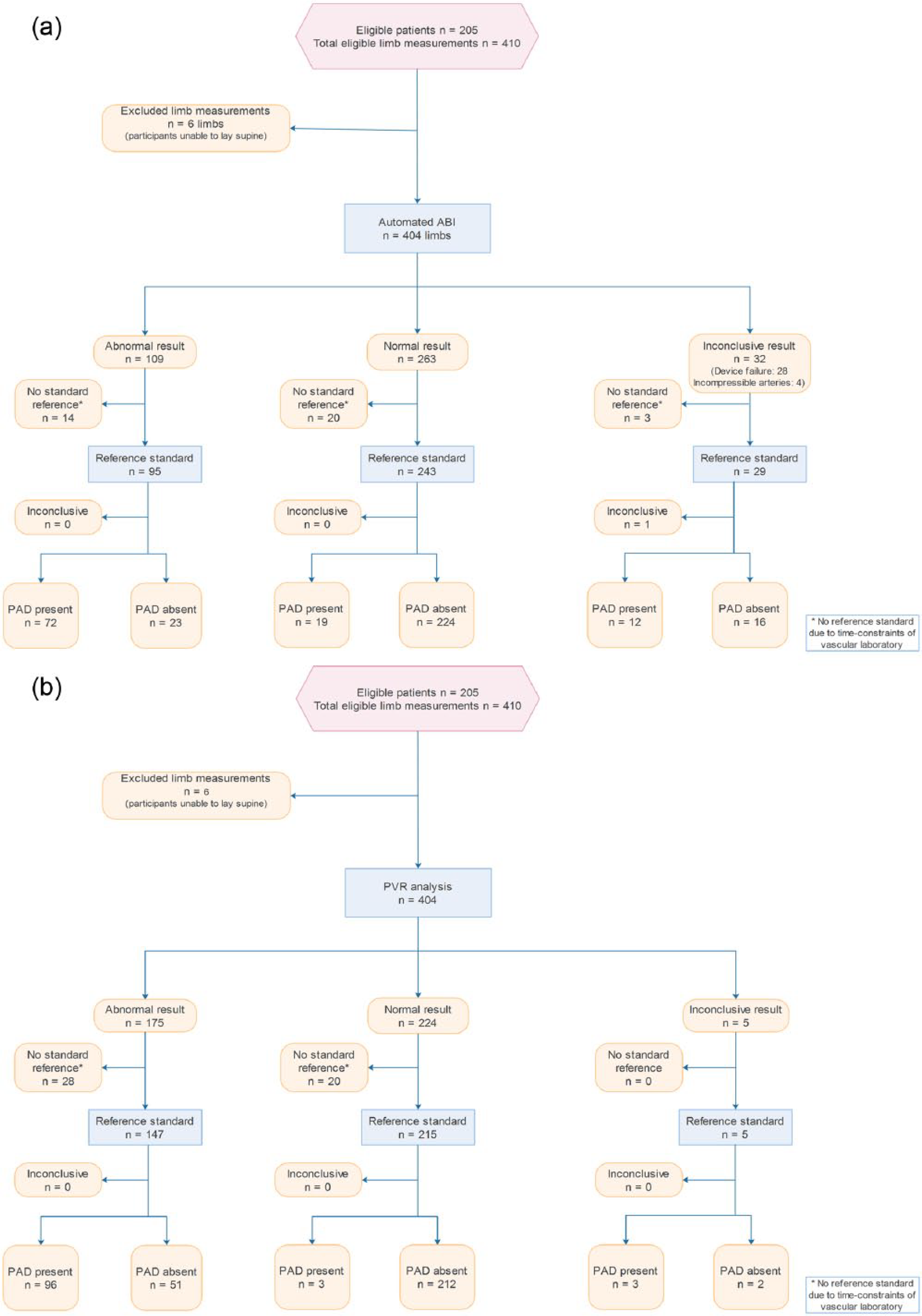
(a) Flow diagram illustrating diagnostic accuracy of ABI as per Standards for Reporting Diagnostic Accuracy (STARD) and (b) flow diagram illustrating diagnostic accuracy of PVW as per Standards for Reporting Diagnostic Accuracy (STARD).
Population demographics.

PAD: peripheral arterial disease; ABI: ankle brachial index; SD: standard deviation; CVA: cerebrovascular accident; CHD: coronary heart disease; DVT: deep vein thrombosis.
Mann–Whitney U test.
Chi-square test.
The sensitivity, specificity, positive predictive value, negative predictive value and overall accuracy of (1) the ABI, (2) PVW analysis and (3) ABI and PVW analysis combined, as compared to the ultrasound duplex scan (UDS) as the reference standard, are presented in Table 3. The distribution of ABI for the study population is shown in Figure 5.
Accuracies of test diagnostic modality.

ABI: ankle brachial index; PVW: pulse volume waveform.

Distribution of ABIs.
Analysis of the ABI ROC curve (Figure 6) revealed an area under the curve (AUC) of 0.88 (95% confidence interval (CI): 0.83–0.93, p < 0.001). The optimal ABI cut-off point for diagnosis of PAD was 0.98 which provided a sensitivity of 87% and specificity of 80%.
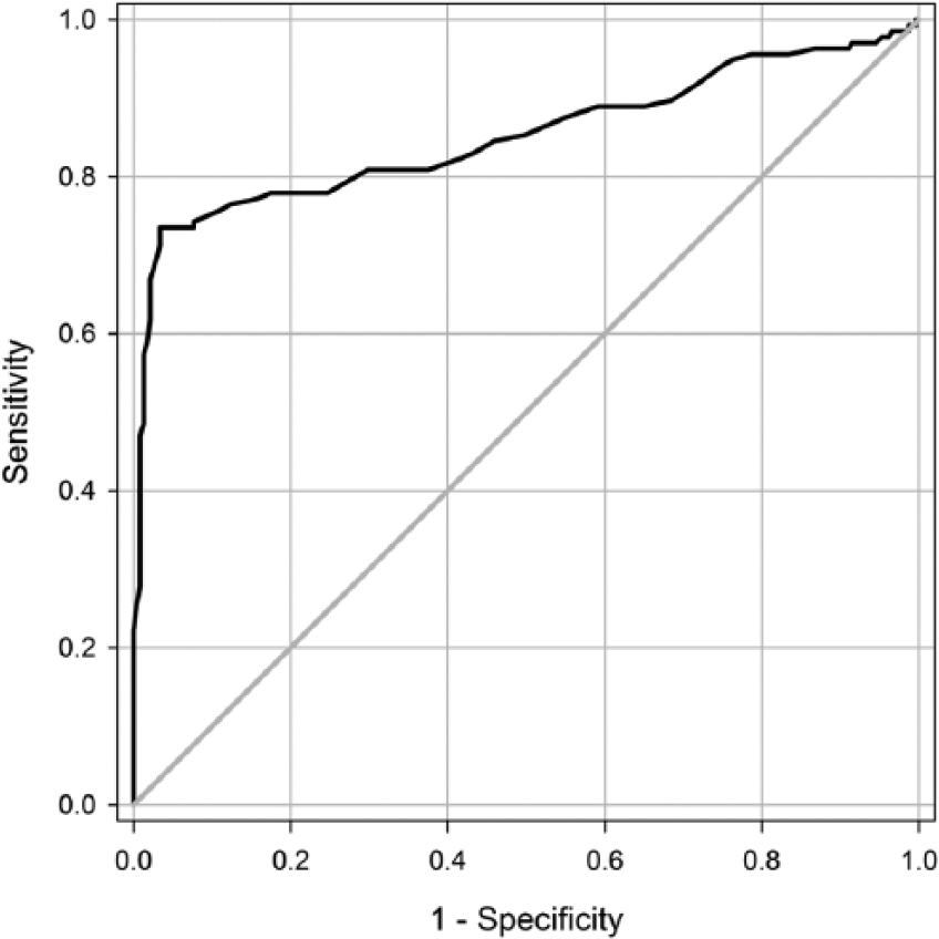
Receiver operating characteristic (ROC) curve for automated ABI device in diagnosing PAD as defined by ultrasound duplex scan. Area under curve=0.88 (95% CI: 0.83–0.93, p<0.001).
Discussion
The ABI
Data suggest that the automated ABI has moderate sensitivity (79%) and good specificity (91%) for PAD diagnosis. ROC curve analysis and AUC of 0.89 also suggest a good degree of accuracy in comparison to duplex ultrasound results as the gold standard. The optimal cut-off point for diagnosis of PAD was 0.98 which is higher than the threshold of 0.9 which is traditionally used for Doppler ABI measurements. However, this appears to be a common finding associated with the use of automated ABI devices; a systematic review and meta-analysis of 25 studies which assessed the usefulness of oscillometric devices for ABI estimation compared to the conventional Doppler method also concluded that to increase the sensitivity for PAD, a higher threshold ABI <1.0 might be preferable. 10
Two factors could have contributed to the reduced sensitivity of the ABI in this study; first, inaccuracies of the automated device itself could have played a part and second, it is possible that the demographics of the study population and the high likelihood of the presence of arterial calcification could have rendered the ABI non-diagnostic in a proportion of participants. In such cases, arterial calcification can artefactually raise ankle systolic pressures of PAD patients, which, in turn, results in the ABI being elevated to within the normal (>0.9–1.3) or high range (>1.3). Studies comparing Doppler ABI with UDS as the reference standard in diabetic populations also reported reduced sensitivities of 71%.16,17
The reported study is novel in design because it has utilised UDS rather than the usual hand-held Doppler ABI method as the reference standard to evaluate the accuracy of an automated ABI device. There are therefore no data to which the current results can be directly compared. However, a recent study compared ABIs attained with the same automated ABI device (Dopplex ABIlity) to ABIs undertaken with a hand-held Doppler. 17 The study population was of similar mean age (64 years) but did not contain any diabetics; it also returned a moderate sensitivity of 70% and good specificity of 96%.
PVW interpretation
The data suggest that analysis of the PVW has excellent sensitivity (97%) and moderate specificity (81%) for PAD diagnosis. Research regarding PVW analysis for the identification of PAD is sparse, hence meaning that, again, there are little data available for comparative purposes. A study by Ro et al. 18 evaluated the sensitivity and specificity of the ABI and subjective PVW analysis derived by photoplethysmography (PPG), with subjective DW analysis compared to the gold standard of computed tomography angiography (CTA) diagnosed PAD. The test results from a total of 97 patients (194 legs) who had coincidently undergone CTA, ABI, PPG and DW were retrospectively reviewed. PVWs and DWs were subjectively interpreted by a single physician. With PVWs, diagnosis of PAD was based on loss of the dicrotic notch, decreased waveform amplitude and/or rounding of systolic peaks. For DWs, diagnosis of PAD was based on loss of triphasic pattern, decreased amplitude and/or loss of reverse flow component. The sensitivity and specificity of PPG PVW analysis compared to the CTA were 82% and 77%, respectively; for DW analysis, sensitivity was 91% and specificity 65%, and for ABI sensitivity was 70% and specificity 97%. The authors concluded that ABI should be combined with PVW analysis or DW analysis in order to improve detection of PAD.
PVW analysis versus DW analysis
Some clinicians may be more accustomed to analysing DWs which can often be viewed on a visual display unit incorporated into the hand-held Doppler; it is therefore useful to make a comparison of this with PVW analysis. The process of obtaining a PVW recording does not require operator skill and merely involves the application of a cuff to the foot or ankle; the device then automatically inflates, obtains and displays the PVW. The process of obtaining a DW is, in contrast, operator dependent where a Doppler probe has to be carefully positioned over an artery, at a specific angle and pressure; the results can vary with the Doppler angle used. 19
Limitations of PVW analysis
There are recognised physiological limitations related to PVW analysis. First, the PVW is dependent on peripheral blood flow and thus may be influenced by factors other than vessel patency such as sympathetic nerve input. 20 Second, severe congestive heart failure may also slow blood flow and mimic inflow disease. 21 Third, the PVW represents the total blood flow through the area being assessed and cannot therefore provide accurate diagnostic information as to what extent a specific artery is diseased.
Combining the ABI and PVW analysis
Combining the ABI and PVW results for each participant, where if either the ABI or the PVW analysis returned a positive result for either leg, then the participant was classed as having PAD, returned a sensitivity of 100%, specificity of 76% and overall accuracy of 85%. The negative predictive value of combining these diagnostic modalities was 100% meaning that the dual diagnostic device (Dopplex ABIlity) utilised within this study can rule out PAD with a high degree of accuracy (as defined by both ABI and PVW analysis returning negative results).
Utility within primary care
Utilisation of this device in the primary care setting, applying the criteria that double negative results (from the ABI and PVW analysis) do not require secondary care assessment would have prevented 46% (93/202) of referrals to the vascular laboratory for the population of this study, and importantly, no cases of PAD would have been missed. An audit by Poots et al. of 451 patients referred to a vascular clinic revealed that a similar proportion of referrals (41%) were deemed inappropriate as subsequent Doppler assessment revealed normal ABIs and normal triphasic Doppler signals. 22
Study strengths and limitations
Strengths
The large sample size and high-risk study population with multiple co-morbidities serve to optimise the clinical relevance of this study. Furthermore, the majority of the existing studies evaluating automated ABI devices utilise Doppler ABI as the reference standard, which itself is operator dependent and with the process susceptible to inherent error. This study has utilised UDS which is recognised as the superior non-invasive modality which can diagnose PAD with a high degree of accuracy. 23
Limitations
This study has evaluated the utility of subjective PVW analysis when undertaken by a single clinician who has experience and a personal interest in the procedure; findings are not therefore representative of how less experienced, non-specialist clinicians would perform.
Conclusion
Within this study population, PVW analysis provided excellent sensitivity for the detection of PAD while the ABI provided very good specificity. Combining these two diagnostic modalities within one device provided a highly accurate method of ruling out PAD. Hence, this suggests that this device could be utilised within the primary care environment to reduce the number of unnecessary referrals to secondary care with concomitant cost savings, reduced patient inconvenience and prioritisation of urgent PAD cases. Future research should investigate ease of use of PVW analysis, along with the cost and training required to achieve reliable results.
Footnotes
Acknowledgements
The authors gratefully acknowledge the assistance of Mrs Elaine Townsend with data collection, and Mr Mike Lewis (Consultant Vascular Surgeon, Cwm Taf University Health Board) and Professor Neil Pugh (Consultant in Vascular Ultrasound, Cardiff and Vale University Health Board) for allowing the study to take part in their Medical Physics Departments. The authors also thank Huntleigh Diagnostics for the loan of equipment used within this study, and Dr Mark Williams (University of South Wales) for his assistance with manuscript preparation. Trial registration: UKCRN 16912.
Declaration of conflicting interests
The author(s) declared the following potential conflicts of interest with respect to the research, authorship, and/or publication of this article: J.H.D. previously undertook a PhD which was part sponsored by Huntleigh Diagnostics. J.E.A.L. and P.W. declare no conflict of interest.
Ethics approval
Ethical approval for this study was obtained from Research Ethics Committee 2 (Cardiff, Wales, REC No: 13/WA/0072).
Funding
The author(s) disclosed receipt of the following financial support for the research, authorship, and/or publication of this article: A proportion of J.E.A.L.’s salary was supported by a Clinical Research Fellowship funded by Health and Care Research Wales, overseen by the Welsh Government.
Informed consent
Written informed consent was obtained from all subjects before the study.