
Abstract
Introduction:
We evaluated the association between two single nucleotide polymorphisms of the vascular endothelial growth factor gene and one of the hypoxia-inducible factor-1α gene and the degree of coronary collateral formation in patients with a coronary chronic total occlusion.
Methods:
Totally, 98 patients with symptomatic coronary artery disease and a chronic total occlusion observed during coronary angiography were recruited. Genotyping of two vascular endothelial growth factor promoter single nucleotide polymorphisms (−152G>A and −165C>T) and the C1772T single nucleotide polymorphism of hypoxia-inducible factor-1α were performed using polymerase chain reaction and restriction fragment length polymorphism analysis. The presence and extent of collateral vessel filling was scored by blinded observers using the Rentrop grade.
Results:
We found no association between the vascular endothelial growth factor −152G>A, −165C>T and hypoxia-inducible factor-1α −1772C>T with the presence and filling of coronary collateral vessels. A history of percutaneous coronary intervention and transient ischaemic attack/cerebrovascular accident were associated with the presence of enhanced collateral vessel formation following binary logistic regression analysis.
Conclusion:
The study findings suggest that coronary collateral formation is not associated with the tested polymorphic variants of vascular endothelial growth factor and hypoxia-inducible factor-1α in patients with symptomatic coronary artery disease and the presence of a chronic total occlusion.
Keywords
Introduction
Collateral coronary vessels provide an important source of blood flow to the myocardium in the presence of an occluded major coronary artery. There is strong evidence that a well-developed pre-existing coronary collateral circulation can protect the myocardium during acute ischaemia1–5 and in patients with established stable coronary artery disease (CAD). 6
In chronic total occlusions (CTOs), coronary collateral vessels vary in extent from patient to patient. While well-developed collaterals can provide sufficient myocardial perfusion at rest, they are rarely as effective as native coronary vessels, which often results in patients experiencing exertional angina.
Collateral vessel formation may be influenced by numerous variables including the patient’s age, 7 body mass index, 8 gender, 9 history of diabetes mellitus, 10 hypertension, 11 hyperlipidemia, 12 smoking status, 13 alcohol consumption, 13 pre-infarction angina 14 and statin use. 15 Furthermore, there is a wide variation in the expression levels of angiogenic and proteolytic enzyme activators and inhibitors (particularly vascular endothelial growth factor (VEGF)). 16 The majority of genes encoding these factors carry single nucleotide polymorphisms (SNPs), with some of these leading to altered gene activity. The VEGF gene consists of eight exons and seven introns and spans a 14-kb segment located on the short arm on chromosome 6p21.3. 17 Many polymorphisms have been described, especially in the promoter region, 5′-untranslated region (UTR) and 3′-UTR. 18 Some of these polymorphisms are associated with VEGF protein expression and disease severity in conditions such as acute renal allograft rejection, 19 psoriasis 20 diabetic retinopathy, 21 cancer, 22 as well as rheumatoid arthritis 23 and sarcoidosis. 24
Hypoxia-inducible factor-1 (HIF-1) is a transcriptional activator for more than 150 genes including VEGF. 25 HIF-1 is a heterodimer that comprises α and β subunits, and HIF-1 activity is controlled by the oxygen-regulated expression of the HIF-1α subunit. The HIF-1α gene is located at chromosome 14q21-q24; furthermore, it has been described that the C1772T (P582S) polymorphism of the HIF-1α gene is associated with an increased expression of HIF-1 mRNA and protein than the wild-type sequence. 26 This polymorphism has also been shown to influence several human phenotypes, possibly leading to greater susceptibility to various forms of cancer.26,27 It has been suggested that the expression of this protein is associated with the presence of coronary collateral vessels in patients with stable CAD. 28
Previous studies have reported an inter-individual difference in the number and extent of collateral vessels in patients with and without CAD.29,30 Furthermore, CAD patients with well-developed coronary collateral circulation are reported to have a 36% reduction in mortality compared with patients with low collateralization. 31
The reasons for this are not fully understood, but genetic factors are suggested to play a role. 30 This study aimed to determine whether two SNPs in the VEGF promoter region (−152G>A and −165C>T) reportedly associated with varying cancer risk, 32 and severity of proliferative diabetic retinopathy 33 and the well-described C1772T polymorphism in the HIF-1α hypoxia response element influence collateral vessel formation in patients presenting with symptomatic CAD.
Methods
Study population
Patients undergoing coronary angiography for the investigation of ischaemic heart disease and found to have a CTO were invited to participate in the study. Clinical details are described in Table 1. Coronary angiography was performed as per contemporary practice in the United Kingdom, and patients were included in the study once the results of angiography were known.
Polymerase chain reaction primer pairs, reaction conditions and restriction enzymes used for each polymorphism.

PCR: polymerase chain reaction.
This study complied with the declaration of Helsinki and was approved by the local research ethics committee. Informed consent was obtained from all patients prior to enrolment in the study.
DNA isolation and genotyping
Genomic DNA was isolated from whole blood samples anticoagulated with ethylenediaminetetraacetic acid (EDTA) using a commercially available DNA isolation kit (Qiagen, Hilden, Germany). Genotyping for HIF-1α −1772C>T, VEGF −152G>A and −165C>T was performed using restriction fragment length polymorphism polymerase chain reaction (RFLP-PCR). All PCRs for genotyping were carried out under the same condition (35 cycles of denaturation at 94°C for 60 s, annealing for 60 s and extension at 72°C for 60 s) with various annealing temperatures for respective amplifications. Table 1 shows all primer sequences for determination of VEGF gene polymorphisms and their melting temperatures. PCR products were digested with appropriate restriction enzymes (Table 1); fragments were separated on a 2%–3% agarose gel and visualised by ethidium bromide staining.
Determination of collateral vessel formation and filling
The presence of collateral flow from the patent vessels to the occluded artery was evaluated using the classification developed by Rentrop et al. 34 by two interventional cardiologists blinded to the genetic results.
The grading is summarised as follows:
Grade 0. No visible filling of any collateral channel;
Grade 1. Minimal recipient filling by collaterals is manifested by minor side branch filling and no epicardial artery or epicardial side branch filling;
Grade 2. Moderate recipient filling by collaterals is manifested by complete filling of epicardial side branches and partial filling of a major epicardial artery (the left main, left anterior descending artery, circumflex, large obtuse marginal, the right coronary artery or the patent ductus arteriosus (PDA)). The collateral filling of the epicardial artery may be obscured and washed out by competitive flow;
Grade 3. Complete filling of epicardial vessel by collateral vessel.
Reproducibility of the Rentrop score between observers was described as good (κ = 0.67, 95% confidence interval 0.50–0.82). Note that 99% of differences between the two observer’s adjudications were with ±1, and 86% had total agreement (no difference). In the event of the two observers not agreeing, a third interventional cardiologist, also blinded to the results of the genetic data, was asked to adjudicate.
In addition to Rentrop grade, the size of the collateral connection (CC) diameter was assessed using the CC grade: CC Grade 0, no continuous connection between donor and recipient artery; CC Grade 1, continuous, thread-like connection; and CC Grade 2, continuous, small side branch-like size of the collateral throughout its course. 35
Data collection and follow up
Baseline variables were collected and recorded in a dedicated database and included age, gender, coronary risk factors, smoking history and drug therapy prior to coronary angiography.
Statistical analysis
Continuous variable data are described as mean ± standard deviation (±SD). Categorical data are expressed as frequencies or percentages. Differences of genotype frequencies between Rentrop grades were evaluated by χ2 analysis. Continuous variables were analysed individually using Students’ independent samples t test. A multivariate analysis was conducted using binary logistic regression analysis to identify which of the continuous and categorical variables identified were able to independently predict collateral vessel formation and filling when entered simultaneously. Values of kappa (statistic) were computed to assess agreement between the two evaluations of collateral vessel formation and filling in the patient population. All statistical calculations were performed using SPSS (IBM, New York, NY, USA) and a two-tailed p value of <0.05 was considered to be statistically significant.
Results
Totally, 98 patients who had at least one CTO of a coronary artery were enrolled in the study and were genotyped (mean age 65.3 ± 10.7 years, female n = 16). Of them, 36 patients had a clinical diagnosis of stable angina, and 62 had non–ST elevation acute coronary syndromes.
The demographic and clinical characteristics of the patients are summarised in Table 2.
Baseline characteristics of patients.

CAD: coronary artery disease; SD: standard deviation; PCI: percutaneous coronary intervention;TIA/CVA: /transient ischaemic attack/cerebrovascular accident; MI: myocardial infarction; CABG: coronary artery bypass graft.
Of the 98 patients enrolled, 2 (2%) patients had a Rentrop Grade 0, 9 (10%) patients had Grade 1 (no epicardial filling), 23 (22%) had Grade 2 (partial epicardial filling) and 64 (64%) had Grade 3 collaterals (complete epicardial filling). The CC grade was distributed as follows: 32 (32.7%) patients with CC Grade 0, 53 (54.1%) patients with CC Grade 1 and 13 (13.3%) patients with CC Grade 2.
The Rentrop collateral score was dichotomised into Group 1 (0–1 = poor collateral) and Group 2 (2–3 = good collateral). We entered the following variables into a binary logistic regression model with backward elimination: age, gender, diabetes, familial history, hypertension, hyperlipidaemia, smoking status, peripheral vascular disease, previous myocardial infarction (MI), previous coronary artery bypass graft (CABG), previous percutaneous coronary intervention (PCI), previous transient ischaemic attack/cerebrovascular accident (TIA/CVA), beta-blocker, angiotensin-converting enzyme (ACE) inhibitor usage, proton-pump inhibitor (PPI) usage, non-steroidal anti-inflammatory drug usage, statin usage, aspirin usage and clopidogrel usage. A history of previous PCI or TIA/CVA were the only variables associated with the presence of enhanced collateral formation on multivariate analysis (Table 3).
Variables associated with enhanced collateral vessel formation.
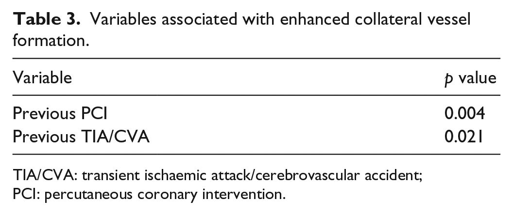
TIA/CVA: transient ischaemic attack/cerebrovascular accident; PCI: percutaneous coronary intervention.
Genetic analysis
The genotype frequencies for all three SNPs investigated are displayed in Table 3. We were unable to demonstrate any association between the Rentrop grades and the three SNPs analysed in this study (Table 4). In addition, no association was established between CC grades and the three SNPs analysed in this study (Table 5).
Genotype distribution of VEGF and HIF-1 gene polymorphisms in patients according to Rentrop grade.

VEGF: vascular endothelial growth factor; HIF-1: hypoxia-inducible factor-1.
Genotype distribution of VEGF and HIF-1 gene polymorphisms in patients according to CC grade.

VEGF: vascular endothelial growth factor; HIF-1: hypoxia-inducible factor-1.
Discussion
At present, there are inconsistencies in the literature in trying to identify reliable predictors of collateral vessel formation, and it is unclear why there are large differences in the number and extent of collateral vessel formation between subjects.9,13,36 Some of these differences may be explained by anatomic variation (e.g. dominance of the right or left coronary tree) or possibly by inter-individual differences in the many processes involved in neovascularisation. Along with well-defined risk factors for CAD, collateral formation may also be dependent on myocardium sensitivity to ischaemia. 37
As it has been suggested that there is a possible genetic cause for the disparity in collateral vessel numbers in human heart, 30 we aimed to determine the possible relationship between two SNPs in the VEGF promoter region and one in HIF-1α in a patient cohort who have a demonstrated CTO of at least one of the coronary vessel.
HIF-1α polymorphisms have been extensively studied in order to determine the association they may have in the appearance or progression of hypoxia-related diseases. There are conflicting reports about the significance of these SNPs in the protein expression or function of HIF-1α.38,39 We studied the C1772T (P582S) polymorphism, which corresponds to the proline to serine amino acid change at residue 582 of the HIF-1α protein. 25 There are reports suggesting that C1772T (P582S) and HIF-1α transcriptional activities are higher than wild type especially under hypoxic conditions. 38 A previous study by Resar et al. 28 (in patients with ischaemic heart disease but not definite CTO) using the Rentrop scoring system has demonstrated that the C1772T polymorphisms may influence the development of coronary collateral vessel development; in contrast to this, Alidoosti et al. 40 found no association with this HIF-1 polymorphism and collateral vessel development. Our current data, in patients with definite CTO, go some way to confirming the results from the Alidoosti et al. study.
VEGF is a major mediator of vascular angiogenesis and is suggested to play a central role in the development of coronary collaterals. 41 Patients with myocardial ischaemia and infarction have elevated levels of VEGF mRNA in myocardial tissues, potentially as an important cardiac response to hypoxia. 42 Furthermore, the −152G>A polymorphism is associated with essential hypertension with the A allele suggesting to have a protective effect on hypertension. Churchill et al. demonstrated the A allele at −152 was significantly associated with proliferative diabetic retinopathy as was the AA genotype. Despite VEGF −152GA/−165CT being associated with varying risk in other disease processes, 32 we were unable to demonstrate a relationship between VEGF −152GA/−165CT and formation of coronary collaterals in this study.
In this study, we did not find any association with the factors that have previously been demonstrated to influence coronary collateral formation.7–15 Instead, we found that a history of PCI or TIA/CVA were associated with enhanced collateral vessel formation. Although this has not been previously reported in the literature, this result should be interpreted cautiously as it is possible that these two factors have no physiological influence on collateral vessel development.
Study limitations
One of the reasons why no associations were identified may be due to the relatively small study size which is a recognised limitation of human genetic studies, which can result in low power to detect differences between genotypes.
Having assessed all of the clinical records of the patients recruited, we were unfortunately unable to estimate the duration of occlusion accurately. Therefore, in this study, we were not able to investigate a possible relationship between the duration of occlusion and Rentrop grade.
The Rentrop method of collateral assessment is a subjective evaluation which is influenced by variables such as the volume of contrast hand injected during angiography and the limited spatial resolution. Novel techniques such as micro–computed tomographic analysis, three-dimensional (3D) reconstruction of tomographic images and magnetic resonance angiography may provide more objective and accurate assessments of collateral vessel formation and should be considered for future studies.
Conclusion
In this study, we were unable to demonstrate an association between coronary collateral formation and the tested polymorphic variants of VEGF and HIF-1α in patients with symptomatic CAD and the presence of a CTO. However, further large-scale studies looking at different VEGF and HIF-1 SNPs are required in the future to determine what role, if any, VEGF and HIF-1 play in coronary collateral vessel formation.
Footnotes
Declaration of conflicting interests
The author(s) declared no potential conflicts of interest with respect to the research, authorship and/or publication of this article.
Ethics approval
Ethical approval for this study was obtained from The Black Country Research Ethics Committee (09/H1202/96).
Funding
The author(s) disclosed receipt of the following financial support for the research, authorship, and/or publication of this article: This research was made possible by generous donation from the Taylor Charitable Trust Foundation and the Rotha Abraham Bequest.
Informed consent
Written informed consent was obtained from all subjects before the study.