
Abstract
Natural biomaterials, such as collagen, gelatin, and chitosan, are considered as promising candidates for use in tissue regeneration treatment, given their similarity to natural tissues regarding components and structure. Nevertheless, only receiving a crosslinking process can these biomaterials exhibit sufficient strength to bear high tensile loads for use in skeletal system regeneration. Recently, genipin, a natural chemical compound extracted from gardenia fruits, has shown great potential as a reliable crosslinking reagent, which can reconcile the crosslinking effect and biosafety profile simultaneously. In this review, we briefly summarize the genipin extraction process, biosafety, and crosslinking mechanism. Subsequently, the applications of genipin regarding aiding skeletal system regeneration are discussed in detail, including the advances and technological strategies for reconstructing cartilage, bone, intervertebral disc, tendon, and skeletal muscle tissues. Finally, based on the specific pharmacological functions of genipin, its potential applications, such as its use in bioprinting and serving as an antioxidant and anti-tumor agent, and the challenges of genipin in the clinical applications in skeletal system regeneration are also presented.
Introduction
Natural biomaterials are applicable for regenerative medicine owing to their excellent biocompatibility. The components of natural biomaterials such as collagen, gelatin, and chitosan are more similar to human tissues than synthetic polymers or metals, as they can positively interact with host tissues to facilitate bioreactions. 1 Multiple functions can be achieved using natural biomaterial as tissue engineering scaffolds, including suppressing inflammation, promoting tissue regeneration, and facilitating differentiation or homing of stem cells.2,3 Nevertheless, there are controversies regarding the natural biomaterials applying in skeletal system reconstruction. Bone, cartilage, intervertebral disc (IVD), tendon, and skeletal muscle are rich in collagen and gelatin, but it is still difficult to create a functional structural scaffold with these natural biomaterials,4 –7 with lack of weight-bearing ability being the pivotal issue.
To address this problem, crosslinking method has been developed to enhance the biomaterials’ mechanical properties by increasing the intermolecular force in materials. Basically, two commonly accepted strategies are created accordingly. The first one is known as “physical crosslinking.” By altering the physical conditions (e.g. temperature, ultraviolet radiation, and pH value), molecules are bond with electrostatic force, hydrogen bond, or physical entanglement, forming a crosslinking network between the tertiary structures of proteins and thereby constructing a solid scaffold.8 –10 But the stability and mechanical properties of physical hydrogel are relatively poor, which are prone to return to sol phase by disturbances. 11 Alternatively, “chemical crosslinking” is a more stable crosslinking strategy to form covalent or ionic bonds between functional groups. Acylation, carboxylation, esterification, alkylation, etherification, halogenation, and Schiff alkalization are all common reaction types in crosslinking natural biomaterials. Chemical crosslinking can establish a stable and irreversible hydrogel network with the help of crosslinking reagents, which is more frequently-used in generating hydrogels for tissue engineering. 12 The carbonyl groups in traditional chemical crosslinking reagents, such as glutaraldehyde and 1-ethyl-3-(3-dimethylamineproply) carbodiimide (EDC), can interact with hydroxyl and amino groups in natural biomaterials.13,14 Based on Schiff base and acetalization reactions, dense three-dimensional hydrogel scaffolds can be formed after crosslinking. 15 However, these artificial chemical crosslinking reagents are more or less cytotoxic, causing cell death after seeding in the hydrogel and leading to low bioactivity or even tissue necrosis around the implantation area. 16
Genipin, a natural component extracted from the fruits of Gardenia jasminoides and Genipa americana L., has been intensively studied with regarding to its multiple pharmacological properties, including the anti-inflammatory, antithrombotic, antiangiogenic, antitumor, antidiabetic, and neurotrophic properties. 17 Additionally, genipin’s ability to induce crosslinking in biological tissues was first discovered in 2000. 18 Since then, genipin has become a star molecule in the field of tissue engineering owing to its promising biosafety and specific crosslinking performances. As a fruit extract, cytotoxicity of genipin is only 0.01% that of glutaraldehyde, and its in vivo biosafety has been extensively demonstrated. 19 During the gelling process, genipin crosslinks amino groups in natural biomaterials (such as lysine units), causing a blue color pigment formation.20,21 Another noticeable feature of genipin crosslinking is the long reaction duration, which can last for >72 h. 22 During this period, the crosslinking degree, color, and mechanical properties continuously change depending on the reacting time. 23 Thus, the long reaction duration provides multiple opportunities to manufacture hydrogel scaffolds with particular specifications, allowing in situ gelling via local injection, in vitro gel casting, and three-dimensional printing.24,25
Collectively, optimal biomaterials with sufficient mechanical property for skeletal tissue regeneration are urgently needed. And the crosslinking capability of genipin has a great potential in tissue engineering. As shown in Scheme 1, the scope of this review is to summarize advances related to the use of genipin in skeletal system regeneration, which have not ever been reviewed. We comprehensively discuss the use of genipin-crosslinked hydrogel for bone, cartilage, IVD, tendon, and muscle tissue regeneration. We also highlight the future directions related to the use of genipin-crosslinked hydrogel in tissue engineering, including those related to its bioprinting, anti-oxidative, anti-tumor functions, and the challenges of genipin in clinical transformation and application.

Schematic illustration of genipin crosslinking characteristics and applications regarding skeletal system regeneration.
Preparation, biosafety assessment, and crosslinking mechanism of genipin
In Chinese traditional medicine, geniposide-containing decoction, which is made from Genipa americana L. or Gardenia jasminoides, is considered to play roles in liver protection, blood pressure control, and inflammation suppression. 26 After oral administration, the genipin in the decoction is enzymatically hydrolyzed by the β-glucosidase produced by intestinal bacteria. 27 However, a series of complex extraction steps is required in order to obtain high purity genipin, as the plants only contain a low level of genipin, or its precursor (geniposide), in the case of Gardenia jasminoides. Before being used as crosslinking reagent, the pharmacological functions of genipin were extensively explored. 28 Additionally, its biosafety has been assessed in a number of studies. 29 After its crosslinking ability was discovered, the underlying crosslinking mechanism was revealed.30,31 Having a full understanding of the genipin crosslinking mechanism allows a better manipulation on the crosslinking results.
Before elaborating on the use of genipin as a crosslinking reagent to aid in skeletal system regeneration, its origin as well as its chemical and biological properties should be introduced. Hence, we present details on the preparation, biosafety assessment, and crosslinking mechanism of genipin (Figure 1) in this section. 32
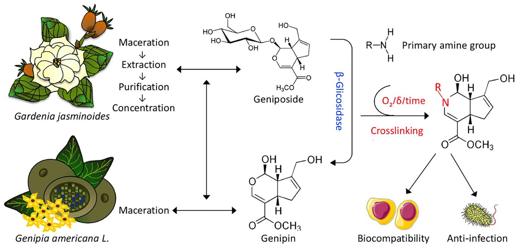
Genipin extraction and preparation from Gardenia jasminoides and Genipa americana L. Genipin can react with amino groups in natural materials and form a chain structure. After crosslinking, genipin exhibits promising biosafety characteristics.
Extraction and purification of genipin
Genipin is obtained from the fruits of Genipa americana L. or Gardenia jasminoides, but the extraction methods are different. 33 Through maceration, genipin can be directly extracted from Genipa americana L. 34 However, a more complicated process is required in dealing with Gardenia jasminoides. First, geniposide (in which the hydroxyl group at the C1 position of the genipin pyran ring is substituted by 1–2 moieties) is isolated by a series of processes including maceration, extraction, purification, and concentration. Then, genipin is obtained from the geniposide by an enzymatic reaction, involving β-glucosidase to alter glucose group into hydroxyl on C3 position of pyran ring. 32 In traditional Chinese medicine, the preparation of genipin (using a modern technique) involves grinding original fruits, drying at 60°C, boiling twice for 30 min, diluting to 1% using ethanol, and heating twice under reflux for 1.5 h, which allows a relatively high concentration of extracted geniposide. Subsequently, genipin can be obtained efficiently from geniposide using β-glucosidase produced by Penicillium nigricans in a microbial reactor.35,36
Biosafety assessment of genipin
The biosafety of genipin is another significant issue that needs to be scrutinized when using in tissue engineering. As mentioned above, genipin has various medical functions that have been verified in a number of studies, and its biosafety has also been fully assessed.28,29 At the cellular level, the cytotoxicity of genipin and glutaraldehyde was compared as gelatin crosslinkers. 37 The survival rate of cell seeded in genipin-crosslinked gelatin was significantly higher than those in glutaraldehyde-crosslinked gelatin when the crosslinking degree was adjusted to the same. Moreover, a quantitative in vitro experiment illustrated that the cytotoxicity of genipin against fibroblasts was approximately 10,000-fold lower than glutaraldehyde, and the proliferation rate of fibroblasts was 5000-fold greater after genipin exposure compared to glutaraldehyde exposure. 38 At the in vivo level, biosafety of genipin before crosslinking was assessed firstly. In rat models of cirrhosis or diabetes, genipin reversed the disease-related biochemical indexes, and it reduced the mortality rate compared to that in the control rats. 39 Moreover, the biosafety of genipin was found to be maintained after crosslinking. Sung et al. 40 subcutaneously implanted genipin-crosslinked chitosan and found no obvious inflammation generated around the implantation area. Yang et al. 41 injected genipin directly into the periodontal pocket, a chronic inflammatory tissue, and found that the inflammation was effectively controlled. The gap between gum and tooth was replaced by fibrous tissue with no sign of tissue inflammatory or necrosis. Thus, genipin has been shown to have a great biosafety profile, being highly compatible with the surrounding host tissue after implantation according to above studies. Additionally, as genipin can suppress inflammation, it is suitable for treating inflammatory diseases such as osteoarthritis and IVD protrusion.42 –44
However, it is irrational to assess biosafety without considering drug concentration. At present, there is still lacking of a systematic evaluation to give a definite safe dose of genipin, but the inhibition effect of cell proliferation was found in line with genipin concentration. In the following text, we emphasize to find a critical concentration of genipin used in skeletal tissue regeneration that inhibits cell proliferation, and hope to give a preliminary biosafety guidance for genipin application.
Crosslinking mechanism of genipin
The diagrammatic sketch of genipin crosslinking with chitosan, collagen, and gelatin was showed in Figure 2. Under basic conditions, nucleophilic attack of genipin by the hydroxyl ions in aqueous solution leads to a ring-opening reaction, and an intermediate aldehyde group is formed. Subsequently, a Schiff reaction occurs between the terminal aldehyde groups on the polymerized genipin and amino groups on the natural materials, forming a crosslinked hydrogel network structure. Under acidic and neutral conditions, the olefinic carbon atom at the C3 position in genipin is attacked by the amino groups in the natural materials, which is followed by the opening of the dihydropyran ring and an attack on the newly formed aldehyde group by a secondary amino group.45,46 Because of the additional condensation reaction, genipin-crosslinked products are more stable than those crosslinked by glutaraldehyde. Moreover, although genipin is a colorless aglycone (derived from the iridoid glycoside, geniposide), a bluish-violet color is formed after reacting with primary amines, which it readily does after exposure to oxygen. Owing to the highly optimized and effective genipin manufacturing technology, the genipin extraction rate has been greatly improved and the price has dropped sharply. Hence, genipin has consequently been used in the food coloring industry, and acting as a chromogenic agent to detect protein concentration.32,47

Reaction equation of genipin reacting with: (a) chitosan and (b) collagen and gelatin.
Use of genipin in skeletal system tissue regeneration
In this part, the use of genipin in skeletal system tissue regeneration, including articular cartilage, bone, IVD, tendon and skeletal muscle, has been summarized (Table 1).
Summary of genipin used for skeletal system tissue regeneration.

Articular cartilage regeneration
The most common application of genipin in tissue engineering field attributes to cartilage regeneration. Collagen, gelatin, and chitosan, which can be crosslinked by genipin, are the basic elements of the articular cartilage. These natural biomaterials can be used to construct an appropriate microenvironment for cells to reside in that is conducive to chondrogenic differentiation. 96 Biomaterials can be transformed from a solution into a hydrogel phase by altering the pH or thermal conditions.97,98 However, the weak mechanical strength limits their application in cartilage regeneration. 99 The load on the native cartilage in the knee joint of adults is >10 GPa, reaching 25 GPa in the weight-bearing area during exercise (such as jumping or sprinting). 100 Hence, a powerful crosslinking reagent is required to enhance the mechanical properties of hydrogels in order to avoid rapid structural failure under high pressure. Recent research advances concerning genipin mean that it has a great potential for use in cartilage regeneration as it can increase the crosslinking degree in the hydrogel while simultaneously maintaining optimal biocompatibility.
The mechanical properties of genipin-crosslinked hydrogels constructed using various genipin concentrations are not always predictable. Commonly, the genipin concentration used in cartilage tissue engineering is <2%. Inducing crosslinking using genipin concentrations >2% led to hydrogels with irregular pore walls, and the mechanical properties decreased accordingly. 101 However, when the genipin concentration was raised from 0.5% to 2%, the compressive strength of the hydrogel increased, while pore size also increased from 180 to 1100 μm3; Mu et al. 102 hypothesized that the larger pore size formation was contributed to ring-opening polymerization of genipin increasing the cross-sectional area of the pores. Although a larger pore size normally causes lower strength (due to less connective substance in the hydrogel), unexpectedly, the compressive strength of the hydrogel increased with the increasing pore size. In this case, the compressive strength was predominantly determined by two other factors besides pore size: pore wall thickness and water content in the hydrogel. 48 An increased genipin concentration from 0.5% to 2% led to a robust reaction that formed strong chemical bonds between the polymers, and the pore wall was thicker dependently. In addition, the hydrogel was slightly swollen by absorbing water in the pores, which also helped with load bearing. Nevertheless, the crosslinking process involves oxidation reactions that form hydrophobic bonds, which repel water from being absorbed into the hydrogel and thus tend to reduce its mechanical strength. 103
Overall, the compressive strength of hydrogel contributes to a trade-off between the three abovementioned factors (pore size, pore wall thickness, and water content). Based on the complexity of the reaction conditions, researchers hope to optimize the compressive strength of genipin-crosslinked hydrogels by adjusting the reaction parameters. The genipin concentration and temperature are two easily modifiable variables. Bi et al. 49 reported that the compressive strength of a chitosan/collagen scaffold increased gradually as the genipin concentration was increased between 0.1% and 1%, and the compressive strength significantly decreased when genipin concentration exceeded 1%. The scanning electron microscope (SEM) images illustrated that the pore size sharply increased when genpin concentration was > 1%, leading to a weak mechanical performance. Likewise, temperature influenced the reaction intensity, and it was found that scaffolds with maximize strength can be formed under 20°C. Notably, the reaction temperature is supposed to be limited in a narrow threshold to keep a stable gelling process, and the mechanical strength of crosslinked hydrogel should be retained under body temperature (which would be required for in vivo implantation). However, the low-temperature gelling condition exerts various influences on the cells seeded into the hydrogel, such as destructing cell membrane and reducing enzyme activity. On these grounds, methods were developed to obtain genipin-crosslinked hydrogels that exhibited good mechanical properties at body temperatures. Lien et al. 50 freeze-dried gelatin solution to form a scaffold, and then crosslinked this scaffold by genipin at 37°C, leading to high mechanical strength and achieving a good biocompatibility. Lu et al. 51 demonstrated that when the collagen scaffold was crosslinked with 10 mM genipin, the compressive modulus was about 16-fold higher than non-crosslinking group. Further, when the carbon dot nanoparticles (CDNs) were added to collagen/genipin hydrogel, the compression modulus could increase to 21-fold higher, indicating the important role of CDNs in photodynamic therapy to promote cartilage matrix expression (Figure 3).

Injectable collagen/genipin/carbon dot hydrogel enhances cartilage regeneration: (a) chondrogenesis is enhanced by combining the collagen/genipin/carbon dot hydrogel with photodynamic therapy and (b) gross view and Safranin O staining of cartilage defect area at 8 weeks after implantation.
The degradation rate and wear resistance related to articular cartilage components also need to be considered when developing treatments for articular cartilage regeneration. Genipin significantly decreased the college degradation rate. After 12 h, non-crosslinked collagen was completely degraded, while approximately 97% of the mass remained after crosslinking using 0.1% genipin. 52 However, it had little effect on the collagen degradation rate when increasing the genipin concentration from 0.5% to 2%. When the concentration was ⩾0.5%, genipin reacted with substances surrounding the scaffold, forming a stable crosslinked layer covering the surface of scaffold. The outer layer hindered the loss of mass and dramatically extended the degradation time. 53 Regarding wear resistance, McGann et al. 104 found that bovine articular cartilage crosslinked using 10 nM genipin showed less wear than that crosslinked using 2 nM genipin. This might be attribute to the enhancement of tissue stiffness because of a high-degree crosslinking, allowing it to withstand more friction. However, the coefficient of friction did not differ, indicating non-significant changes in the smoothness of the crosslinked bovine articular cartilage.
Mesenchymal stem cells (MSCs) are the most commonly seeded cells used in cartilage tissue engineering because of their accessibility and differentiation potential. MSCs seeded in genipin-crosslinked hydrogels retained a satisfactory level of bioactivity. Additionally, the chondrogenic differentiation of MSCs could be induced by mechanical stimulations when covering in the hydrogels, with chondrogenic markers clearly upregulated. 54 Among the markers, aggrecan (which is a major component of the cartilage matrix) was most highly expressed, which indicated the differentiation of MSCs into chondrocytes and the continuous secreted cartilage matrix.55,56 In addition, adipose-derived mesenchymal stem cells (ADSCs) have been integrated into spherical genipin-crosslinked chitosan/chitin nanocrystal scaffolds for cartilage tissue engineering. Hypoxia culture and chondrogenic induced medium were then used for induction, and the viability of the ADSCs was found to be maintained after culturing for 21 days. The histological assessment demonstrated that ADSCs transformed into chondrocyte-like cells, and cartilage matrix components were also secreted (Figure 4). 57 Autogenous tissue, such as plasma-derived fibrin gel, is also one of the common used materials for cartilage regeneration. As a host-derived material, using plasma-derived fibrin gel avoids allograft rejection, and various growth factors, such as transforming growth factors (TGFs) and fibroblast growth factors (FGFs), contained in fibrin gel are beneficial to chondrogenic differentiation. 58 To prevent enzymatic degradation, fibrin gel was crosslinked with genipin via covalent coupling. After in vivo implantation into ribbit knee articular cartilage, the genipin-crosslinked fibrin gel maintained its structural integrity and promoted cartilage regeneration by integrating with adjacent tissue. 59 Another autogenous tissue that can be crosslinked by genipin is acellular cartilage matrix. The essential component in acellular cartilage matrix is collagen II, which can be effectively crosslinked by genipin to reinforce its mechanical properties. Genipin-crosslinked acellular cartilage matrix has advantages in repairing large-scale articular defects and exhibits increased wear resistance, which makes it suitable for load-bearing cartilage repair.60,105

Genipin-crosslinked spherical porous chitosan/chitin nanocrystal scaffolds for cartilage tissue engineering. Live/dead staining of ADSCs cultured for 21 days in spherical scaffolds: (a) under hypoxic and normoxic conditions, (b) in control medium and induction medium, and (c) histological assessment comprising hematoxylin and eosin (H&E), Masson’s trichrome, Safranin-O, and Alcian blue staining of ADSCs incorporated into spherical scaffolds and cultured under hypoxic and normoxic conditions for 21 days.
Bone regeneration
The elastic modulus of human cortical bone is >20 GPa, and normal polymers are too weak to support bone (unlike metals and inorganic materials). 61 Generally, polymers are incorporated in hard porous scaffolds or act as coatings to enhance the cell adhesion rate. Natural polymers are beneficial for promoting bone formation. The tripeptide Arg-Gly-Asp (RGD) sequence found in collagen and gelatin can specifically bind to integrin on the surface of cells and promote cell adhesion to scaffolds.62,106,107 Chitosan has auxiliary effects as it can stimulate the expression of osteogenic factors and promote osteogenic differentiation.108,109 Unlike cartilage regeneration, the bone healing process involves three steps, taking about 3 months. 108 In the phases of repair (callus formation) and remodeling, inflammatory cells assemble and increase blood flow to accelerate the enzymatic degradation of the natural polymers.110,111 However, genipin crosslinking can help to maintain the stability of the soft materials to ensure sustained stimulation of MSCs and the release of beneficial growth factors. 112
The inorganic elements in bioceramics (such as hydroxyapatite and β-tricalcium phosphate (β-TCP)) are similar to those in native bones, and they help to induce osteogenic differentiation. Genipin crosslinking, as an alternative to sintering, serves as an adhesive to bond bioceramic powder together. The physical properties and biocompatibility of materials consisting of bioceramics in genipin-crosslinked hydrogels have been assessed. Vozzi et al. 63 constructed composite scaffolds involving collagen/gelatin/genipin/hydroxyapatite particles (HAps). HAps are highly stiff, and adding genipin led to a homogeneous mixture of stiff particles in soft materials. The addition of genipin increased the elastic modulus of the hydrogel, preventing HAp sedimentation. Moreover, human primary osteoblasts were seeded into the scaffold, and osteogenic markers (including alkaline phosphatase, osteopontin, and osteocalcin) were significantly upregulated. However, soft material-based scaffolds cannot be used to repair large bone defects. Other research focused on creating porous chitosan/nano β-TCP scaffolds.64,65 Unlike in traditional sinter methods (in which the bioactive substances are inactivated by high temperature), the nano β-TCP was wrapped in a stiff hydrogel composed of genipin-crosslinked chitosan, which braced against the pressure that the scaffold was subjected to after in vivo implantation. When adding 0.1% genipin, the compressive strength increased by 4%, and the swelling rate decreased by 29% due to hydrophobic bond formation, with a clear increase in sturdiness. 112 An in vivo study demonstrated the drug delivery potential of genipin-crosslinked porous gelatin/β-TCP composite scaffolds by adding lumbrokinase (an earthworm extract used for osteogenic induction) to the scaffolds. The lumbrokinase was continuously and smoothly released over 8 weeks, promoting osteoblasts assembly and repairing skull defects in rats. 66 In addition, Ren et al. 67 created a membrane for guided bone regeneration using electrospun polycaprolactone (PCL)/gelatin nanofiber to mimic periosteum. To avoid phase separation of nanofiber, materials were crosslinking with 2% genipin, and the tensile modulus was increased at the same time. MC3T3 cells (osteoblast precursor cells) could tightly adhere to the membrane after implantation, and alizarin red staining showed that the membrane guided osteogenic differentiation. The osteogenic potential increased with an increasing gelatin ratio in the membrane (Figure 5).

Electrospun polycaprolactone (PCL)/gelatin nanofiber membranes used to guide bone regeneration: (a) scanning electron microscope (SEM) images of membranes with different PCL/gelatin ratios, (b) atomic force microscope (AFM) images and 3D morphology of the different membranes, (c) SEM images of MC3T3 cells (osteoblast precursor cells) adhering to the membranes, and (d) Alizarin red staining of MC3T3-seeded electrospun PCL/gelatin nanofiber membranes after culture for 7 and 14 days. Semi-quantitative measurements of calcium nodule deposition (based on absorbance at 545 nm).
Another strategy for treating bone defects is to coat scaffolds with gels, such as genipin-crosslinked hydrogel (which can tightly adhere to the scaffold surface), leading to an increased stiffness and viscosity of coating gels to enhance biocompatibility. Titanium (Ti) alloys have been widely used for bone tissue engineering owing to their superior mechanical strength and corrosion resistance. However, Ti alloys are basically bioinert and not conducive to cell adherence. 113 After immobilizing genipin on the surface of Ti2448 scaffolds, the contact angle of scaffold surface was reduced by 1/4, which greatly increased MSCs adherence and proliferation. Additionally, MSCs in stiff genipin-treated hydrogel have been shown to exhibit tendencies toward mineralizing and differentiating, which was attributed to the mechanical signals from the stiff hydrogel stimulating MSCs toward osteogenic differentiation. 68 To demonstrate that the bone integration of load-carrying Ti implants could be further enhanced, another study placed a double-layered chitosan/gelatin/genipin nanosphere coating on Ti surfaces using layer-by-layer electrophoretic deposition along with genipin crosslinking (Figure 6). 69 BMP-2 was loaded into the chitosan/gelatin/genipin coating and released sustainably to promote osteogenic markers expression and aid bone regeneration. Additionally, the release rate could be regulated based on the coating thickness, in order to meet requirements in various therapeutic conditions.

Workflow showing the fabrication of metallic surfaces coated with chitosan/gelatin/genipin nanospheres by electrophoretic deposition, in order to create a scaffold for bone regeneration.
There are also some specific factors that need to be considered in bone regeneration. Acute bone fracture or defect caused by trauma is always accompanied with rapid blood loss. To control the blood loss more effectively, genipin-crosslinked chitosan has been introduced as a spacer to fill bone defect areas, where it promoted bone formation as well as stopping bleeding. The positive charge in the chitosan triggered platelet activation, adhesion, and aggregation, promoting an initial hemostatic process. 114 The genipin then crosslinked with the fibrinogen in the blood to form fibrin clots, which further reduced blood loss; and 5% genipin exhibited the optimum coagulant activity. 70
Another noticeable issue is the infection of open fractures, as bacteria can come into direct contacting with and colonizing the inner bone tissue. Conventional debridement involves removing all infected tissue, which is followed by fixing the fractured bone; however, the infection risk remains, as thoroughly eliminating pathogens is difficult and bacteria can adhere to metallic prostheses if they are used. 115 Interestingly, genipin was found to possess a sterilization function and it now offers a new approach for promoting bone regeneration when there is an infection risk. Reich et al. 71 reported that 6% genipin can penetrate into the cortical bone and eliminate deep Bacillus subtilis populations in 48 h, while the cortical bone retained good bioactivity after sterilization by genipin. Furthermore, genipin-crosslinked gelatin has been used to coat porous bioglass scaffolds, and the coating was shown to have broad-spectrum antibacterial effects, killing both B. subtilis and Escherichia coli. Thus, antibacterial and bone formation functions were present in a single scaffold, as the bioglass material aided the bone reconstruction. 72
IVD regeneration
IVDs are discoid elastic structures that connect adjacent vertebral bodies and undertake vertical load bearing, lateral bending, and rotating functions. IVDs consist of three parts, comprising the outer annulus fibrosus (AF), inner nucleus pulposus (NP), and a pair of endplates that attach to the upper and lower surfaces of adjacent vertebral bodies. 116 Low back pain associated with IVD degradation affects approximately 80% of people over their lifespans. In the terminal phase of IVD degradation, collagenous fibers in the AF become weak or even ruptured, and the degraded NP matrix can herniate through the weakened point in the AF, stimulating and/or constrict the spinal cord. 117 Traditional treatment for IVD degradation involves removing the herniated tissue followed by fusion surgery on the adjacent vertebrae to stabilize the segment and limit motion. However, the operation sacrifices segmental flexibility while still retaining the risk of re-herniation. Using novel tissue engineering techniques, a biomimetic artificial IVD can be constructed which is able to restore the biological and biomechanical properties of the operating segments.118,119
Different from the cases of articular cartilage or bone damage requiring regeneration, IVD damage requiring regeneration always involve tissues near several vital structures, including the spinal cord, major blood vessels, and thoracic and abdominal organs. 120 Open surgery increases the injury risk to these complex structures. However, percutaneous injection is deemed as a safer and effective method to deliver biomaterials to the IVD. In brief, a syringe needle is used to access the IVD directly via a tube in the epidural lateral interspace, and sol state biomaterials are then perfused into the damaged area of the IVD, and in vivo gelling creates a stiff hydrogel that seals the damaged area. 121
The crosslinking property of genipin makes it appropriate for IVD regeneration delivered via percutaneous injection. First, the gelling duration associated with genipin crosslinking lasts for hours, allowing a sufficient time for injection. Additionally, genipin-crosslinked hydrogel is hydrophobic and the swelling rate is low. This is beneficial for sealing in the extruded tissue tightly, and separating the NP from vertebral canal to avoid immune interference. By adjusting the genipin concentration and altering the crosslinking conditions, the physicochemical properties can be controlled in order to fit the distinct requirements for restoring the AF and NF tissues.
The AF tissue bears most of the vertebral load in the standing position. Hence, ensuring optimal mechanical properties during AF regeneration is a priority. The AF tissue is rich in collagen, which genipin can directly crosslink with. Kirking et al. 73 attempted to enhance the mechanical properties of AF tissue by soaking in 20 nM genipin solution for 4 h. They found that the resistance to interlamellar debonding was significantly increased because of strengthened connections between the concentric cartilaginous lamellae. The amino groups in adjacent lamellae were crosslinked tightly by genipin instead of simply being linked by physical cohesion involving proteoglycan, which ensured maintenance of the structural integrity. Additionally, the yield strength of the AF was increased after genipin treatment, as isotropic collagen fibers in adjacent lamellae were linked to form a three-dimensional fiber network, which improved the vertical compressive strength. When the AF structure is severely damaged, biocompatible scaffolds are required to repair the defect. Fibrin gel is one of the most commonly used biomaterials in AF regeneration as it can be efficiently crosslinked by genipin and is associated with low immune rejection. 122 The use of genipin-crosslinked fibrin gel for repairing partially ruptured AF tissues considerably reduced the herniation risk, even preventing herniation at a super-physiological torsion level.
The genipin concentration used for AF regeneration tends to be higher than that used for cartilage regeneration.74 –76 Cruz et al. 77 investigated the mechanical properties of genipin-crosslinked fibrin gel formed using genipin concentrations from 1% to 36%. The results showed that the compressive strength increased from 25 to 150 kPa and the shear modulus increased from 10 to 110 kPa, which are much higher than the values of natural AF tissue. However, there was a negative relationship between fibrin gel stiffness and seeding cell survival. Fibrin gel crosslinked using 11% genipin was too stiff to promote cell proliferation as over-stiffness could block nutrition supply. However, the use of 6% genipin maintained a good cell survival rate and achieved a sufficient strength. Consistently, Guterl et al. 78 obtained similar results by comparing fibrin crosslinked with 6% and 11% genipin. The shear modulus associated with 6% genipin was about 1/3 of fibrin crosslinked by 11% genipin, suggesting a similar shear modulus to that of native AF tissue. Adding fibrinogen to the genipin-crosslinked fibrin hydrogel increased the shear modulus, so that they were even higher than the values associated with human AF tissue. Subsequently, the research team investigated the ratio of genipin and fibrinogen to identify the optimal ratio for AF regeneration. The highest survival rate of AF cells was achieved in fibrin by adding with 35mg/mL fibrinogen and 1% genipin. While, the group of 140 mg/mL fibrinogen and 6% genipin combination exhibited the highest shear modulus. Meanwhile, the combination of 70 mg/mL fibrinogen and 1% genipin was demonstrated with balanced biological function and mechanical performance. 79
In addition to ensure AF structural reconstruction, eliminating the pathogeny, inflammation generally, underlying IVD degeneration is also urgently required. Therefore, the ability to inhibit inflammation should be given weight when considering AF repair materials. During AF defect repair, genipin-crosslinked fibrin was found to suppress nitric oxide release, which is deemed as a key factor underlying the inflammation. Additionally, because of the drug delivery ability of hydrogels, anti-inflammatory reagents can be loaded in hydrogels for sustained release during tissue repair. Infliximab, an anti-tumor necrosis factor (TNF)-α drug, was loaded into fibrin/genipin hydrogel for AF defect sealing and was stably released for >20 days, which effectively suppressed the inflammation during the initial stage of AF regeneration. 80
The NP is a highly hydrated gelatinous tissue which is surrounded by AF rings. Its homogeneous structure plays roles in buffering vertical pressure, transmitting forces, and acting as a central shaft to control movements between segments. Accordingly, the characteristics that are key in NP regeneration are different from those that are pivotal in AF regeneration. The mechanical properties of the regenerated NP tissue are not central. Instead, maintaining structural integrity and ameliorating NP cell degradation are the main tasks. The genipin concentrations applied in NP regeneration approaches are lower than those used for regenerating other tissues, because retaining biological functions is more important than mechanical strength. Zhou et al. 81 used 0.02% genipin to crosslink a type II collagen/chondroitin sulfate composite hydrogel for injection into the NPs in the coccygeal vertebrae of rats. The crosslinking degree was only around 37%, and more than half of the material was degraded at 24 days after in vivo implantation. However, the seeded ADSCs exhibited the NP phenotype, and high levels of NP matrix proteins were secreted to repair the NP defects. Similarly, genipin-crosslinked chitosan/fibrin has been used for NP regeneration, and it showed satisfactory results regarding restoring disc height. In addition, collagen II, which accounts for >20% of the NP dry weight, can be directly crosslinked by genipin. 82 A study showed that injecting genipin alone into damaged NP tissue reversed the tissue damage by normalizing the levels of chondroitinase and glycosaminoglycan, which are beneficial for disc stability. 83 Moreover, to treat severely damaged NP tissue, a decellularized nucleus pulposus scaffold loaded with ADSCs was used for NP reconstruction. The scaffold was integrated with the host tissues and induced specific NP gene expression in the ADSCs. The microstructure of the decellularized scaffold was similar to that of native NP tissue, and provided the appropriate physical stimuli to promote ADSC differentiation (Figure 7). 84

Constructing an injectable nucleus pulposus scaffold based on decellularized nucleus pulposus (dNP): (a) decellularized nucleus pulposus (dNP)/chitosan/ADSCs/genipin composite material for rabbit nucleus pulposus regeneration, (b) expression of chondrogenic genes (Acan, Col2 and Sox9) at 7 and 14 days of incubation, and (c) hematoxylin and eosin (H&E) and safranin O staining of repaired nucleus pulposus tissue at 16 weeks after implantation.
Tendon regeneration
Tendon tissue is a pliable connective tissue that transmits power generated by muscle contraction to the bones, ensuring joint movements. Notably, the tendon is the only physical tissue that links soft and stiff substances, and its special components and structure endow simultaneous flexibility and stiffness.123,124 Tendon injury or even rupture can occur due to direct trauma, excessive load, tendon degeneration, and tendinopathies. Tendon injuries affect >10 million individuals worldwide and even more general in athletes (8.3–24 per 100,000), which pose a huge financial burden on the associated medical costs.125,126 Surgical reconstruction is currently the only treatment method for severe tendon injuries, because the tendon tissue lacks self-healing ability, and the high loads that the tendons tend to be subjected to may inhibit the generation of new tendon tissue.127,128 Nevertheless, surgical reconstruction of the tendon still remains a great challenge. First, the tendon is an avascular tissue, so it is difficult to ensure nourishment of bioactive transplants and sufficient complex organization to form mature tendon tissue. Second, the mechanical requirements associated with tendon reconstruction are particularly rigorous, as the stiffness and ductility in the repair area must be restricted in a narrow range to ensure smooth mechanical conduction, otherwise improper force concentration in the repair area may lead to failure.129,130
Hence, current research is focused on enhancing toughness and stiffness of generated tissue. Genipin has been found to offer unique advantages when used in tendon regeneration. The tendon is composed of linearly arranged collagen fibers, which can be crosslinked by genipin (by forming Schiff base bonds) to effectively enhance the mechanical performance. 131 Additionally, genipin, as a natural small molecular reagent, can be released slowly and reacted with adjacent tissues. When placing genipin in the injured area of the tendon, the genipin concentration is gradually spontaneously distributed, with a concentration gradient formed. 132 Thereafter, the crosslinking degree and mechanical properties of the collagen fibers in the tendon are essentially determined by the local genipin concentration. Thus, a cross-linked mechanical strengthened structure for tendon regeneration can be created by spontaneous diffusion of genipin.
Intratendinous injection of genipin into the injury area in repairing an incompletely ruptured tendon can increase the linearity and rigidity of collagen fibers. This increases the tensile strength of the injured tendon so that it can resist traction and further damage. 133 However, the effective dose of genipin required to significantly improve the mechanical properties of the tendon is extremely high. Genipin at a concentration >20 mM/L was shown to stop tendon tear propagation, though it was also shown to cause side effects such as tissue swelling. 85
Nevertheless, complete tendon rupture is more common than incomplete rupture. Serious destruction and contracture of a ruptured tendon decreases the tissue length and precludes the possibility to suture directly. Accordingly, the two main strategies including tendon allograft or the use of synthetic biomaterials are commonly used. The former requires harvesting of a non-critical tendon (the patellar or hamstring tendon in most cases) followed by transplantation into the damaged area to restore the tendon length. To reduce graft contamination, the harvested tendon generally needs to be sterilized, and gamma radiation is one of the effective methods to eliminate infection.134,135 However, high-energy radiation damages the collagen fibers in the graft tendon and attenuates its mechanical properties. 136 Ng et al. 86 used 0.625% genipin to enhance the radiation resistance of bovine patellar tendon, which increased the tensile modulus of the bovine patellar tendon before irradiation by 2.4-fold and, after exposure to 5 Mrad gamma radiation, the tensile modulus was equivalent that of the untreated tendon.
Regarding these uses of synthetic biomaterials for tendon repair, this approach can solve the issue of lack of donor tendon tissues. Collagen fibers are widely used owing to their promising functions to integrate with native tendon tissue. Electronically aligned collagen (ELAC) has been synthesized to mimic native tendon tissue, which greatly improved mechanical properties compared to the properties of randomly oriented collagen. Additionally, the stress and Young’s modulus of dehydrated tissue were found to be 6-fold higher for 2% genipin-treated ELAC than untreated ELAC, and the swelling ratio was decreased by a half after adding genipin, which reduced the risk of graft deformation and degradation after implantation. 87 In addition, the cytotoxicity of genipin needs to be considered, as the viability of tendon cells was shown to progressively decrease when the concentration of genipin used to treat the ELAC increased from 0.5 to 5 nM. However, Fessel et al. 88 reported that the use of 5 nM genipin to crosslink tendon tissues was benefit for maintaining the viability of the tendon cells while also maintaining the mechanical properties (Figure 8).

Dose- and time- dependent genipin crosslinking in tendon tissue and cells: (a) color transformation of tendon tissues treated with various genipin concentrations over various time periods, (b) cell viability, metabolic rate, and explant viability of tendon cells treated with various genipin concentrations, and (c) expression of tissue degradation-related genes after treatment of tendon tissues with 0, 0.1, or 1 nM genipin.
Apart from directly crosslinking tendons tissues or collagen fibers, genipin has also been used as a coating to cover tendon suture lines. These coatings gradually released genipin to improve the crosslinking degree at the suture point, decreasing the in vitro tendon failure rate. 89 However, the repair effect was dependent on the physical condition of patients. After suturing involving genipin-coated suture lines, age-related degenerated tendon tissues exhibited a lower level of blue fluorescence than healthy tendon tissues, which was contributed to the insufficient collagen matrix in the degenerated tendon tissues. Hence, genipin-coated suture lines are more suitable for repairing tendon injuries involving trauma rather than age-related tendon degeneration. 90
Skeletal muscle regeneration
Skeletal muscle consists of muscle belly and tendon. As tendon regeneration has been discussed above, we aim to illustrate regeneration strategy using genipin for muscle belly in this part. Skeletal muscle is the initial tissue responsible for body movements by drugging attached two adjacent bones via muscle contractions. 137 Skeletal muscle is constituted of a bundle of skeletal muscle cells with anisotropy organization. When stimulations transmit from neuromuscular junction, excitation-contraction coupling occurs via myofibers sliding to complete a contraction action. 138
Volumetric skeletal muscle loss generally occurs after severe trauma, which lacks of self-restorability. Thus, biomaterials are necessary to reconstruct skeletal muscle structurally and functionally. And an optimal biomaterial for skeletal muscle regeneration should meet three criteria: possesses a laterally arranged structure to withstand traction; integrates with host muscle after implantation; conducts electrical signals, and contracts spontaneously mimicking native skeletal muscle function. 139
Natural and synthesis materials are both selected for skeletal muscle regeneration, and genipin crosslinking endows them great biocompatible and enhanced mechanical properties. Collagen I-fibrin gel, as a natural material, was initially tested for its influence on myoblasts. Myogenic markers including MyoD and desmin were highly expressed after culture for 7 days. An optimal mechanical property of scaffold can be obtained when genipin concentration was 10%, but the apoptotic rate was also elevated at 80%. Therefore, it was a trade-off between mechanical strength and cell survival rate using this scaffold. 91 Then, the correlation of mechanical property and materials structure was noted, and alignment structure was proved to enhance ductility and elasticity. 0.2% genipin crosslinked gelatin was placed in micro-scale parallel strips and formed a buddle-like structure. The Young’s modulus of the gel bundle can reach 13kPa which was equivalent to native muscle. By culturing C2C12 cells into this alignment gel for 6 weeks, oriented myotubes tissue was formed which was highly similar to muscle fascicle. 92
In addition, an alignment structure can be obtained via artificial approaches. A covalently modified polydimenthylsiloxane (PDMS) membrane presented an arranged aligned structure after crosslinking by 5% genipin. Subsequently, H9c2 cells were seed on PDMS membrane and formed linear-morphology myotubes after 28 days’ culture, demonstrating a myogenic differentiation was induced on PDMS membrane. Electrospinning is another method to fabricate scaffolds constructed with oriented fibers. 93 Kim et al. 94 developed a genipin-crosslinked PCL/gelatin nanofiber for skeletal muscle regeneration. With the genpin concentration raised, the diameter and tensile strength of PCL/gelatin nanofiber elevated as well. On the contrary, the contact angle increased with genipin concentration raised from 0.5% to 2%. In this study, the ratio of PCL and gelatin was another key factor influencing myogenic behaviors. By comparing the ratio of 7:3, 5:5, 3:7 of PCL and genipin, the ratio of 5:5 showed a highest myogenic differentiation capability.
Conductivity is one of significant requirements for muscle repair materials, which allows transduction of electronic signals to control muscle contraction. Chitosan is an optimal material with great biocompatibility and conductivity. An artificial muscle was developed by genipin-crosslinked chitosan with a series concentration. Contrary to mechanical strength, the conductivity as well as conductive velocity of this artificial muscle decreased with genipin concentration raised from 0.1% to 0.4%. However, the conductive speed on the artificial muscle was far less than natural muscle tissue. 95 The following study are continuously required to explore suitable muscle-repair materials with higher conductivity and autonomy contraction capability.
Future directions
In the current study, genipin was used as a crosslinking reagent to enhance performances of biomaterials for skeletal system regeneration. Nevertheless, genipin has unique functions that enables it to play roles in tissue engineering under special circumstances. In this part, we discuss the possibility and prospect of three future applications of genipin in skeletal system regeneration, including its combination with bioprinting technique, antioxidative effect in alleviating arthritis, and anti-tumor function in reconstructing bone integrity after bone tumor resection. Moreover, the challenges of genipin crosslinked biomaterials in the clinical applications is also demonstrated in this part.
Bioprinting
Bioprinting is an additive manufacturing method used to construct three-dimensional bioactive tissues or organs by distributing cell-loaded bioink in specific arrangements. Extrusion-based bioprinting has been verified to be suitable for constructing skeletal system tissues that maintained a high cell survival rate in cell-laden structures. 140 It is necessary to crosslink bioprinted tissue in order to enhance the connection strength of overlapping points and maintain structural stability after the structures have been created by stereolithography or chemical crosslinking. Additionally, ultraviolet crosslinking or the use of traditional crosslinking reagents cause cytotoxicity, resulting in <40% cell survival after the completion of the corresponding bioprinted tissues. 141
Genipin has been shown to be adequate for bioprinting. In comparison to glutaraldehyde-crosslinked hydrogel, the viability of cells seeded in genipin-crosslinked hydrogel was almost doubled. 142 However, several problems hinder the application of genipin in bioprinting. First, the crosslinking duration of genipin is relatively long, even lasting >72 h, which means that rapid batch production is difficult. Moreover, the contamination risk increases with the prolonged incubation time. Second, the high price of genipin could lead to high costs comparing with traditional chemical crosslinking reagents associated with clinical applications.
Two genipin-crosslinking strategies have been put forward. One is to submerge bioprinted tissue in genipin solution. However, the cost is high as a large amount of genipin solution is required, and the soaking solution needs to be renewed once the genipin concentration decreases. Additionally, the genipin solution only contacts and crosslinks the surface of the bioprinted tissue, and the internal structure is not fully gelled, which can lead to mechanical instability. The other method is to directly add genipin into the bioink and print genipin-containing tissue, leading to self-gelling. By this means, genipin consumption is effectively saved and the internal structure can be crosslinked. However, genipin begins increasing the crosslinking as soon as it is added to the bioink (with the mechanical properties of the bioink continuously changing from that point until equilibrium is reached), which means that there is only a narrow time span in which to complete the bioprinting. Additionally, as the mechanical properties of the bioink change over time, this method requires a bioprinter with high-quality performance that is able to extrude homogeneous and precise hydrogel frameworks. Therefore, two issues must be investigated regarding using genipin in bioprinting, First, the issue of how the genipin crosslinking duration affects the bioink printability, and the consequent most suitable duration threshold for bioprinting, should be investigated. Second, bioprinters must be improved to ensure reliable printing performance, and parameters must be optimized to ensure fluent printing.
Anti-oxidative effects
Oxidative stress is an imbalance between oxidation and anti-oxidation processes when the body experiences harmful stimuli. Reactive oxygen species (ROS) can accumulate in cells, which cause bone and cartilage deterioration in five major ways: promotion of inflammatory progression and direct damage to tissues; disruption of chondrocyte maturation and deterioration of the mechanical properties of cartilage tissue; interference with chondrocyte apoptosis and autophagy and disruption of the metabolic equilibrium in bone and cartilage tissue;143 –146 DNA and microRNA damage; and prevention of repair, regeneration, and differentiation. 147 Oxidative stress is deemed as an important factor in the progression of osteoarthritis (OA) and rheumatoid arthritis (RA). Activated macrophages in OA and RA secrete superoxide anions, which are the source of ROS.145,148 In addition, ischemia–reperfusion injury in the synovia caused by joint abrasion also leads to ROS production. 149
In traditional Chinese medicine, Gardenia jasminoides is used to relieve pain and reduce inflammation. 150 Genipin has been found to diminish oxidative stress, and it is used to attenuate oxidative injury after chemotherapy.151 –153 However, no studies have explored the effects of genipin on OA or RA currently. In the previous study, our group constructed MSCs/hydrogel/porous titanium scaffolds to repair osteochondral defects and simultaneously ameliorate the RA inflammatory state, which is an example of the use of a bioactive material to simultaneously achieve tissue regeneration and symptomatic treatment. 154 Genipin, which has both crosslinking and anti-oxidative abilities, is considered an optimal candidate to implement these functions. In future studies, it is worth putting more focus on investigating the use of genipin-crosslinked hydrogel for treating OA or RA osteochondral defects. As its cartilage and bone regeneration ability has been demonstrated, priority should now be given to the study of the anti-oxidative effects of genipin related to regulating OA or RA.
Anti-tumor function
Traditional synthetic chemotherapeutic drugs in cancer treatment have sort of adverse effects such as gastrointestinal reactions, myelosuppression, and hepatorenal toxicity. 155 Genipin as a natural low-toxic component played roles in anti-cancer treatment. Uncoupling protein 2 (UCP2) is highly related to the occurrence of cancer, and participates in energy metabolism, cachexia, and drug tolerance in cancer progress. 156 Notably, genipin can inhibit the expression of UCP2 to restrain the proliferation, invasion and migration of cancer cells. Besides, apoptosis-related proteins including Bcl2, Bax, and caspase-3 are also demonstrated to be controlled by genipin, which lead to autography and apoptosis of tumor cells. 157 Angiogenesis is identified in promoting cancer progression, and genipin is an effective antiangiogenic reagent by inhibiting NOS production. 158 The anti-tumor effect of genipin can also be contributed to its anti-angiogenic capability. However, the current in vivo anti-tumor function of genipin was assessed through oral administration or intraperitoneal injection, making it difficult to predict the actual local drug concentration in target tissue, especially after first pass elimination by liver.159,160
So far, there is no relative study to investigate the anti-tumor function of genipin crosslinked biomaterials. The effective concentration of genipin in local anti-tumor administration and whether the degradation products of genipin crosslinked biomaterials can influence on the outcomes of anti-tumor still remain unclear. If the anti-tumor function of genipin crosslinked biomaterials was confirmed, it can certainly possess a prolonged meaning in dealing with the tissue defect after osteosarcoma resection. At this point, using genipin-crosslinked materials to rebuild defective tissue after tumor resection is deemed as an ideal strategy. Beyond recovering the integration of operated limbs, genipin also play roles in devitalizing residual tumor cells, achieving the double effect of a complete curation in osteosarcoma.
Challenges of genipin crosslinked biomaterials in the clinical applications
Whereas the genipin crosslinked biomaterials have gained a great attention and their tissue repair function has been extensively studied, there are still barriers hindering the clinical applications. In this part, we will discuss the challenges and bottlenecks of genipin crosslinked biomaterials in the clinical and translational applications, and look forward to provide enlightenments for further studies.
Firstly, although the biosafety of genipin has been verified through a large number of in vivo and in vitro tests, there is still lack of a standardized clinical safety experiment and approval to eliminate potential safety hazards of genipin. Secondly, the color changed reaction of genipin during crosslinking can decrease its acceptance in clinical application. Especially in the superficial tissue repairing, the dark blue color can be observed through the skin. Finally, the price of genipin is about $150/g, which has been reduced by the advances of pharmaceutical engineering. However, the price of genipin in medical grade will be even higher.
Generally speaking, there is still a long way to realize clinical application of genipin crosslinked biomaterials. However, every journey begins with the first step, and we are still quite confident in their clinical transforming prospect in the near future.
Conclusion
Since the crosslinking ability of genipin has been discovered over two decades, genipin has attracted much attention in the tissue engineering field owing to its natural origin, promising biocompatibility, and unique crosslinking properties. Interestingly, the crosslinking characteristics of genipin indicates that it has immense potential in aiding skeletal system regeneration. In this review, we summarized the advanced strategies for restoring cartilage, bone, IVD, tendon, and skeletal muscle by using genipin as a crosslinking reagent. The hydrogel strength can be readily adjusted by altering the genipin concentration, and biomaterials crosslinked with 2% genipin have been suitable for cartilage and IVD repair, as the it possessed sufficient mechanical strength and promoted chondrogenic differentiation of the seeded cells. As for bone regeneration, genipin-crosslinked hydrogel has been used as coating on the surfaces of stiff scaffolds to promote sustainable release of osteogenic factors (by nearby cells) or acting as an anti-infection agent (as genipin has broad-spectrum antibacterial effects). Additionally, genipin-crosslinked hydrogel has a promising water-absorbing function, which allows to absorb blood coming from bone defects. In tendon repair, genipin can directly interact with collagen fibers in tendon tissue to enhance the tensile strength. In addition, genipin has also been used to coat tendon suture lines in order to improve the suture performance, avoiding stress concentration and consequent suture failure. As for skeletal muscle regeneration, it is vital to restore the aligned structure of artificial muscle. By strip modeling process or crosslinking fibrous material directly, can aligned structure be formed. Lastly, future directions of research and challenges regarding using genipin to aid skeletal system regeneration were discussed. Bioprinting using genipin-crosslinked bioink can be used to fabricate customized implants for skeletal tissue regeneration, but this requires a higher-quality printing performance by bioprinters. Additionally, the anti-oxidative and anti-tumor functions of genipin were also emphasized, and we expect that genipin-crosslinked bioactive materials may be useful for treating arthritis and bone tumor systemically while simultaneously repairing local cartilage or bone defects.
Footnotes
Declaration of conflicting interests
The author(s) declared no potential conflicts of interest with respect to the research, authorship, and/or publication of this article.
Funding
The author(s) disclosed receipt of the following financial support for the research, authorship, and/or publication of this article: This study was supported by the National Natural Science Foundation of China (81772456, 81671804, 51861145311, 82001971 and 21174048), Scientific Development Program of Jilin Province (20200802008GH, 20200404202YY, 20200404140YY, 20200403088SF, 20190304123YY, 20180201041SF, and 20180623050TC), Program of Jilin Provincial Health Department (2019SRCJ001 and 2019SCZT001), and Youth Talents Promotion Project of Jilin Province (192004).