
Abstract
Novel sources of replacement sinews are needed to repair damaged tissue after injury. The current methods of repair ultilise autografts, allografts or xenografts, although each method has distinct disadvantages that limit their success. Decellularisation of harvested tissues has been previously investigated for sinew repair with the long-term aim of repopulating the structure with autologous cells. Although this procedure shows promise, the demand for donor scaffolds will always outweigh supply. Here, we report the fabrication of fibrin-based tissue-engineered sinews, which can be decellularised, dehydrated and stored. The sinews may then be rehydrated and repopulated with an autologous cell population. In addition to enabling production of patient-specific implants, interestingly, the process of combined decellularisation, dehydration and rehydration enhanced the mechanical properties of the sinew. The treated sinews exhibited a 2.6-fold increase in maximum load and 8-fold increase in ultimate tensile strength when compared with the control group (p < 0.05 in both cases).
Introduction
Injury to the tendinous and ligamentous tissues of the musculoskeletal system is a frequent occurrence in the young, active population.1,2 Common soft tissue injuries include rupture of the Achilles’ tendon (~11 per 100,000), rupture of the anterior cruciate ligament (ACL) (~8 per 100,000) and tears of the rotator cuff tendons (~3 per 100,000). 3 Rupture of the Achilles’ tendon can occur at the bony interface or, more commonly, in the midsubstance of the tendon. 2 Unlike bone, which is highly vascularised and repairs to restore the original structure and function, tendon and ligament injuries heal slowly and can take up to 1 year to restore function. 4 Despite an eventual restoration of function, the mechanical properties of the tissue are rarely restored to the level of that before injury. 5 Due to the poor healing capacity of tendinous tissues, the most successful methods of repair ultilise autograft tissues to restore mechanical properties and function after injury. For example, Achilles’ tendon rupture can be managed with surgical reconstruction using tendons excised from various locations in the patient (e.g. the flexor hallucis longus tendon,6–8 the peroneus brevis tendon, 7 the flexor digitorum longus tendon 8 and the peroneus longus tendon). 9 Despite the initial success of these procedures, complications arising from pain or donor site morbidity at the explant site can occur, predisposing the patient to further complications. For this reason, other options for tissue implants have been explored.
One alternative option to autografting is the decellularisation of allograft tissues. Removing the cell population from a tissue or organ eliminates the potential for immunogenic rejection while retaining the complex three-dimensional matrix that is critical to tissue and organ function. 10 To date, research has been conducted on many tissue and organ types to provide an alternative option to autografts, from liver, 11 heart valves, 12 cartilage 13 and of particular interest here, in tendon14,15 and ligament16,17 tissues. Despite the potential success of decellularised tissues for implantation following tendon or ligament injury, the supply of suitable allograft material from cadavers, or indeed, xenografts from animal sources, will always be the main obstacle in providing an ‘off-the-shelf’ ligament or tendon replacement.
To overcome donor shortage, tissue-engineered options are being explored to provide an adequate supply of tissues for implantation. We have previously reported the manufacture of tissue-engineered ligaments in vitro for soft tissue repair. 18 These artificial sinews are manufactured from a fibroblast-seeded fibrin gel and, once matured in vitro, are macroscopically similar to native tendons and ligaments.18,19 Despite their macroscopic appearance and endogenous matrix containing aligned collagen fibres, there are several steps to overcome to make these a viable option for tendon or ligament repair. First, the sinew-like constructs take around 6 weeks to form and mature in vitro, 19 which is an unrealistic time frame for clinical application. Second, the mechanical properties of these sinews are poor and, at present, are several orders of magnitude lower than what would be required to meet the demands required for in vivo application.18,20
To overcome these problems, we have devised a method whereby patient-specific constructs could be manufactured in vitro. Here, we propose that tissue-engineered structures could be manufactured in vitro using a generic fibroblast cell type and matured until the tissue is formed (Figure 1(a)). Once formed, the construct would undergo decellularisation and dehydration procedures where the resulting scaffold could be stored dehydrated until required (Figure 1(a)). Then, once required, the decellularised scaffold could be reconstituted with patient-specific cells to produce an ‘autologous’ construct for implantation (Figure 1(a)). The aim of this study was to evaluate the possibility of using this procedure in our sinew model and to provide evidence that this could lead to the production of bespoke tissue-engineered structures for implantation following injury.

(a) Schematic diagram of the proposed procedure to produce a patient-specific graft for tendon or ligament repair. Artificial sinew constructs are formed in culture using a generic cell population. Once matured, the sinew construct is decellularised and undergoes dehydration. When required, the dehydrated construct can be reconstituted with an autologous cell population to produce a patient-specific ‘autograft’. (b) Image of sinew construct manufactured in this study, with dimensions. Each division on scale is 1 mm.
Materials and methods
Sinew construct formation
35 mm Petri dishes (BD Bioscience, Oxford, UK) were coated with 1.5 mL of Sylgard (Dow Corning, Midland, MI, USA) and left to polymerise for at least 1 week. Cotton thread sutures were cut to approximately 5 mm in length and two sutures were pinned to the Sylgard surface of the Petri dishes, 15 mm apart, using stainless steel Minutien pins (Interfocus, Cambridge, UK). The dishes and sutures were then sterilised by soaking in 70% ethanol for 20 min before the ethanol was aspirated and the plates were left to air-dry in a laminar flow cabinet. Once dry, a solution of Dulbecco’s Minimum Essential Media (DMEM; Sigma-Aldrich, UK) supplemented with 10% foetal bovine serum (PAA Laboratories, UK), 2.4%
Decellularisation
After 3 weeks of culture, sinew constructs were removed from culture and decellularised using an established decellularisation protocol.21,22 Briefly, constructs were soaked in 0.1 wt% ethylenediaminetetraacetic acid (EDTA; Sigma-Aldrich) in deionised water for 4 h at room temperature. Next, the constructs were washed in 0.1 wt% sodium dodecyl sulphate (SDS; Sigma-Aldrich) in 0.1% EDTA for 24 h with a single change of solution at 12 h. The constructs were then washed for 1 h in phosphate-buffered saline (PBS) with a single change of PBS after 30 min. Constructs were stored in PBS until tensile testing.
Dehydration
The sinew constructs were removed from culture after 3 weeks and washed with sterile PBS. Dehydration was achieved by vacuum drying for at least 24 h.
Fluid uptake
Sinew constructs were scaled down and formed in 12-well plates using half the amount of DMEM + thrombin solution and fibrinogen described previously. Each fibrin gel was coated with 50,000 CTF cells and sinew constructs were formed over 3 weeks as described above. Once formed, sinew constructs were weighed to obtain the initial wet mass. Then the constructs were washed in PBS before decellularisation and subsequent dehydration. The dry mass of sinew constructs was obtained and then wet mass was ascertained by rehydrating (RH) in PBS or DMEM (n = 6 in each group) and weighing at set intervals over the course of 5 h, by which time both groups were seen to equilibrate.
Mechanical properties
The mechanical properties of the sinew constructs were determined by tensile testing using an Instron microtester (model 5848; Instron, UK) equipped with a 10-N load cell. All constructs used for mechanical testing were manufactured in 35-mm Petri dishes as described in section ‘Sinew construct formation’. Constructs were unpinned from the Sylgard layer of the Petri dish and each end was glued between waterproof silicon carbide sandpaper (P240; 3M, UK) using cyanoacrylate glue (Bostik, UK) to provide a non-slip surface for gripping the sinew construct during testing. The sandpaper/construct ends were inserted into custom made aluminium grips and the constructs were tested in tension at a 0.4 mm/s extension rate until failure occurred. The sampling rate was set at 12 measurements/s. Constructs were submerged in PBS maintained at 37°C for the duration of the experiment. Load–extension data were collected and converted to stress–strain data using the construct dimensions (gauge length and width measurements) determined by digital images of the constructs taken prior to testing (see section ‘Construct size measurement’). Stress was calculated by dividing the load by the circular cross-sectional area of each sinew construct, and maximum modulus was calculated from the linear portion of each stress–strain curve. For all tensile tests, each group contained between four and six constructs per group.
Construct size measurement
The size of each construct was measured using digital images taken prior to mechanical testing, or after treatment with EDTA and/or SDS. For mechanical testing, once mounted into the sandpaper grips, a digital image was taken of the construct, with a scale included in the image. The scale was used to calibrate the imaging software (Image J; NIH, USA) for each individual image, and measurements were taken of the gauge length (distance between the two sandpaper pieces). Three width measurements were taken (centre and two ends) to obtain an average width along the construct. Following EDTA and SDS treatments, digital images were taken as described above, and the area of each construct was measured (Image J; NIH).
4′,6-Diamidino-2-phenylindole (DAPI) staining
To observe the presence or the absence of cells in the constructs and, therefore, the success of the decellularisation protocol, constructs were stained with 4′,6-diamidino-2-phenylindole (DAPI), a cell nucleic acid stain. Following decellularisation, constructs were washed three times in PBS and submerged in a solution of 300 nM DAPI (Invitrogen, UK) in PBS for 5 min in the dark. Constructs were then washed three times in PBS and mounted on a glass slide in ProLong® Gold mountant (Sigma-Aldrich). Control samples were prepared in exactly the same way (but without decellularisation steps) and both sample types were viewed by confocal fluorescence microscopy to observe the presence or the absence of cells (n = 3 in each group).
Rehydration following dehydration
After 3 weeks in culture, sinew constructs were decellularised and dehydrated as described previously and following dehydration were sterilised by 30 min exposure to ultraviolet (UV) light. Following this, individual constructs were rehydrated with S-DMEM and were incubated at 37°C, 5% CO2 and re-fed with S-DMEM every 2–3 days. The mechanical properties of the rehydrated constructs were tested after 1 week of culture, as described previously. Prior to tensile testing, another group was added, consisting of decellularised, dehydrated and sterilised sinew constructs rehydrated in S-DMEM. In this additional group, sinew constructs were rehydrated for 3 h before testing.
Statistical analysis
The results are presented as mean ± standard error of mean (SEM). Statistical analysis was performed using one-way analysis of variance (ANOVA) using BrightStat software. 23 Significant differences between groups were determined using a post hoc Tukey’s honestly significant difference (HSD) test. 23 The significance level was set at p < 0.05.
Results
Effectiveness of the decellularisation procedure
As expected, control sinew constructs that had not been subjected to a decellularisation protocol displayed numerous cell nuclei throughout the body of the construct (Figure 2, control). In contrast, sinew constructs that had been decellularised showed a lack of cell nuclei (Figure 2, decellularised). From this, it can be concluded that the decellularisation protocol was effective in removing cellular material from the sinew constructs.

Confirmation of the decellularisation procedure. Artificial sinew constructs subjected to decellularisation display a lack of cell nuclei in the construct body, whereas numerous nuclei can be identified in the control samples.
Effect of decellularisation alone
The mechanical properties of the constructs following the decellularisation procedure alone were assessed. Decellularisation of the sinew constructs caused a significant reduction in their ultimate tensile stress, reducing from 90.56 ± 11.72 to 16.69 ± 5.31 kPa (p = 0.0001; Figure 3(a)) and from 0.34 ± 0.03 to 0.10 ± 0.04 MPa in maximum modulus (p = 0.0004; Figure 3(b)). In this instance, there was also a significant reduction in maximum load of the constructs following decellularisation (0.17 ± 0.04 to 0.03 ± 0.02 N, p = 0.0005). Due to the large difference in stress and maximum modulus determined following decellularisation, the size of the ligament constructs following each stage of the decellularisation procedure was assessed via digital imaging (Figure 3(c)). The size refers to the total area of the construct when imaged from above. Prior to soaking in EDTA, there was found to be no significant difference in construct size between groups (Figure 3(c); p = 0.82). Following treatment with EDTA, no significant difference was observed between groups (Figure 3(c); p = 0.08). Similarly, following treatment with SDS, no significant difference was observed between the treatment and non-treatment group (Figure 3(c); p = 0.136). Furthermore, comparison of the cross-sectional area following decellularisation (Table 1) revealed no significant difference between groups, and therefore, construct size alone cannot explain the large drop in mechanical properties following the decellularisation procedures.

Effect of decellularisation only on sinew constructs: (a) ultimate tensile stress and (b) maximum modulus values for sinew constructs following decellularisation alone (n = 5/6 in each group). (c) Artificial sinew construct size following treatment with EDTA and SDS as measured by digital imaging. Size refers to the total area of the construct when viewed from above (n = 6 in each group).
Properties of sinew constructs following either decellularisation or no decellularisation.

p < 0.05 when compared to control for each experiment.
Effect of dehydration alone
The mechanical properties of the constructs were also assessed following dehydration and rehydration. This process led to a significant increase in the ultimate tensile stress of the sinew constructs from 87.74 ± 15.79 to 159.31± 22.71 kPa (p = 0.03; Figure 4(a), Table 2). This was coupled with a significant increase in the maximum modulus of the constructs from 0.56 ± 0.11 to 1.01 ± 0.16 MPa following the dehydration/rehydration step (p = 0.04; Figure 4(b), Table 2). Despite this, the maximum load of the dehydrated/rehydrated constructs did not differ from the non-dehydrated controls (0.13 ± 0.05 N vs 0.12 ± 0.02 N, p = 0.6; Table 2).
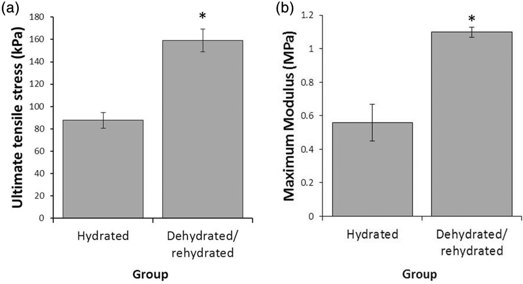
Effect of dehydration only on artificial sinew construct mechanics: (a) ultimate tensile stress and (b) maximum modulus values for sinew constructs following dehydration alone (n = 5/6 in each group).
Properties of sinew constructs following either dehydration and rehydration or no treatment (hydrated control).

p < 0.05 when compared to control for each experiment.
Rehydration following combined decellularisation and dehydration
To test the effectiveness of the full procedure (decellularisation, dehydration and rehydration), the properties of the sinew constructs were assessed by two methods: (1) the fluid uptake over time and (2) the mechanical properties after full rehydration. Decellularisation and dehydration of the sinew constructs resulted in a significant reduction in the fluid uptake of constructs when compared to their initial wet mass (Figure 5) with only 73.3 ± 8.3% of the wet mass reached when rehydrated in PBS and 86.1 ± 9.39% of their original wet mass when rehydrated in DMEM. Despite observing a difference between swelling behaviour in the two different media, there was no statistically significant difference between the PBS group and the DMEM group (p = 0.32).
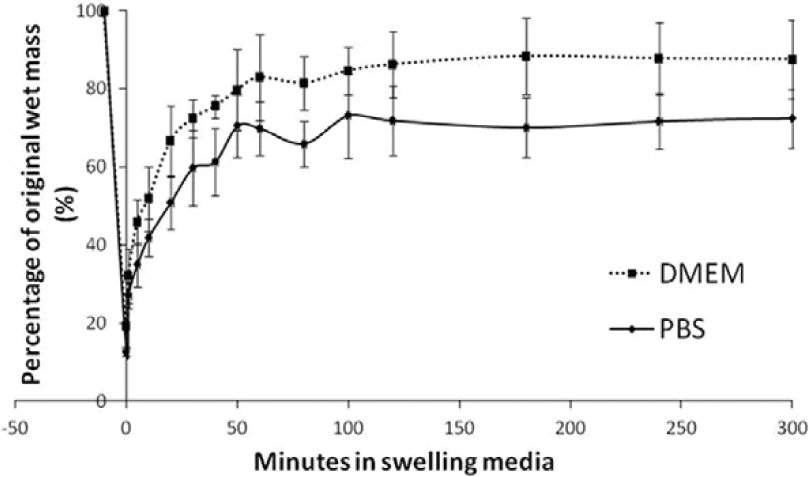
Fluid uptake in decellularised and dehydrated sinew constructs. Artificial sinew constructs subjected to decellularisation and dehydration were rehydrated in DMEM and PBS. Their wet mass was recorded at set time points up to 300 min (n = 6 in each group).
The mechanical properties of rehydrated sinew constructs that had been previously decellularised and dehydrated were assessed after rehydration in DMEM for only 3 h and after 1 week of rehydration in DMEM. Decellularising (DC) the sinew constructs and then dehydrating (DH) and rehydrating (RH) with DMEM for 3 h prior to testing resulted in no significant increase in maximum load (0.10 ± 0.02 to 0.18 ± 0.03 N, p = 0.115; Figure 6(a)), stress (65.02 ± 24.12 to 312.76 ± 72.14 kPa, p = 0.106; Figure 6(b)) or maximum modulus (0.18 ± 0.09 to 2.32 ± 0.97 MPa, p = 0.127; Figure 6(d)) when compared to sinew constructs that had not been subjected to any treatments. Although a trend to increasing mechanical properties was evident, none of these results was considered statistically significant when compared to the non-treatment group (p > 0.05 in all cases). Despite this, the trend was in agreement with the previous results and, importantly, suggested that the decellularisation-induced reduction in mechanical properties of sinew constructs (Figure 3(a) and (b)) is, in part, rescued when used in combination with dehydration. Furthermore, rehydration of sinew constructs in DMEM and then culturing for 1 week led to a trend to increase in maximum load (0.18 ± 0.02 to 0.23 ± 0.03 N), stress (312.76 ± 72.14 to 482.23 ± 91.43 kPa) and maximum modulus (2.32 ± 0.97 to 2.69 ± 0.64 MPa), although these were not considered statistically significant when compared to the group rehydrated 3 h prior to testing (p > 0.05 in all cases; Figure 6, Table 3). What is clear, however, is that the act of combined procedures of decellularisation, dehydration and rehydration in media resulted in sinew constructs that could withstand 2.6 times the maximum load (p = 0.013) and 8 times the stress (p = 0.023) of the sinew constructs without any treatments. This demonstrates a useful method of improving the strength of the constructs produced by our sinew model and also a potential method for producing bespoke patient-specific implants.

Effect of combined DC, DH and subsequent RH for 3 h or 1 week on artificial sinew construct mechanics: (a) maximum load, (b) ultimate tensile stress, (c) strain and (d) maximum modulus values for sinew constructs following the combined procedures of DC, DH and RH for either 3 h or 1 week prior to testing (n = 4/5 in each group).
Properties of sinew constructs subjected to DC, DH and RH for 3 h or 1 week prior to testing.

DC: decellularisation; DH: dehydration; RH: rehydration.
The non-treatment group did not experience any of the above procedures.
p < 0.05 when compared to control for each experiment.
Discussion
With tendon and ligament injuries frequently occurring in the young, active population, an important research focus is establishing a method whereby replacement tissues can be implanted quickly and with high functional success. Current methods of repair rely on the transplantation of autograft material from the patient, which can predispose the patient to further pain, donor site morbidity and infection. Allografting is also a possibility, especially with the rapid increase in the potential success of decellularised tissues and organs. Nevertheless, the availability of donor organs and overall suitability of xenograft transplantations remains a significant problem. To overcome these issues, we have investigated the use of the method of formation of tissue-engineered sinews in vitro, followed by decellularisation and dehydration procedures. The combination of these procedures allows a sinew template to be fabricated and stored, which then has the potential to be repopulated with an autologous cell population, providing an ‘autograft’ option for implantation following injury.
The choice of decellularisation regime used in this study was selected due to its previous success in decellularisation of flexor tendons without reducing their biomechanical properties and biocompatibility. 21 As anticipated, the use of this decellularisation regime resulted in a reduction in visible cell nuclei within the sinew-like construct body when compared to the control (non-decellularised) samples (Figure 2). However, decellularisation alone had marked effects on the mechanical properties of the engineered sinews, significantly reducing their maximum load, maximum stress and maximum modulus (Figure 3(a) and (b), Table 1). This was unexpected since previous work on native tendons did not report differences in mechanical properties following decellularisation. 21 There are many different methods of decellularisation reported in the literature, which use mechanical, chemical or enzymatic factors (or a combination of these) to remove cellular material from matrices. 10 For example, for tendons, other successful decellularisation protocols involve combinations of enzymes such as trypsin15,24,25 or nucleases14,15 or detergents such as SDS,14,15,21,22,26–28 EDTA,21,22,26 Triton-X21,24,25 or tri(n-butyl phosphate). 29 The use of mechanical disruption to aid cell removal can also be employed by techniques such as freeze–thawing, 15 agitation or mechanical force. 10 Therefore, there are many other options that could be used to decellularise the sinew constructs described here, which may not have such a detrimental effect on sinew mechanical properties. It is also important to note that despite the widespread use of SDS in decellularisation procedures, and many in vitro studies reporting no reduction in mechanical properties with native tendon tissue,15,21,28 an in vivo study that used SDS in their decellularisation regime has reported that the mechanical properties of the SDS-treated group were reduced following implantation. 27 This may be an important consideration in future work with this technique and may preclude the use of SDS if other options are available.
Although collagen content of our tissue-engineered sinew constructs was not measured here, previous work suggests that at the 3-week time point, sinew constructs contain approximately 10% endogenous collagen. 19 It is therefore assumed that a significant proportion of the resulting sinew construct remains as the initial fibrin gel. Since calcium has an important role in fibrin stabilisation and polymerisation of fibrinogen into fibrin, 30 it was thought that the use of EDTA (a calcium chelator) may result in the premature breakdown of the clot. However, seminal work by Okada and Blomback 31 states that while EDTA will reduce the clotting ability of fibrinogen into fibrin (by binding calcium), it has no effect on the structure of fibrin once clotted. Therefore, further work is needed to establish the mechanism behind the reduction in mechanical properties following decellularisation. Also, since we have previously reported the increase in collagen content of the tissue-engineered sinews with increasing culture time 19 and mechanical stimulation, 32 it is likely that a combination of these factors would result in a shift in the balance between endogenous collagen and original fibrin scaffold to a point where there is no noticeable effect on construct mechanical properties following decellularisation, as observed in native tendon structures. Achieving this step may also decrease the level of variability found within the heterogeneous structure at this stage.
Dehydration of the sinew constructs has several advantages. First, dehydration of the sinews allows a tissue scaffold to be stored, for future reconstitution with an autologous cell population. This means that the original sinews can be formed in culture, far in advance of being required, therefore significantly reducing the time required to produce a patient-specific implant. Second, the act of dehydration alone significantly improved the mechanical properties of the sinew constructs, resulting in an increase in the maximum stress and maximum modulus of the constructs (Figure 4, Table 2). From Figure 5, it can be seen that rehydration of the sinew constructs in DMEM resulted in sinew constructs weighing only 86.1 ± 9.39% of the original wet mass. This reduction in fluid content (Figure 5) and significant reduction in cross-sectional area (Table 2) are likely reasons for this positive change in mechanical properties. Furthermore, during dehydration, the polymer chains move closer together and begin to interact. Since the sinew constructs were not completely rehydrated (Figure 5), it is likely that this interaction remains and acts to stiffen the structure (Figure 4). Since our previously published work has highlighted the need to increase the mechanical properties of the sinews to achieve a level suitable for clinical implantation,18,32 this could be a promising method to further explore and may be an important factor to investigate across the wider tissue engineering field.
One of the most important findings in this study is that the decellularisation-induced reduction in mechanical properties was rescued when used in combination with dehydration (Figure 6). A trend to increasing maximum load, maximum stress and maximum modulus was observed where comparing the non-treated group to the decellularised, dehydrated and rehydrated group, which was rehydrated 3 h prior to tensile testing (Figure 6, Table 3), although none of these were considered statistically significant in this case. Furthermore, when the constructs were maintained in S-DMEM for 1 week, the mechanical properties improved further and resulted in a significant increase in the load and stress of the sinews when compared to the non-treated group (Figure 6, Table 3). This is a significant finding since it verifies that the rehydrated scaffolds are stable in culture, following the decellularisation and dehydration steps of the procedure. The next important steps are to characterise the re-seeding potential of these scaffolds and investigate the viability and matrix production of donor cells in the decellularised scaffolds.
To consider these constructs as potential tendon or ligament replacements, it is important to highlight some current limitations. For example, as a replacement for the ACL, replacement tissue would need to match the anatomical dimensions, which range from 22 to 41 mm in length and from 7 to 12 mm in width in humans. 33 At approximately 20 mm in length and only 1 mm in width, these constructs are currently too small to be considered as a replacement. Similarly, the mechanical properties of the constructs differ from human ACL mechanics, which possess failure stresses of approximately 13–14 MPa. 34 Despite this, we have previously manufactured a construct with dimensions more akin to the native tissue 18 but cost is the limiting factor with regard to manufacturing multiple constructs of this size for data collection. We are also currently investigating methods whereby we can reinforce the strength of the constructs to approach levels more appropriate for clinical use.
Although many groups have attempted the fabrication of acellular scaffolds for tendon regeneration with autologous cells, the majority of these have used either human cadaveric allografts14,22,26,35,36 or xenografts16,37,38 to manufacture the initial acellular scaffold. For this to become a realistic therapy for human tendon repair, there needs to be high availability of donor organs and tissues. The use of an in vitro cell-derived decellularised matrix is a relatively new technique, but has already found some use in the orthopaedic field. 39 Cell-derived decellularised matrices have been used to force the differentiation of stem cells down specific lineages for bone40 –42 and to culture cartilage cell types43,44 with promising results. To our knowledge, the work we have described here is the first report of using a tissue engineering approach to manufacture a scaffold from a cell population to then remove the initial cell population to produce templates for tendon and ligament tissue regeneration with autologous cells. This proposed method will greatly reduce the time taken to generate bespoke implants for patients since the scaffold will already be produced and stored ready for recellularisation with patient-specific cells. This has far-reaching implications for tendon and ligament regeneration and also as an approach in the manufacture of acellular scaffolds for the regeneration of other tissues and organs. Furthermore, the fact that the dehydration process also leads to a significant improvement in the mechanical properties of the scaffolds is a finding that may have significant impact across the wider tissue engineering community.
Footnotes
Acknowledgements
The authors thank Dr K. Baar for the provision of CTF cells used in the study.
Declaration of conflicting interests
The authors declare that there is no conflict of interest.
Funding
The authors would like to thank Orthopaedic Research UK (project # 497) and the Biotechnology and Biological Sciences Research Council (BBSRC project # BB/G022356/1) for funding this work. The Instron microtester used in this research was obtained through Birmingham Science City: Innovative Uses for Advanced Materials in the Modern World (West Midlands Centre for Advanced Materials Project 2), with support from Advantage West Midlands (AWM) and part funded by the European Regional Development Fund (ERDF). C.L. was supported by an internship from the ISIFC.