
Abstract
Purpose
This study aimed to establish a combined histological assessment system of neo-cartilage outcomes and to evaluate variations in an established rat defect model treated with human juvenile cartilage-derived chondrocyte (JCC) sheets fabricated from various donors.
Methods
JCCs were isolated from the polydactylous digits of eight patients. Passage 2 (P2) JCC sheets from all donors were transplanted into nude rat chondral defects for 4 weeks (27 nude rats in total). Defect-only group served as control. Histological samples were stained for safranin O, collagen 1 (COL1), and collagen 2 (COL2). (1) All samples were scored, and correlation coefficients for each score were calculated. (2) Donors were divided into “more effective” and “less effective” groups based on these scores. Then, differences between each group in each category of modified O’Driscoll scoring were evaluated.
Results
(1) Modified O’Driscoll scores were negatively correlated with %COL1 area, and positively correlated with %COL2 area and COL2/1 ratio. (2) Four of 8 donors exhibited significantly higher modified O’Driscoll scores and %COL2 areas. JCC donors were divided into two groups by average score values. Significant differences between the two groups were observed in modified O’Driscoll categories of “Nature of predominant tissue,” “Reconstruction of subchondral bone,” and “Safranin O staining.”
Conclusion
The combined histological evaluation method is useful for detailed in vivo efficacy assessments of cartilage defect regeneration models. Variations in histological scores among juvenile cartilage-derived chondrocyte donors were correlated to the quality of regenerated cartilage hyaline structure and subchondral bone remodeling observed in the nude rat defect model.
Keywords
Introduction
Chondral defects in human knees are prevalent in both general and athletic populations from multiple etiologies. Frequently incurred with osteological disorders from acute trauma, increasing participation in athletic activities is especially associated with increasing prevalence.1,2 Articular cartilage defects have a limited capacity to regenerate after injury and may lead to accelerated wear, worsening pain, and potential arthritis progression.3-5 Many methods have been described to treat chondral defects in the knee: microfracture or drilling,6-8 osteochondral autograft transfer (OAT),9-11 osteochondral allograft (OCA),12-14 autologous chondrocyte implantation (ACI), matrix-assisted autologous chondrocyte implantation (MACI),15-17 and particulated juvenile allograft.18-20 However, fibrocartilage after microfracture, 21 fragmentation and resorption of allografts after OAT and OCA, and disturbed fusion of the regenerative cartilage and the healthy surrounding cartilage after ACI and MACI have been reported, while low product scalability is another problem. 22
Scaffold-free cell-dense implants, termed “cell sheets,” are routinely fabricated using temperature-responsive cell culture surfaces (
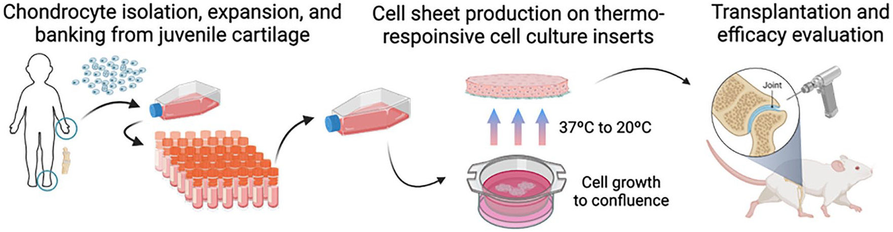
Schema of the experiment. Chondrocytes were expanded from an established JCC bank, cultured in thermo-responsive cell culture inserts, and transplanted into nude rats. Created with BioRender.com.
Allogenic cell source variability is known to affect cell-based therapeutic results. 31 Differences in secreted paracrine regenerative and immuno-modulatory factors from mesenchymal stromal cell (MSC) and JCC sheets derived from different human donors have been reported.30,32,33 These data suggest that JCC sheet characteristics relevant to their regenerative potency are affected by variations in donors. To improve JCC sheet therapeutic efficacy and reliability, extents to which therapeutic effects vary with human cell sourcing must be determined, and key factors that influence this donor variation must be elucidated to optimize donor selection criteria using relevant animal models.
Histological analysis of cartilage tissue to assess structural and biomolecular changes in animal defect models remains a primary strategy to describe regenerative progress in injured cartilage during treatment and attempt to extrapolate treatment results to clinical feasibility. O’Driscoll scoring was established to assess cartilage repair in rabbit models. 34 This score has frequently been used in modified versions 33 ( Table 1 ) and remains among the few scoring systems in which integration of the repair tissue with native cartilage is assessed. 36 Structural characteristics, for example, subchondral bone assessments, are also included in one of O’Driscoll score categories. Separate analyses of in situ changes of key collagen expressions by immunohistochemistry are more sensitive and accurate for specific healing-related markers. 37 Therefore, combinations of these assessments are expected to better stratify in vivo JCC sheet implantation results based on multi-component assessments of cartilage regeneration.
Modified O’Driscoll Score.

The purpose of this study was 3-fold: (1) to stratify observed human JCC sheet in vivo therapeutic effects from an established nude rat cartilage defect-implant model using modified O’Driscoll scoring and specific collagen immunohistochemistry (IHC), (2) to examine relationships between these in vivo scores, and (3) to identify specific characteristics that define significant in vivo differences in cartilage regeneration using different human donors.
Materials and Methods
Cartilage Sourcing
Primary human cartilage cells were isolated from 8 amputated polydactylous fingers and a toe from 8 juvenile patients aged between 6 and 30 months (5 males, 3 females; 14.6±6.9 months; Caucasian; Suppl. Table S1). These patients underwent polydactyly surgery, and tissue discards from these surgeries were collected at Intermountain Primary Children’s Hospital (Salt Lake City, USA). Institutional Review Board oversight from the University of Utah and Intermountain Primary Children’s Hospital was waived due to the use of de-identified routine surgical discards for research purposes.
Fabrication of Juvenile Chondrocyte Sheets
Cartilage tissues from polydactyly surgical discards were dissected, enzymatically digested, and cultured for cryogenic cell banking, and JCC sheets were fabricated according to methods described by Kondo et al. 27 and as summarized below. Briefly, human cartilage tissue was cut into <4 mm2 pieces by scalpel and incubated with 5 mg/mL Type 1 collagenase (LS004197, Worthington Biochemical, Lakewood, USA) at 37°C for 1.5-3.0 h. Resulting cells were filtered through a 100-µm cell strainer, washed with saline, and then resuspended in chondrocyte culture medium (DMEM-F12, 11320082, ThermoFisher Scientific, Waltham, USA) containing 1% antibiotic-antimycotic (15240062, ThermoFisher) and 20% fetal bovine serum (FBS) (16000044, ThermoFisher). Isolated chondrocytes were seeded at 5,000–10,000 cells/cm2 on polystyrene dishes (CELLTREAT, Pepperell, USA) in chondrocyte culture medium. At the first medium change on day 4, 100 μg/mL L-ascorbic acid phosphate magnesium salt n-hydrate (013-19641, Fujifilm Wako Pure Chemical, Osaka, Japan) was added and the medium was replaced. Cells were passaged with this medium thereafter. Sub-confluent cells were suspended in STEM-CELLBANKER GMP grade media (Zenoaq, Fukushima, Japan) and cryopreserved at the end of P0. Serial subculture was performed with thawed cells at an initial density of 10,000 cells/cm2 passaged every 3-5 days. To create passage 2 (P2) JCC sheets, P1 thawed cryopreserved cells were seeded and sub-confluent P1 cells were collected, then seeded at a density of 10,000 cells/cm2 on 23-mm diameter temperature-responsive cell culture inserts (CellSeed, Tokyo, Japan). Cell culture medium was changed every 2-4 days. After 2 weeks of culture, confluent cell sheets were harvested with forceps after incubation at room temperature. P2 JCC sheets from all human donors were used for in vivo transplantation as described below.
Nude Rat Cartilage Defect Model and Human Cell Sheet Transplantation
All in vivo implant procedures were approved by the Institutional Animal Care & Use Committee (IACUC, University of Utah, assigned ID: 20-12001). A total of 30 nude rats (6-week-old), both male and female, were purchased from Charles River Laboratories (Wilmington, USA). After a week of acclimatization at the animal facility, rats were randomized to each experimental condition by comparable body weights. Under anesthesia using isoflurane and O2 gas, medial parapatellar incision was made on the right knee, and the patella was laterally dislocated. A focal chondral defect (diameter 2 mm; depth 300-400 μm) was created on the patellofemoral groove using an electric grinder with minimal damage to the subchondral bone. We have previously published histological findings immediately after defect creation, confirming the accuracy of this technique.
29
Freshly prepared JCC sheets were washed with saline, then cut in half with a sterile razor blade, and single sheet halves were transplanted into the defect (
Transplantation of human juvenile cartilage-derived chondrocyte (JCC) sheets on nude rat knee cartilage defect model. (
Harvested rat knee tissue was fixed in 4% paraformaldehyde for 4 days and decalcified in RapidCal Immuno (BBC Biochemical, Mount Vernon, USA) for 1 day at RT. Samples were embedded in paraffin blocks and then cut into 5-µm transverse sections with a microtome. P2 JCC cell sheets from donor 1 were implanted in 3 female rats, cell sheets from donor 2 into 2 male and 1 female rat, cell sheets from donor 3 into 3 female rats, cell sheets from donor 4 into 3 male rats, cell sheets from donor 5 into 3 male and 3 female rats each, and cell sheets from donor 6, 7, and 8 into 3 male rats. Defect models served as controls using 3 male rats.
Histological Evaluation of Harvested Cartilage
Allocation and assessment were done by multiple investigators for randomization. Histological samples from each rat defect site were prepared separately for safranin O, collagen type 1 (COL1), collagen type 2 (COL2), and human-specific vimentin (hVIM) staining. Slides were deparaffinized by baking in an oven at 65°C and subsequent washes with xylene and ethanol. Safranin O staining was conducted using standard methods. 38 Briefly, samples were stained for 5 min with Weigert’s Iron Hematoxylin (MilliporeSigma), 5 min with 0.5 g/L Fast Green (MilliporeSigma), and 5 min with 0.1% Safranin O (MilliporeSigma). After safranin O staining, slides were randomized and scored with modified O’Driscoll scoring, and the observer was blinded regarding JCC donor information.
For immunostaining, histology samples were rehydrated and protease K (S3020, Agilent Technologies, Santa Clara, USA) was used for antigen retrieval for COL2 staining. Peroxidase blocking was performed with 3% hydrogen peroxide (216763, MilliporeSigma). After blocking with 5% donkey serum and 0.1% Triton-X in PBS for 1 h, samples were treated with primary antibodies to COL1, COL2, and hVIM at 4°C overnight. Polyclonal goat anti-COL1 (1:200, SouthernBiotech, Birmingham, USA), monoclonal mouse anti-COL2 (1:200, 2B1.5, ThermoFisher, USA), and monoclonal rabbit anti-hVIM (1:200, SP20, Abcam) were used as primary antibodies. Normal goat IgG (1:50, NI02, MilliporeSigma), normal mouse IgG2a (1:100, X0943, Agilent), or normal rabbit IgG (1:1,500, X0903, Agilent) were used as isotype controls. Horseradish peroxidase (HRP)-conjugated donkey anti-goat antibody (1:1,000, 705-035-147, Jackson ImmunoResearch, West Grove, USA) was used for type 1 collagen. HRP-conjugated goat anti-mouse antibody (1:1,000, 115-035-166, Jackson) was used for type 2 collagen. HRP-conjugated goat anti-rabbit antibody (1:1,000, 111-035-144, Jackson) was used for hVIM staining. ImmPACT DAB Peroxidase (HRP) Substrate (SK-4105, Vector Laboratories, Burlingame, USA) was used as a chromogen. Brightfield images were taken with a BX41 microscope and processed with AmScope Software. For quantitative IHC, the positive cell area across the entire cell sheet was measured and expressed as percent using ImageJ software (NIH) (%COL1 and %COL2 areas,
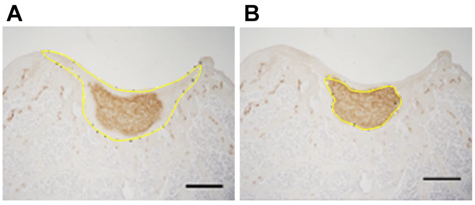
Analysis of image stained with anti-type 2 collagen antibody using Image J. The area of the entire implanted cell sheet regenerative area (
Correlation coefficients for each modified O’Driscoll score and %COL1 area, %COL2 area, and COL2/1 ratio were calculated. For donors treated with P2 JCC sheets, scores were compared between donors and the defect-only group and divided into “more effective” and “less effective” groups based on average scores. Significant differences between each group and the defect-only group in each category of modified O’Driscoll score were evaluated. A summary of the methodological process is described in Figure 4 .
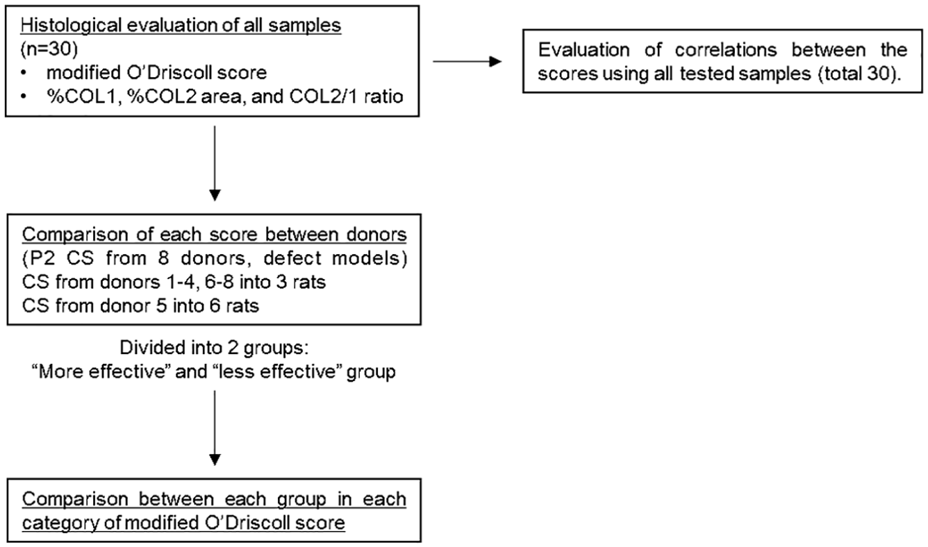
Study methodology. P2 cell sheets (CS) from donors 1-4, 6-8 were transplanted into 3 rats and CS from donor 5 into 6 rats. 3 defect models were also prepared as a control. Pearson’s correlation coefficient was used to evaluate the correlations between scores. ANOVA with post hoc Tukey-Kramer testing was used to compare each score between donors and each category of modified O’Driscoll score between groups.
Statistical analysis
Numerical results are expressed as a mean and standard deviation. Pearson’s correlation coefficient was used to identify significant relationships between modified O’Driscoll scores and %COL1 area, %COL2 area, and COL2/1 ratios. ANOVA with post hoc Tukey-Kramer testing was used to identify significant differences between donors in each score and between groups in each category of modified O’Driscoll score. All tests were performed at a significance level of p
Results
Assessment of in vivo cartilage defect regeneration capacity is essential to improve cartilage regenerative medicine products. We employed a published nude rat knee cartilage defect model
29
as a platform to implant and test human juvenile chondrocyte sheets. JCC-regenerated histological samples were confirmed positive for hVIM immunostaining,
29
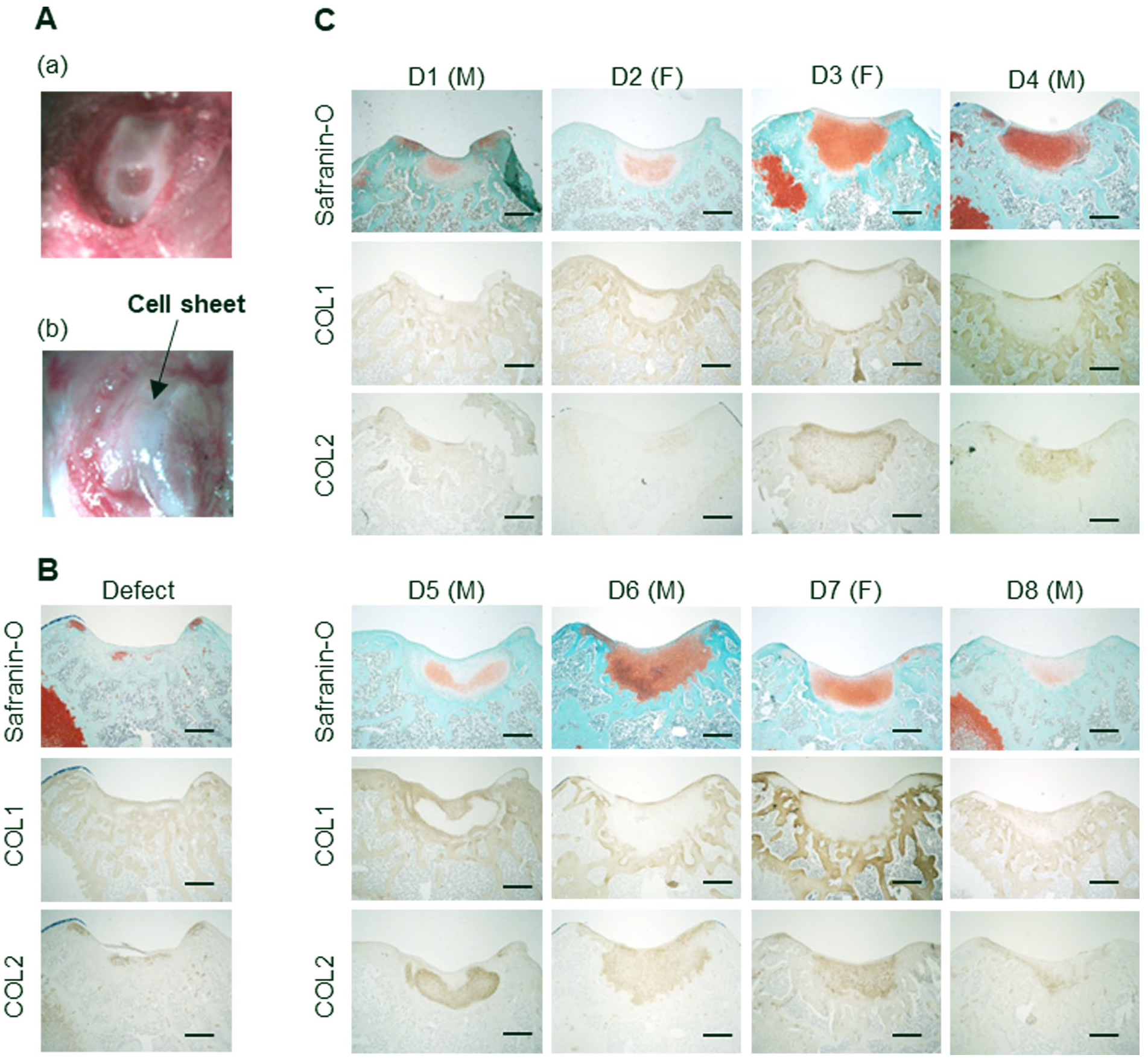
supporting that new cartilage originates from implanted JCC human cells (Suppl. Fig. S1). Representative histological samples are shown in
Histological Scores and Their Correlations in the Cell Sheet Regeneration Model
Modified O’Driscoll scores, %COL1 and %COL2 areas, and COL2/1 ratios varied among the 8 donor JCC sheet implant groups (
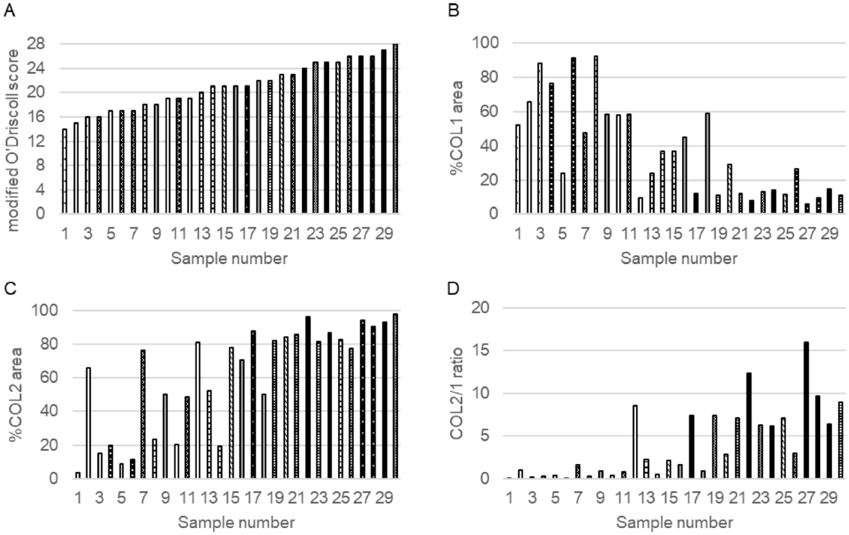
Scores of each system in all samples. The scores were arranged in order of modified O’Driscoll score. Bars sharing the same pattern represent samples from the same donor. Sample numbered 1, 3, and 5 represent the defect groups. (
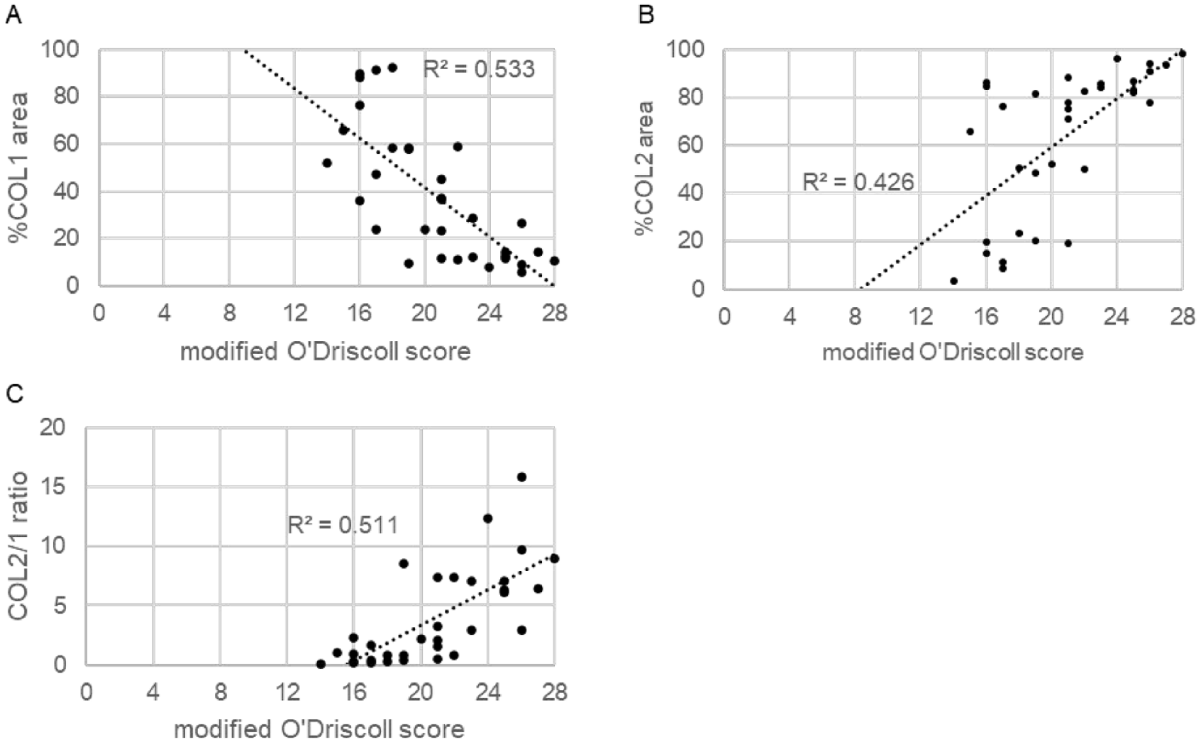
Scatter plot of the correlation between modified O’Driscoll score and %COL 1 area (
Comparisons of modified O’Driscoll score, %COL1 area, %COL2 area, and COL2/1 ratios between JCC donors
Four human donor-derived JCC sheet-transplanted groups showed significantly higher modified O’Driscoll scores compared to the defect-only group (p<0.01). %COL1 area was not significantly different from control due to high intrinsic variation within this assessment. %COL2 area of donor 3 was significantly higher than that of donor 8 and the defect-only group (p=0.047 and p<0.01, respectively), and those of donors 4, 6, and 7 were significantly higher than that of the defect-only group (p<0.01). The COL2/1 ratio has been used for assessing cartilage matrix quality in previous gene expression assays.
40
In our study, the COL2/1 ratio distinguishes donor matrix differences; for example, the ratio for donor 6 was significantly higher than that of donors 2, 5, 8, and the defect-only group (p<0.01); donor 3 and 4 were both significantly higher than that of the defect-only group (p<0.05). Using these assessments and histological scoring, JCC donors were divided into two groups: groups 3, 4, 6, and 7 (group “more effective”) and groups 1, 2, 5, and 8 (group “less effective”) (
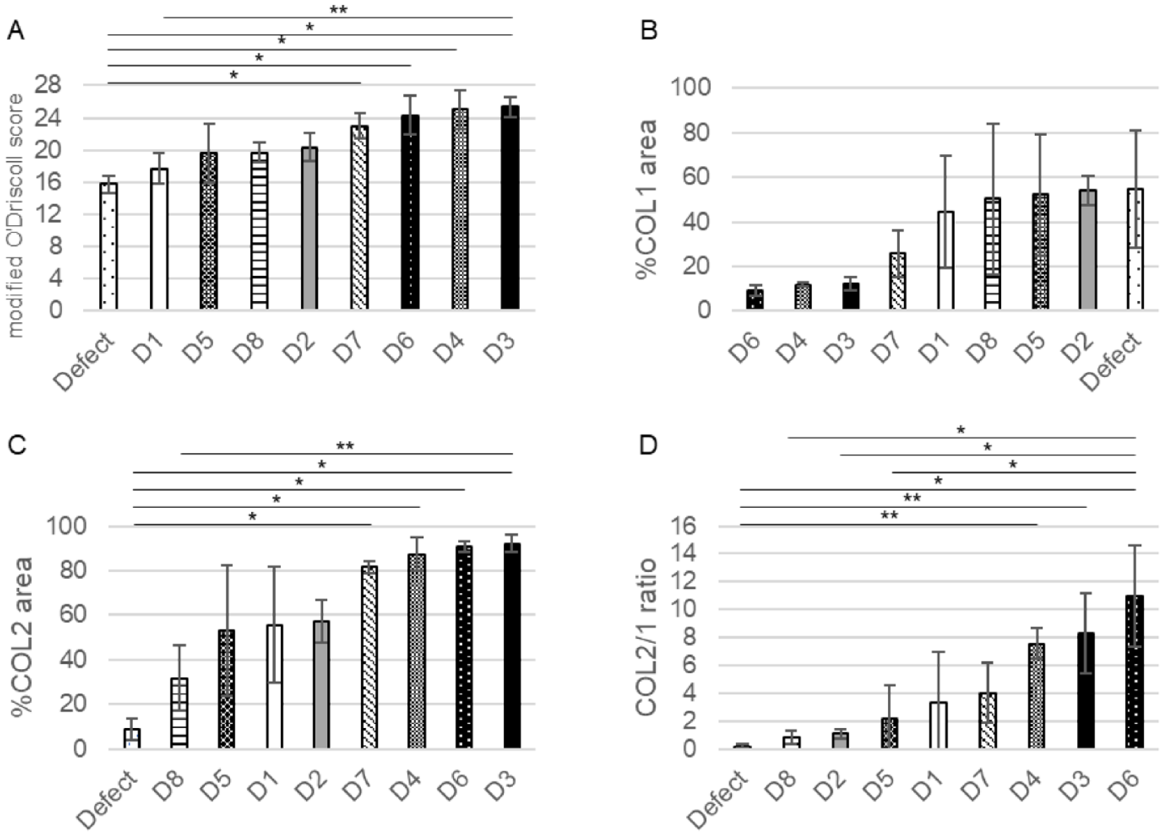
Comparison of each score between groups. (
Comparing each group in each modified O’Driscoll score evaluation category
To further identify differential regenerative effects by JCC sheets from 8 donors in nude rat cartilage defects, modified O’Driscoll score subcategories were compared between knees bearing “more effective” sheet implants, “less effective” sheet implants, and defect-only group. Significant differences between group “more effective” and group “less effective” were observed in the modified O’Driscoll score categories of “Nature of predominant tissue” (I), “Reconstruction of subchondral bone” (V-A), and “Safranin O staining” (VI) (p<0.01). No significant differences were seen between these two groups in the categories “Structural characteristics” (II), “Freedom from cellular changes of degeneration” (III), and “Freedom from degenerative changes in adjacent cartilage” (IV) (
Discussion
Development of improved, validated, and scalable cartilage regeneration strategies remains an urgent clinical challenge. Assessment of reliable human cell-derived regenerative approaches is integral to assuring progress. The potential for human juvenile cartilage as an allogeneic cell source for regenerating cartilage is long recognized.41-43 Although multiple histological scoring systems have been introduced as part of regenerative outcomes analysis,
44
correlations of these systems to human cell-based approaches are not fully investigated. Modified O’Driscoll scoring is widely used to assess in vivo cartilage repair in defect models and has been reported to be superior to other scores in terms of coefficient of repeatability.34,45,46 This study showed variations in O’Driscoll scores, %COL1-and %COL2-positive stained defect histological section areas, and COL2/1 ratios for human JCC sheets derived from various donors in an established nude rat defect model.
29
Modified O’Driscoll scores correlated negatively with %COL1 areas and positively with %COL2 areas and COL2/1 ratios (see
In contrast, our in vivo data with human juvenile-sourced cell sheet treatment shows >80% COL2 and ~10% COL1 stained areas after 4 weeks of treatment with “more effective” donors in our model (
Based on the scoring shown, juvenile donors could be divided into “more effective” and “less effective” groups (
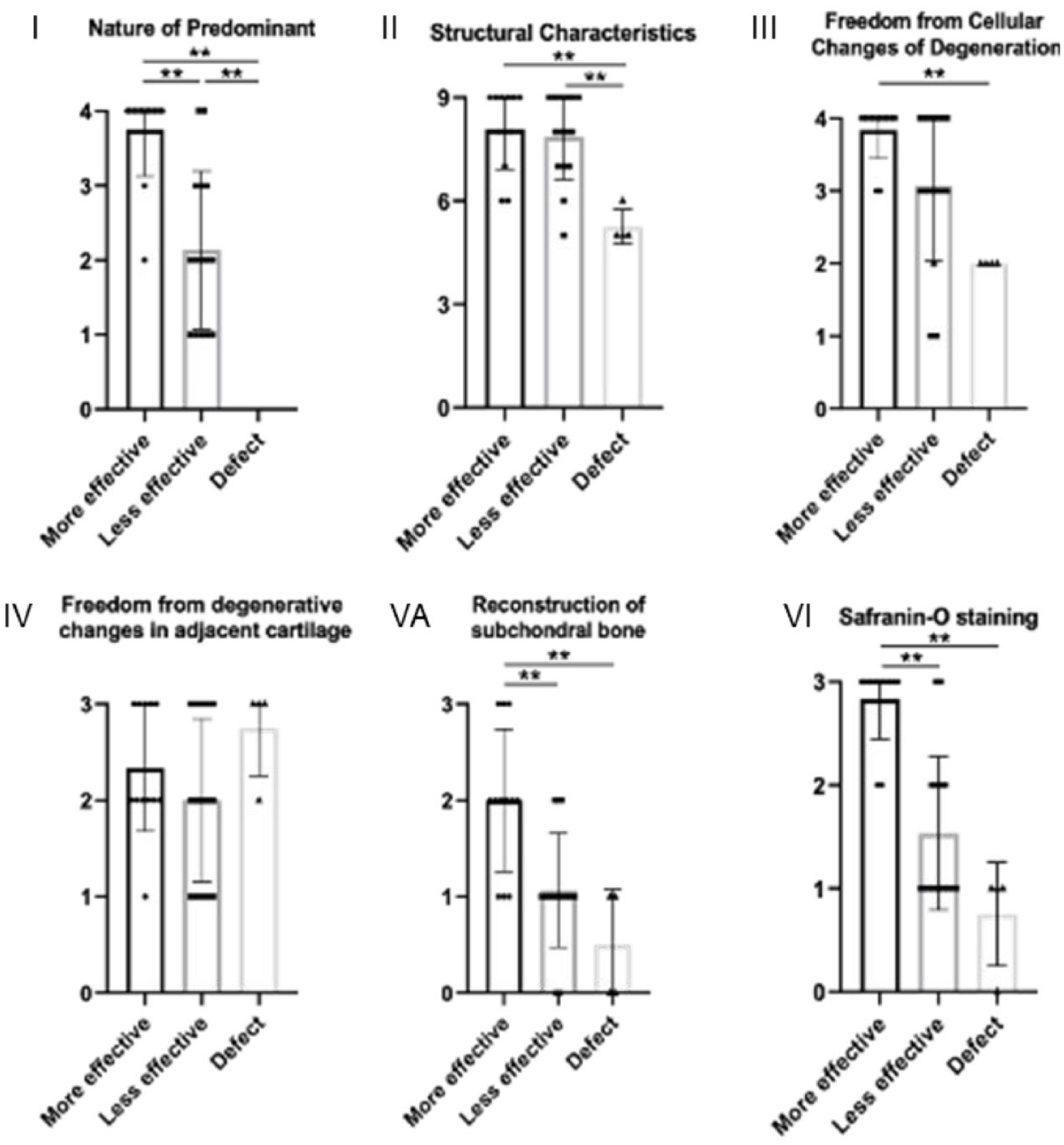
Each category of modified O’Driscoll score in each group. (1) Nature of predominant, (II) Structural characteristics, (III) Freedom from cellular changes of degeneration, (IV) Freedom from degenerative changes in adjacent cartilage, (VA) Reconstruction of subchondral bone, and (VI) Safranin O staining. “More effective” group consisted of donors 3, 4, 6, and 7. “Less effective” group consisted of donors 1, 2, 5, and 8. *<0.01.
Safranin O staining showed structures that appear to be blood vessels in the subchondral layer in the “less effective” group at the 4-week time point, not observed in the “more effective” group at 4 weeks (Suppl. Fig. S2A and B). Angiogenesis is a necessary process for bone remodeling, and its presence and staging in subchondral bone in joints with OA is recognized. 51 Thus, subchondral bone remodeling and associated neovascular dynamics post-injury may be completed earlier in the “more effective” group than in the “less effective” group as part of defect repair. While details to describe the defect healing process within 4 weeks remain tentative, at 2 weeks post-implantation with a “more effective donor,” blood vessels are found in the subchondral bone and subchondral bone remodeling is not yet completed (Suppl. Fig. S2C).
The scoring category “Structural characteristics” produced high scores in sheet-transplanted groups (see
Analogous allogenic cell sheet transplantation studies can be performed in outbred animals, but cell properties, especially chondrogenic potential, are highly variable among different animal species. 54 Hence, predictive outcomes extrapolated to human potential will be difficult and speculative without direct correlations. The athymic rat model employed here enables the safety and efficacy testing of human cell products in the in vivo knee synovial articulating joint environment. A rabbit xenotransplantation model using immunosuppressing drugs is also reported for this purpose, although acute pharmacological immuno-suppression could affect natural healing cascades. 55 Future studies should consider intrinsic limitations of these xenogeneic models: biomechanical loading of the articulating surfaces and its influences on cartilage healing are distinct between species, healing processes are likely distinct from that of allogeneic transplants, and essential involvement of all host immune cells in cartilage repair is lacking in immune-incompetent models. Nonetheless, we believe that the nude rat cartilage implant model employed here provides useful mechanistic and potency screening of human cell transplantation products for cartilage regeneration. Host immune response to allogeneic JCC sheet transplantation in inflamed and post-surgery conditions to validate engraftment, safety, and efficacy of this potent human juvenile cell-based therapy is warranted.
Conclusions
This study establishes a combined histological assessment system for human cell products for cartilage repair in a nude rat knee cartilage defect model. 29 The system stratifies regenerative effects of human juvenile cartilage-derived chondrocyte (JCC) sheets derived from various juvenile donors. Scoring differences identified in histological evaluations of various juvenile JCC donor cartilage repair potentials are attributed to safranin O staining and subchondral bone remodeling. Most JCC sheet implant outcomes exhibit excellent “Structural characteristics” scoring in this model. The model outcomes extend and support previous work, 29 demonstrating the benefits of human juvenile chondrocyte sourcing to yield potent cell sheet constructs that spontaneously adhere to chondral defects and rapidly integrate to regenerate full thickness hyaline-like cartilage. While allogeneic transplantation studies are vital for evaluating immune response and rejection, leveraging this implant regenerative model to screen human cell sources for critical quality attributes is essential for product development. This process enables reliable cartilage regeneration and facilitates donor selection, banking, scaling, and achieving the desired cost-effectiveness for allogeneic living cartilage implants.
Supplemental Material
sj-docx-1-car-10.1177_19476035241277946 – Supplemental material for Regenerative Variability of Human Juvenile Chondrocyte Sheets From Different Cell Donors in an Athymic Rat Knee Chondral Defect Model
Supplemental material, sj-docx-1-car-10.1177_19476035241277946 for Regenerative Variability of Human Juvenile Chondrocyte Sheets From Different Cell Donors in an Athymic Rat Knee Chondral Defect Model by Keisuke Matsukura, Makoto Kondo, Nicolas F. Metzler, Adam J. Ford, Travis G. Maak, Douglas T. Hutchinson, Angela A. Wang, Masato Sato, David W. Grainger and Teruo Okano in CARTILAGE
Supplemental Material
sj-docx-2-car-10.1177_19476035241277946 – Supplemental material for Regenerative Variability of Human Juvenile Chondrocyte Sheets From Different Cell Donors in an Athymic Rat Knee Chondral Defect Model
Supplemental material, sj-docx-2-car-10.1177_19476035241277946 for Regenerative Variability of Human Juvenile Chondrocyte Sheets From Different Cell Donors in an Athymic Rat Knee Chondral Defect Model by Keisuke Matsukura, Makoto Kondo, Nicolas F. Metzler, Adam J. Ford, Travis G. Maak, Douglas T. Hutchinson, Angela A. Wang, Masato Sato, David W. Grainger and Teruo Okano in CARTILAGE
Footnotes
Acknowledgments and Funding
The author(s) disclosed receipt of the following financial support for the research, authorship, and/or publication of this article: This research was supported in part by the University Technology Acceleration Grant (UTAG) from the Utah Science, Technology and Research (USTAR) program to T.O. and D.W.G. and American Orthopedic Society for Sports Medicine (AOSSM)-JRF Allograft Research Grant to M.K.
Declaration of Conflicting Interests
The author(s) declared the following potential conflicts of interest with respect to the research, authorship, and/or publication of this article: Teruo Okano is a shareholder of CellSeed, Inc. and is an inventor/developer designated on the patent for CellSeed’s commercialized thermos-responsive cultureware. Travis Maak discloses the following possible competing interest: Arthrex Inc. (paid speaker and consultant). All other authors declare no competing interests.
Ethical Approval
All in vivo implant procedures were approved by the Institutional Animal Care & Use Committee (IACUC) at the University of Utah (ID: 20-12001). For the use of human tissue, Institutional Review Board (IRB) oversight from both the University of Utah and Intermountain Primary Children’s Hospital was waived, as the research involved de-identified routine surgical discards.
Data Availability
Data sets generated and analyzed during the current study are available from the corresponding author upon reasonable request.
References
Supplementary Material
Please find the following supplemental material available below.
For Open Access articles published under a Creative Commons License, all supplemental material carries the same license as the article it is associated with.
For non-Open Access articles published, all supplemental material carries a non-exclusive license, and permission requests for re-use of supplemental material or any part of supplemental material shall be sent directly to the copyright owner as specified in the copyright notice associated with the article.