
Abstract
The working group, “Clinical Tissue Regeneration” of the German Society of Orthopedics and Traumatology (DGOU) issues this paper to update their guidelines.
Methods
Peer-reviewed literature was analyzed regarding different topics relevant to osteochondral lesions of the talus (OLTs) treatment. This process concluded with a statement for each topic reflecting the best scientific evidence available for a particular diagnostic or therapeutic concept, including the grade of recommendation. Besides the scientific evidence, all group members rated the statements to identify possible gaps between literature and current clinical practice.
Conclusion
In patients with minimal symptoms, OLT progression to ankle osteoarthritis is unlikely. Risk factors for progression are the depth of the lesion on MRI, subchondral cyst formation, and the extent of bone marrow edema. Conservative management is the adaptation of activities to the performance of the ankle joint. A follow-up imaging after 12 months helps not to miss any progression. It is impossible to estimate the probability of success of conservative management from initial symptoms and imaging. Cast immobilization is an option in OLTs in children, with a success rate of approximately 50%, although complete healing, estimated from imaging, is rare. In adults, improvement by conservative management ranges between 45% and 59%. Rest and restrictions for sports activities seem to be more successful than immobilization. Intra-articular injections of hyaluronic acid and platelet-rich plasma can improve pain and functional scores for more than 6 months. If 3 months of conservative management does not improve symptoms, surgery can be recommended.
Keywords
Introduction
Osteochondral lesions of the talus (OLTs) affect the talar dome with varying involvement of the articular cartilage and subchondral bone. In 2017, the working group “Clinical Tissue Regeneration” of the German Society of Orthopedics and Traumatology (DGOU) published the first recommendation for treating OLTs. 1 Much further research has been done within the last 5 years. The rationale behind this update was to include recent results and the latest knowledge on the treatment algorithms and update the guidelines with the new literature. Due to a lack of evidence, in 2017, many of the recommendations were based on expert opinion. Meanwhile, more concepts are supported by an increasing number of scientific studies. Besides the continuous discussion within the working group, the development was also driven by several consensus meetings, including the “International Consensus Meeting on Cartilage Repair of the Ankle” which took place in Pittsburgh in 20172-12 and Dublin in 2019. 13
The working group on “Clinical Tissue Regeneration” of the DGOU issues the present paper. It represents the best evidence available for managing OLTs and updates the guidelines published in 2017. 1 This paper focuses on the etiology, classification, diagnostics, and conservative management of OLTs. Abbreviations are defined in Table 1 .
Abbreviations and Definitions.
Method
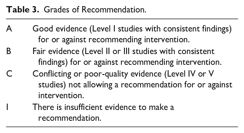
The working group “Clinical Tissue Regeneration” of the DGOU brought together 60 orthopedic and trauma surgeons with a particular interest in treating articular cartilage lesions. According to their subspeciality, a subgroup of 29 focused on foot and ankle surgery. Under the leadership of the first author, literature was analyzed regarding different topics relevant to OLT treatment (PUBMED, Cochrane, Web of Science, Scopus, MEDLINE, University Library Munich). The following keywords were used in combination: ankle, talus, cartilage, damage, chondral, osteochondral, articular, injury, chondropathy, focal, defect, pain, classification, platelet-rich plasma (PRP), hyaluronic acid (HA), arthritis, degenerative, clinical examination, imaging, MRI, computed tomography (CT), weightbearing computed tomography (WBCT), single photon emission computed tomography in combination with a computed tomography scan (SPECT-CT), x-ray, progression, injection, immobilization, shock wave, extracorporeal shockwave therapy (ESWT), and magnetic field. Papers were collected for each topic, and the main conclusions were brought together. This process concluded with a statement for each topic reflecting the best scientific evidence available for a particular diagnostic or therapeutic concept. The level of evidence for the studies was analyzed, and a grade recommendation was given for each statement ( Tables 2 and 3 ).14 -16
Grades of Evidence.
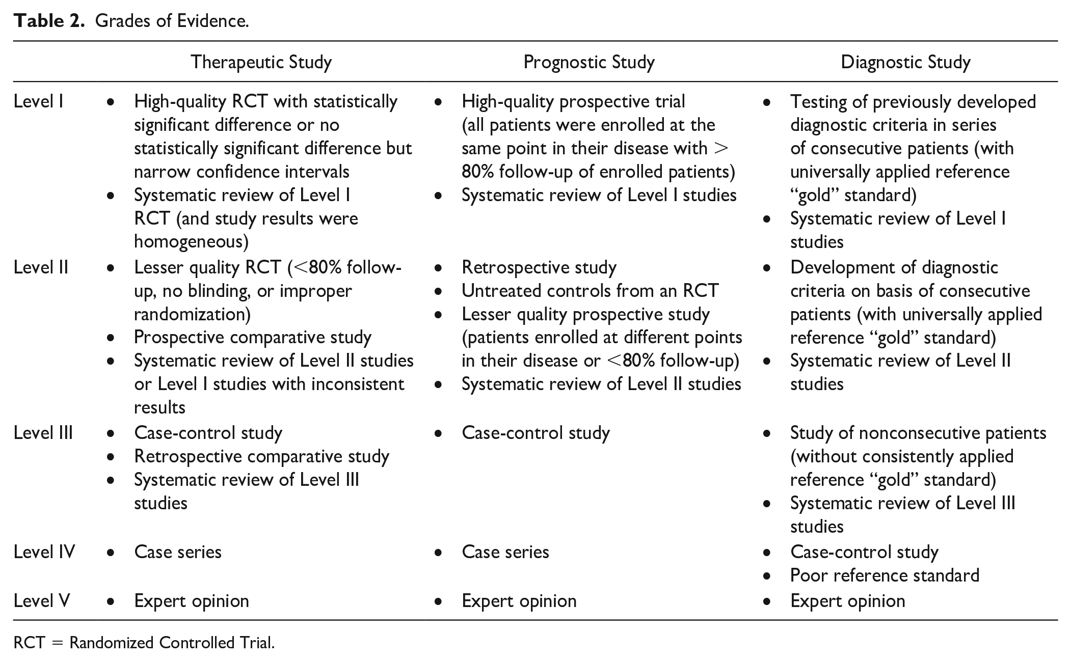
RCT = Randomized Controlled Trial.
Grades of Recommendation.
In the second step, the group members were asked to rate the different statements according to their clinical practice. The goal was to identify possible gaps between clinical experience and evidence in the literature. Blinded electronic surveys were distributed to all group members. The participants could agree or disagree with the statements, comment on the statements, and provide additional references. Based on the participants’ input, statements were revised if additional literature was provided and sent for a second vote. The process ended with a statement on the different topics, based on the best evidence available, together with a grade of recommendation based on the quality of the studies supporting each statement. In addition, agreement among the 29 experts was given, reflecting the current clinical practice and experience. 17
Etiology and Location
OLT summarizes the large variety of pathologies at the talus. As the abbreviation implies, the constant factors are bone and cartilage involvement. However, there is no single etiology of OLTs. In the active pediatric and young adult population, osteochondritis dissecans is the predominant presentation. It is considered an atraumatic, idiopathic phenomenon. Etiopathogenetic theories include local ischemia, aberrant endochondral ossification of the secondary subarticular physis, repetitive microtrauma, and genetic predisposition.18,19
In the adult population, OLTs can be caused by avascular necrosis, 20 systemic vascular diseases, trauma,21,22 chronic microtrauma, 23 endocrine or metabolic factors, 24 Vitamin D deficiency, 25 degenerative joint disease, 26 and malalignment. 27 There seems to be also a genetic predisposition, as some patients present nearly identical pathologies in both ankle joints.28 -30 Flick and Gould 23 reported a history of trauma in 98% of the lateral and 70% of the medial lesions.
van Dijk et al. 31 hypothesized the progression of a cartilage lesion to a subchondral cyst. Elias et al. 32 investigated data from 424 patients characterizing the location of the OLT and reported that 96.2% of all lesions affected either the medial (62.8%) or the lateral (33.4%) talar dome, with 53% of the lesions located at the central third of the medial talar dome.
There is only a minor focus on different etiologies in papers dealing with OLT treatment. If an underlying cause can be improved, it should be included in the treatment concept.
Statement: The causes of OLTs are diverse. The predominant location is the central third of the medial talar dome. Many of the causative factors cannot be changed. However, remediating any causative factors should be part of the treatment concept, if possible.
Grade of Recommendation: B
Diagnostics
Clinical Examination
Typically, the patients report deep pain in the ankle joint, usually increasing with activity. They may present with swelling and tenderness to palpation. The location of the pain is not necessarily related to the location of the OLT. Pain is typically felt in the ankle joint during or after weightbearing.12,33,34 Especially in unstable lesions, locking of the joint can also be a symptom. 33
The clinical examination in patients suspecting an osteochondral lesion of the talus should include hindfoot alignment, lower extremity alignment, the ankle’s range of motion, and ankle stability. These factors are essential for deciding the final treatment concept. 27 ,35 -37
Statement: The evaluation of hindfoot alignment, ankle stability, and range of motion should be part of the clinical assessment.
Grade of Recommendation: B
Imaging
Various diagnostic imaging modalities have been described in the literature. These include radiography (weightbearing, non-weightbearing, anteroposterior (AP), mortise, lateral, heel rise), CT, WBCT, MRI, CT arthrography, MRI arthrography, scintigraphy, ultrasound, SPECT, and SPECT-CT.
Weightbearing x-rays of the ankle (AP, mortise) help evaluate the leg’s and the hindfoot’s mechanical axis. Weightbearing x-rays are more critical for planning the treatment strategy rather than for detecting a cartilage lesion. X-ray has a low sensitivity to cartilage defects. Cartilage defects can only be suspected if they have already caused secondary bony pathologies like cysts, loose bodies, or osteochondral fragments. 38 Therefore, about 50% of OLTs cannot be identified on plain x-rays. 39
MRI is the best imaging modality to visualize cartilage defects and bone edema. MRI imaging has significantly improved since the first papers were published in the early 1990s. 40 The development of the last years was driven by the introduction of 3 Tesla scanners where even routine (2D) sequences with an in-plane resolution of less than 0.5 mm can be acquired. 41 In a study on cadaver specimens, the sensitivity of cartilage lesion images varied from 50% at 1.5-Tesla and 75% at 3-Tesla. 42
For the ankle examination, the patient is placed supine with the ankle in a neutral position. High-resolution imaging is ideally achieved using dedicated multichannel coils. A small image field of 12 to 16 cm and slice thicknesses of maximum 3 mm in 3 spatial directions is essential. In particular, proton density (PD) or intermediate-weighted, fat-suppressed sequences are used in the sagittal, axial, and coronal slice planes. 43 These sequences are supplemented by a coronal T1-weighted sequence and a sagittal planned 3-dimensional (3-D) sequence. A routine intravenous contrast agent is not needed to assess post-traumatic cartilage damage. 41
Current 3-D sequences promise a further gain in spatial resolution and secondary reconstruction possibilities.44 -47 T2- or PD-weighted, fat-suppressed SPACE sequence (Sampling Perfection with Application Optimized Contrast with Different Flip Angle Evolution) or a T2*-weighted MEDIC (Multi-Echo Data Image Combination) sequence IS excellent for cartilage imaging. 48 Many of those sequences have not been introduced in daily routine imaging. However, these sequences can be recommended as a supplement, especially in complex or unclear cases.
The orthopedic surgeon should be aware that for cartilage lesions, the sensitivity of MRI was 91% and specificity was 55% for the Outerbridge grading scale in a study performed by Staats et al. 49 For the Berndt and Harty classification system, sensitivity was 91% and specificity was 28%. An intact cartilage surface reported by the radiologist in MRI may not necessarily represent the truth. This is important as some treatments (e.g., retrograde drilling) are only indicated in patients with intact cartilage surfaces.
CT gives the best image of any bony pathology. With new detector technology, the radiation for a high-quality CT scan has been significantly reduced during the last few years. Regarding bone pathologies, MRI tends to overestimate the defect size due to the bone edema pattern, whereas CT gives a precise picture of the width and depth of a bony defect and any other bony pathology. 12 Moreover, a CT scan in maximum plantar flexion can be used to assess the accessibility of the lesion with arthroscopy, ventral arthrotomy, or osteotomy. 50 Ultra-high resolution axial slices with an increment of 0.3 mm and a thickness of 0.6 mm produce a high-quality primary data set with an axial and secondary reconstruction of 1.0 mm. CT arthrography with the injection of a contrast agent into the joint space improves detection and direct visualization of cartilage defects at the ankle and can be a relevant tool for treatment decisions in unclear cases. 51
A very recent development is the introduction of WBCT.52,53 The information on WBCT is not limited to the bony pathology as in traditional CT scans but also includes information about hindfoot mechanics and axis. It, therefore, can replace standard weightbearing x-rays and traditional CT scans. An additional advantage is the low radiation dose compared with a routine CT scan. 54
Statement: The standard assessment for evaluating cartilage lesions includes MRI to visualize bone edema and the cartilage in combination with weightbearing x-rays or WBCT. Traditional CT visualizes the bony pathology more precisely than MRI; however, it does not provide information on the mechanical axis and foot position under weightbearing conditions. Although many new MRI protocols have been published within the last few years, the sensitivity and specificity of MRI regarding cartilage lesions have limitations.
Grade of Recommendation: B
Classification
The most popular classification system for OLTs, based on plain x-rays of the ankle, was published by Berndt and Harty in 1959. 21 The classification system was based on plain x-rays of the ankle. In 1993, Loomer et al. 39 added the type with the subchondral cyst based on CT imaging ( Fig. 1 ).
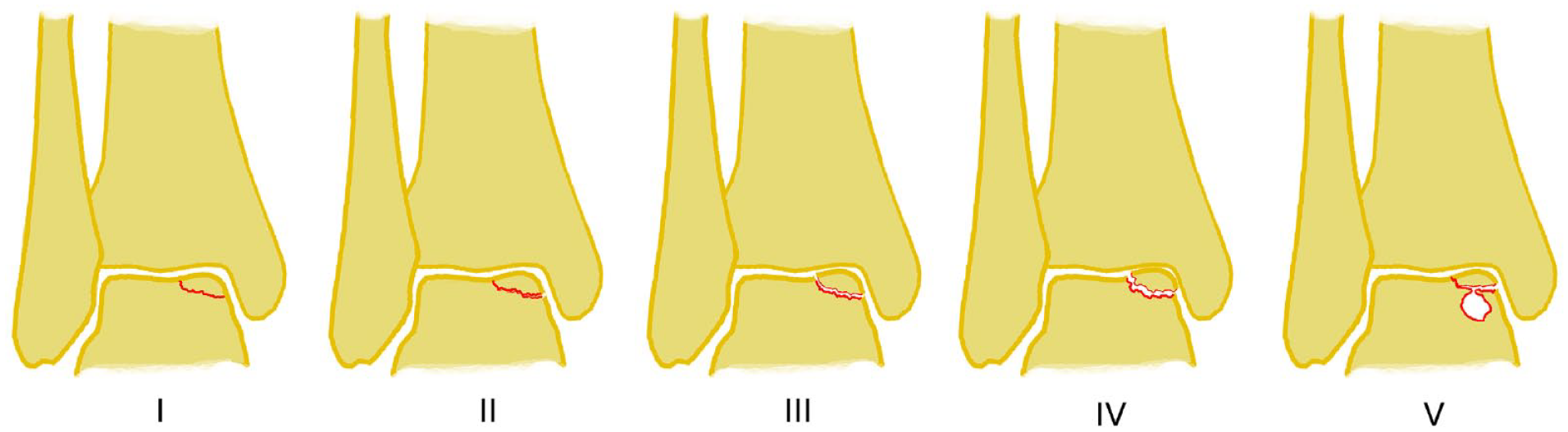
Classification by Berndt and Harty (stages I-IV), modified by Loomer (stage V).
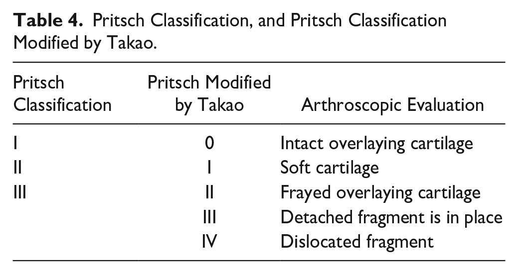
Two other grading systems frequently found in the literature are based on the arthroscopic appearance of the cartilage surface. The arthroscopic grading system was primarily published by Pritsch et al. 55 and later modified by Takao et al. 56 ( Table 4 ).
Pritsch Classification, and Pritsch Classification Modified by Takao.
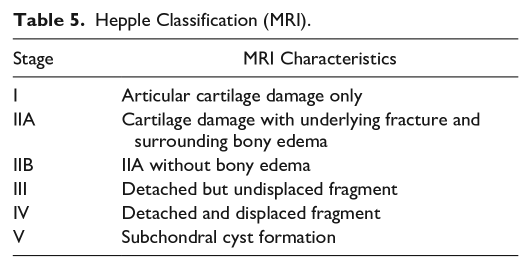
With MRI becoming more accessible in the 1990s, MRI-based classification systems have become increasingly popular. The Hepple et al. 57 classification is based on the initial classification system of Berndt and Harty but introduces additional traumatic, cystic, or idiopathic subtypes of OLTs ( Table 5 ). The Mintz et al. 58 classification correlates the MRI with arthroscopic findings.
Hepple Classification (MRI).
Today, the ICRS (International Cartilage Regeneration and Joint Preservation Society) 59 Classification and the Outerbridge 60 classification are used to grade cartilage lesions independent from a particular joint ( Fig. 2 ).
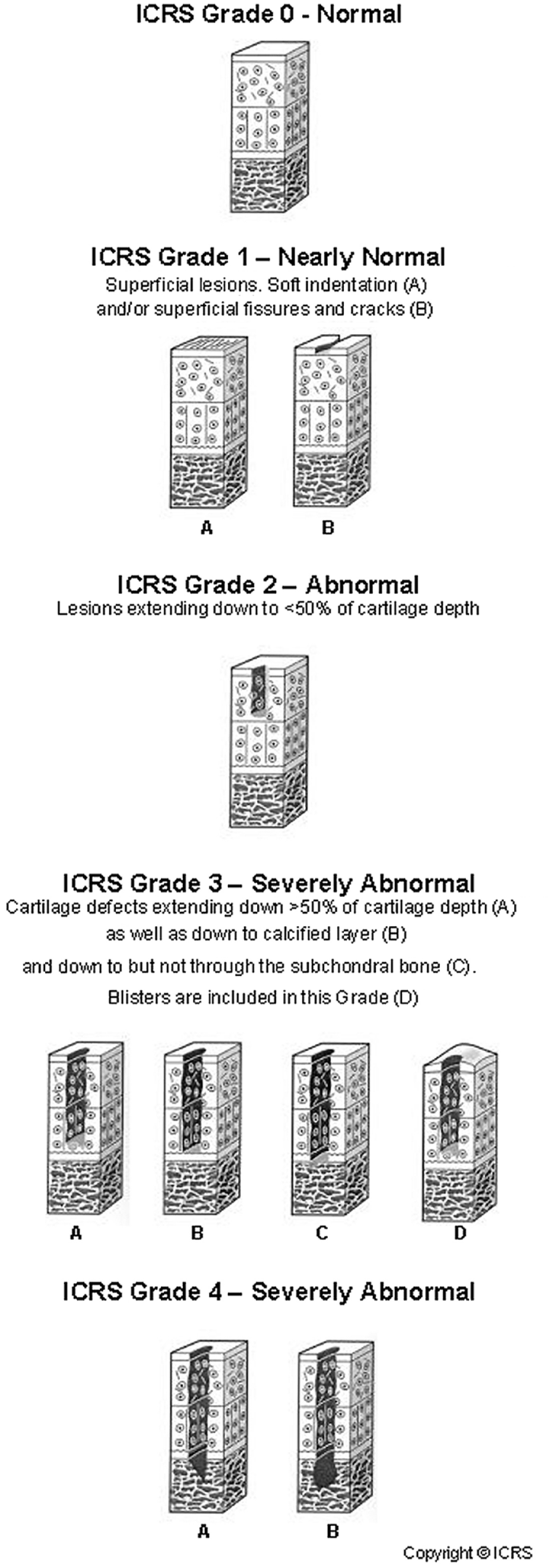
ICRS classification. ICRS = International Cartilage Regeneration and Joint Preservation Society.
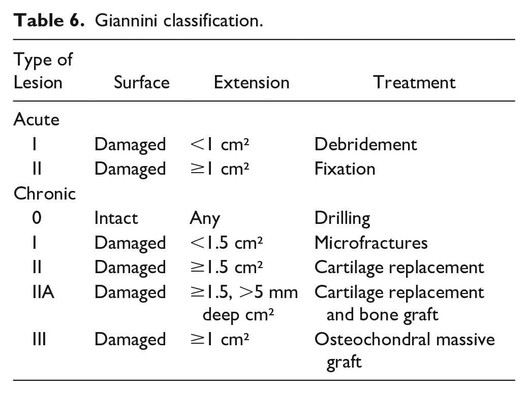
The Giannini et al. 61 classification was developed specifically for the ankle joint. This comprehensive classification distinguishes between acute and chronic lesions, intact, or damaged cartilage surface, and includes the size of the lesion and the depth of a bony defect. Subchondral cysts with intact cartilage surfaces can also be classified. Massive OLTs are characterized by a lesion larger than 3 cm² and a depth of more than 1 cm ( Table 6 ).
Giannini classification.
Besides the size of the lesion, the localization may influence the treatment options. Therefore, any documentation of the localization is recommended. Typically, location and size are documented by the imaging of a patient. For scientific studies, the location can be categorized using a 9-zone anatomic grid scheme (medial/central/lateral and anterior/central/posterior), as described by Elias et al. 32 The lesion size can be estimated in 3 planes, including surface area and depth of the lesion. 12 It is mandatory to document the source of the measurement as findings in arthroscopy, open surgery, MRI, and CT may differ.38,51,58,62,63
Statement: The descriptive classification system of Berndt and Harty, modified by Loomer, can still be recommended, as well as the Giannini Classification, ICRS Classification, and Outerbridge Classification. Especially for scientific studies, location and size should be documented, including the source of the measurements.
Grade of Recommendation: B
Conservative Management Strategies
Natural Progression
OLTs are not necessarily related to clinical symptoms. Bezuglov et al. 64 reviewed a cohort of 37 asymptomatic professional soccer players and found osteochondral lesions in 42% of the athletes. So far, there is no evidence that OLT progresses to ankle osteoarthritis (OA). Klammer et al. 65 followed 48 patients with OLT for a minimum of 2 years (mean = 52 months; range, 27-124 months) and noted that the minimally symptomatic OLT did not progress over time when treated non-operatively. Weigelt et al. 66 followed 22 patients for 11 to 20 years, in whom the progression of OA was analyzed based on plain ankle radiographs at the initial presentation and the final follow-up (van Dijk et al. 67 classification). At the final follow-up, 11 cases (73%) showed no progression of OA while 4 (27%) showed progression by 1 grade, and 38% of the patients reduced their sports activities because of sports-related symptoms. However, it is unlikely that OLT will disappear, and OLT may progress with increasing symptoms. Risk factors for progression are the depth of the lesion on MRI, subchondral cyst formation, and the extent of bone marrow edema. 65 A follow-up imaging is recommended after 12 months, even in asymptomatic patients, to avoid missing any progression.
Statement: OLTs with minimal symptoms are unlikely to progress to OA, especially if the modification of activities leads to a symptom-free situation. Based on MRI findings alone, there is no need to urge patients with minimal or no symptoms for prophylactic surgery. After 12 months, a follow-up imaging with MRI prevents missing any asymptomatic progression of the lesion.
Grade of Recommendation: B
Immobilization
There are 2 systematic reviews on the conservative management of OLTs. Zengerink et al. 68 calculated a success rate of 45% among patients who had conservative management with rest. Patients who underwent cast immobilization ranging from 3 weeks to 4 months had a success rate of 53%. Tol et al. 69 reported good or excellent results in 59% of patients with rest and restrictions for sports activities with or without nonsteroidal anti-inflammatory drugs (NSAIDs). The cohort with cast immobilization for 3 weeks to 4 months presented with good or excellent results in 41%. Shearer et al. 70 analyzed conservative management in chronic Stage V OLTs (Loomer classification). 39 They found a minimal relation between the morphological findings and clinical symptoms. They concluded that the conservative strategy should be limited to 3 months in patients with persistent symptoms.
In an investigation of OLTs in children, Perumal et al. 71 reported on 32 patients with an average age of 12 years. After 6 months of conservative management with cast immobilization and unloading, 77% continued to have persistent lesions on radiographs, 16% demonstrated complete clinical and radiographic healing, and 6% had severe pain after cast removal that required surgery. In those patients with persistent radiographic lesions and after an extra 6 months of nonoperative treatment, 42% had to undergo surgery for unhealed lesions and pain, whereas 46% had no symptoms despite persistent lesions on radiographs.
Statement: Reduction of activity is a strategy to reduce symptoms in OLTs. There is no evidence that cast immobilization leads to better results than reducing activities to a level with minimal or no symptoms. The overall success rate seems to be about 50%. There is only a limited correlation between the morphological appearance in imaging and the clinical symptoms. Surgery can be recommended if patients do not improve within 3 months of conservative management.
Grade of Recommendation: B
Injections
There is only limited literature on the effect of injections in OLTs. 72 Mei-Dan et al. 73 performed a prospective study on the effect of HA injection. They followed 15 patients, 60% of whom presented with stage III lesions (Cheng-Ferkel classification), 74 for 26 weeks after receiving 3 weekly intra-articular HA injections. They reported a significant improvement in pain and functional scores, with the effect of the injection lasting for more than 6 months with minimal adverse events. A second study compared injections of HA and PRP. Although both injections improved pain and functional scores, PRP led to a significantly better outcome than HA. However, further scrutiny of the data in a meta-analysis revealed that the study was underpowered. Although there is strong evidence of an improvement, it is impossible to conclude the superiority of PRP over HA. 72
In a retrospective cohort study of 49 patients with OLT grade I to III (Berndt and Harty 21 classification), Akpancar and Gul 75 compared PRP injections and prolotherapy, noting that both the injection of PRP and prolotherapy had a similar effect on pain and functional scores, extending 1 year. They reported excellent or good outcomes in 88.8% of the patients in the prolotherapy group, while 90.9% of the patients in the PRP group reported excellent or good outcomes. As we could not find any research on the potentially harmful effect of the 25% dextrose solution for intra-articular injection on the cartilage, further research is needed until prolotherapy can be recommended as a treatment for OLTs.
Statement: Injection therapy can improve pain and function in OLTs with PRP and HA demonstrating a similar positive effect on pain and functional scores. The effect extends approximately 6 months after injecting HA and 12 months after injecting PRP.
Grade of Recommendation: B
Extracorporeal Shockwave Therapy
After some promising results in animal studies, 76 there is increasing discussion of how ESWT might be used to treat OLTs. Zhang et al. 77 evaluated the efficiency of focused ESWT in patients with persistent pain 3 months after arthroscopic microfracture for OLTs and reported that the visual analog scale and American Orthopaedic Foot and Ankle Society (AOFAS) score significantly improved 12 weeks after ESWT and at the last follow-up (mean follow-up: 27.8 months). In addition, areas of lesions (sagittal plane MRI) were distinctly reduced at the last follow-up. Gao et al. 78 reported combined therapy with focused ESWT and retrograde bone marrow–derived cell transplantation for OLT Hepple grade I to III with a follow-up for more than 2 years. They saw a better reduction in pain and a significantly higher AOFAS score in the group that received additional focused ESWT. Thus, the authors concluded that the combined technique is a highly effective therapeutic option in OLTs with intact cartilage. In both studies, the ESWT was applied transmalleolar to medial talar lesions.
Statement: So far, there are no studies on shockwave as a standalone treatment option for OLTs. Focused ESWT can be considered a complementary treatment in patients with persistent pain after surgical treatment or adjunctive to stimulate tissue regeneration.
Grade of Recommendation: C
Magnetic Field
Some recent studies examined the effect of electromagnetic fields on enhancing osteochondral repair in rabbits. 79 Based on basic science research, there might be an effect on the healing in OLTs, 80 but there is no data available on the application in humans.
Statement: Currently, no data are available to support the use of an electromagnetic field in treating OLTs in humans.
Grade of Recommendation: I
Discussion
The etiology of OLTs covers a wide range of possible causative factors. Many potential risk factors are intrinsic, with limited possibilities for therapeutic improvement. While one causative factor might be chronic overloading of the talar dome, in most papers published on treatment outcomes, etiology is of minor relevance. Hopefully, this may change over the coming years with a better understanding of the underlying problems. For example, it can be estimated that a blood supply problem at the talar dome with bone necrosis may trigger other treatment algorithms than an acute traumatic fracture of the talar dome. Increasing histologic probes taken during OLT treatment may further enlighten possible categories of causes. Other causative factors might be chronic overloading of the talar dome.
Imaging has improved with new MRI sequences and WBCT, and while many new MRI imaging sequences are of scientific interest, they have not been widely established in the daily diagnostic routine. The standard MRI of the ankle requires 3 planes, including sagittal, coronal, and transversal planes with T1 and T2 sequences. 3T systems with high-resolution coil systems provide the best image quality, while open, low-field MRIs are insufficient to provide the information needed to plan the treatment.
There has been no relevant change in the classification systems over the last few years, so the descriptive classification system of Berndt and Harty, modified by Loomer ( Fig. 1 ), can still be recommended. Also, the Giannini Classification ( Table 6 ), ICRS Classification ( Fig. 2 ), and Outerbridge Classification are helpful tools. Especially for scientific studies, location and size should be documented, including the source of the measurements, as findings in arthroscopy, open surgery, MRI, and CT may differ significantly.38,51,58,62,63
The published literature on the conservative management of OLTs shows increasing evidence for the different treatment options. However, the maximum grade of recommendation never exceeds level B—fair evidence. Studies supporting a grade of recommendation A are extremely difficult to perform, requiring level I RCTs with similar findings or a meta-analysis.
The treatment algorithm based on the findings of the study is shown in Figure 3 . There are several key messages regarding conservative treatment, which are in general also supported by the voting of the expert group ( Table 7 ):
In asymptomatic patients and patients with minimal symptoms, OLT progression to ankle OA is unlikely.64 -66 Risk factors for progression are the depth of the lesion on MRI, subchondral cyst formation, and the extent of bone marrow edema. 65 To some extent, conservative management is the adaptation of activities to the performance of the ankle joint. 69 A follow-up imaging after 12 months helps to monitor any progression.
It is impossible to estimate the probability of success of conservative management from initial symptoms and imaging. 70
Cast immobilization is an option in OLTs in children, 71 with a success rate of approximately 50%, although, based on imaging, complete healing is only seen in 6% of the patients.
In adults, the success rate of conservative management ranges from 45% to 59%.68,69 Rest and restrictions for sports activities seem to be more successful than immobilization in adults, contrary to the findings in children where cast immobilization is an option.
Intra-articular injections of HA and PRP can improve pain and functional scores for more than 6 months.72,73,81
If 3 months of conservative management does not improve symptoms, it is unlikely that further conservative management will be successful, and surgery can be recommended. 70
Focused ESWT can be considered a complementary treatment in patients with persistent pain after surgical treatment or adjunctive to stimulate tissue regeneration. So far, no studies support ESWT as a standalone treatment for OLTs.76,78
The literature does not support the use of an electromagnetic field in the treatment of OLTs in humans.
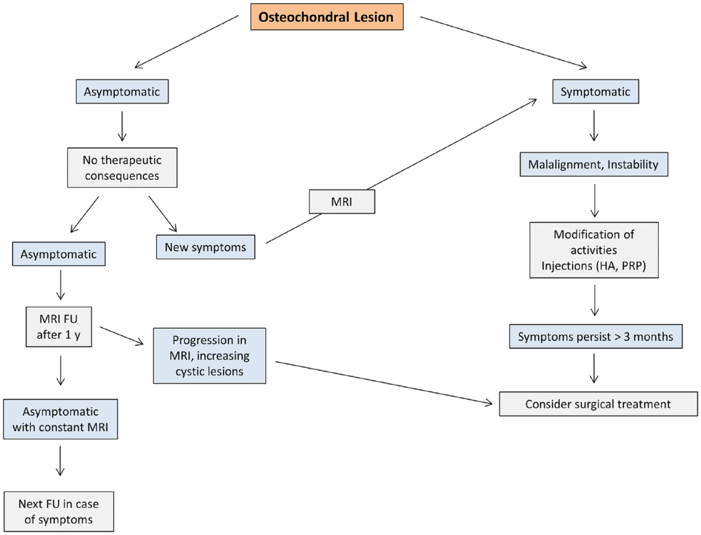
Suggested conservative treatment algorithm for osteochondral lesions of the talus. HA = hyaluronic acid; PRP = platelet-rich plasma; FU = Follow-up.
Statements, Grade of Recommendation, LOE of the Best Study on the Topic, and Agreement on the Statements Among the Experts.
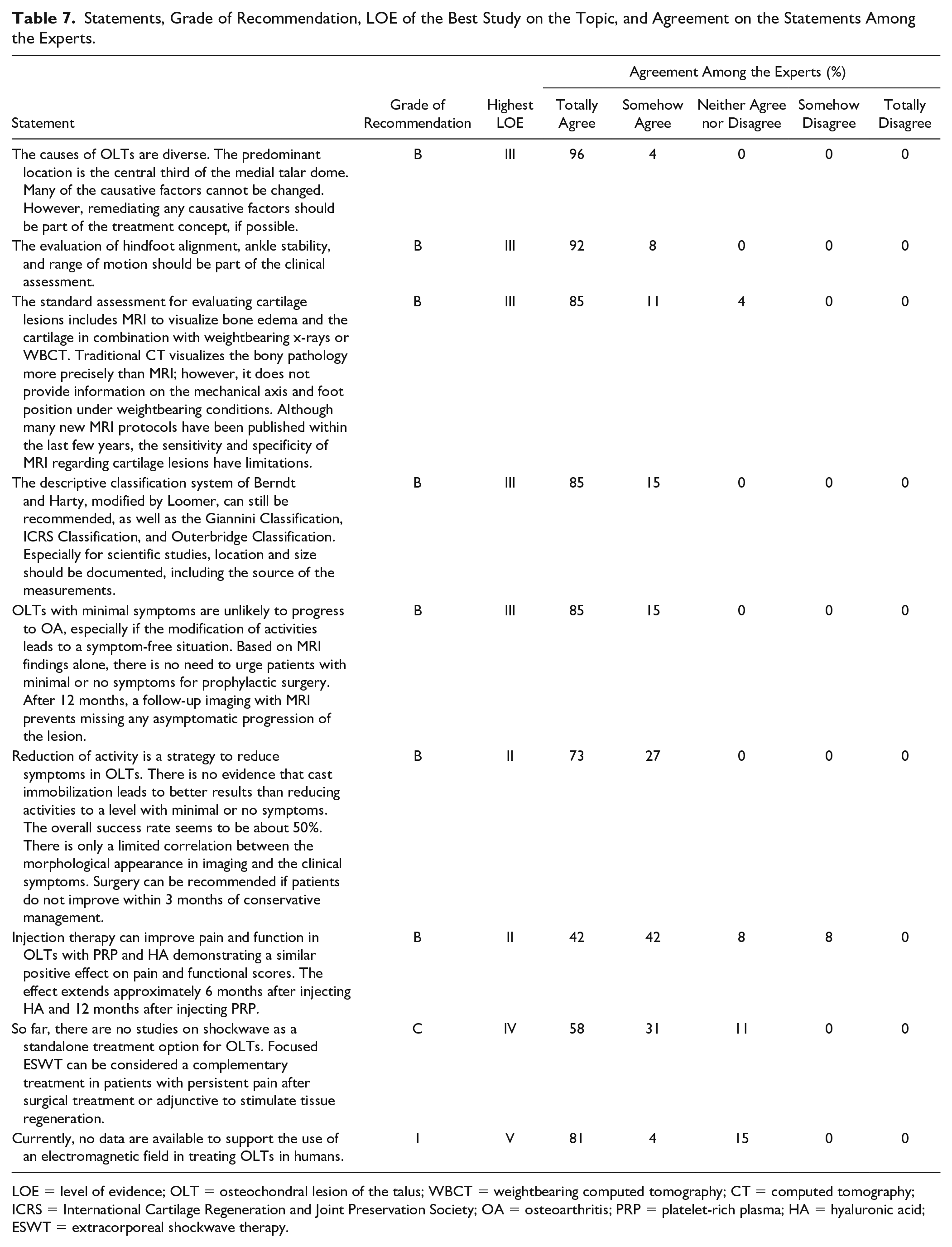
LOE = level of evidence; OLT = osteochondral lesion of the talus; WBCT = weightbearing computed tomography; CT = computed tomography; ICRS = International Cartilage Regeneration and Joint Preservation Society; OA = osteoarthritis; PRP = platelet-rich plasma; HA = hyaluronic acid; ESWT = extracorporeal shockwave therapy.
Footnotes
Acknowledgments and Funding
The publication funds of the Martin-Luther-University Halle-Wittenberg covered the open access fees.
Declaration of Conflicting Interests
The author(s) declared the following potential conflicts of interest with respect to the research, authorship, and/or publication of this article: Peter Angele: Consultant, Arthrex, Aesculap; Christoph Becher: Consultant, Plasmaconcept; Oliver Gottschalk: Paid speaker, Geistlich; Daniel Günther: Paid speaker, Geistlich, Arthrex, Codon; Peter Müller: BBraun Aesculap; Markus Walther: Paid speaker, Geistlich. The remaining authors have nothing to disclose.
Ethical Approval
Not applicable.