
Abstract
Objective
The purpose of this study was to determine the preventive effects of treadmill exercise or physiological loading on disuse atrophy in the rat knee joint cartilage and bone during hindlimb suspension.
Design
Twenty male rats were divided into 4 experimental groups, including the control, hindlimb suspension, physiological loading, and treadmill walking groups. Histological changes in the articular cartilage and bone of the tibia were histomorphometrically and immunohistochemically evaluated 4 weeks after the intervention.
Results
Compared with the control group, the hindlimb suspension group showed thinning of cartilage thickness, decreased matrix staining, and decreased proportion of noncalcified layers. Cartilage thinning, decreased matrix staining, and decreased noncalcified layers were suppressed in the treadmill walking group. The physiological loading group exhibited no significant suppression of cartilage thinning or decreased noncalcified layers, but the decreased matrix staining was significantly suppressed. No significant prevention of bone mass loss or changes in subchondral bone thickness were detected after physiological loading or treadmill walking.
Conclusion
Disuse atrophy of the articular cartilage caused by unloading conditions could be prevented by treadmill walking in rat knee joints.
Introduction
Normal metabolism in articular cartilage requires physiological loads.1,2 Articular cartilage is highly sensitive to mechanical stress and responds adaptively.3,4 For example, moderate loading promotes the anabolism of the articular cartilage, increases the number of chondrocytes, and induces cartilage thickening.5 -7 However, when loading is absent or low, articular cartilage thins, and the matrix decreases without surface damage. 8 These atrophy-like histological changes in articular cartilage occur in both humans and animals,9 -15 and Vincent and Wann 8 called this condition disuse atrophy of the articular cartilage. We previously showed that disuse atrophy in articular cartilage results in decreased chondrocyte numbers, cartilage thickness, and matrix staining, but the chondrocyte density does not change and the surface is intact. 16 The effects of unloading extend to the subchondral bone and bone, causing decreased subchondral bone thickness and bone mass. 17 In clinical practice, disuse atrophy of the articular cartilage frequently can occur as a complication of bed rest associated with treatment. 11
In our previous studies, we clarified two clinically important findings regarding disuse atrophy of the articular cartilage. First, disuse atrophy of the articular cartilage can be recovered by reloading. 17 This histological finding suggests that the articular cartilage possesses a certain plasticity, similar to skeletal muscle and bone. Second, disuse atrophy of the articular cartilage contributes to the onset and severity of osteoarthritis (OA). 18 The prevalence of OA is increasing worldwide and is a major medical and economic problem.19,20 Also, OA is a major cause of sarcopenia and negatively affects healthy life expectancy.21,22 Therefore, the prevention and control of OA progression are increasingly important.23,24 In addition, OA progression is closely linked to the anatomical properties of the articular cartilage. The ability of the articular cartilage to repair is poor due to the absence of blood vessels and nerves. 25 In result, OA often advances substantially before clinical symptoms appear. 26 Therefore, preventing disuse atrophy of the articular cartilage may help prevent OA onset and progression.
In view of the above, we hypothesized that physiological loading and treadmill walking during the unloading period can inhibit disuse atrophy of the articular cartilage in an experimental animal model. The reasons for this hypothesis are as follows: Physiological loading during an unloading period inhibits atrophy in disused muscle atrophy27,28 and physiological loading maintains cartilage thickness,1,4 and exercise, such as treadmill exercise, increases cartilage thickness. 7 Thus, this study aimed to determine the inhibitory effects of physiological loading and treadmill exercise on articular cartilage disuse atrophy induced by unloading conditions, using a rat hindlimb suspension model. The disuse atrophy induced by unloading affects the cartilage, subchondral bone, and bone. 17 Thus, these tissues were included in the histological and immunohistochemical analyses.
Method
Experimental Animals and Animal Care
The study protocol was approved by the Animal Research Committee of the Graduate School of Medicine of Kanazawa University (Kanazawa, Japan; approval nos. 204125 and 214293) and conducted in accordance with the ARRIVE guidelines29,30 and Guidelines for the Care and Use of Laboratory Animals of Kanazawa University.
Twenty male Wistar rats (8 weeks old) were purchased from Japan SLC (Shizuoka, Japan) and housed under normal conditions for 1 week before starting the experiments to acclimatize the animals. One rat was housed per cage in a sanitary ventilated room under controlled temperature and humidity conditions and a 12-hour light-dark cycle with ad libitum access to food and water. The health status of the animals was monitored 2 to 3 times per week, including general food and water intake, surgical wound condition, gait, and hindlimb suspension.
Grouping
The experimental protocol is shown in Figure 1 . Rats were divided into the following 4 groups with 5 rats per group: control group (CON), hindlimb suspension group (HS), physiological loading during hindlimb suspension group (PL), and treadmill walking during hindlimb suspension group (TW). Rats in the CON group were kept in a physiological environment. Rats were subjected to hindlimb suspension for 4 weeks in the HS, PL, and TW groups, as described in our previous study.31 -33 During hindlimb suspension, rats were allowed to walk freely in their cages using only their forelimbs. In the PL and TW groups, the rats were removed from the hindlimb suspension device and allowed to walk freely in a physiological environment using all limbs during the loading or exercise time. Rats in the PL group underwent physiological loading by removing the hindlimb suspension for 1 hour per day, 5 days per week for 4 weeks. In the TW group, rats were subjected to treadmill exercise on a motor-driven treadmill (SN-460, Shinanoseisakusyo, Tokyo, Japan). The rats ran for a total of 10 minutes, increasing their speed by 2 m/min every 2 minutes from 0 m/min to 10 m/min to warm-up. Then, the rats ran 12 m/min for 20 minutes per day, 5 days per week, for 4 weeks. The exercise regimen was set to a low-intensity protocol based on previous study,7,34 because low-intensity exercise has been reported to increase normal articular cartilage thickness and chondrocyte density 7 and low-intensity exercise has protective and positive effects of inhibiting OA progression. 34 In addition, this study considered the effect of non-weightbearing on the hindlimb’s disuse muscular weakness. No further interventions were performed during the experimental period, and no analgesics or anti-inflammatory drugs were administered.
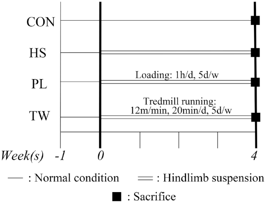
Schema of experimental schedule. Rats in the CON group were subjected to normal conditions, and rats in the HS, PL, and TW groups were subjected to tail suspension. Rats in the PL group were subjected to self-weighted physiological loading for 1 hour per day, 5 days per week. Rats in the TW group performed treadmill walking (12 m/min), 30 minutes per day (including warm-up), 5 days per week. Bone and articular cartilage were collected after the intervention and histological and immunohistochemical analyses were performed. (n = 5 per group). CON = control; HS = hindlimb suspension; PL = physiological loading; TW = treadmill walking.
Sample Size Calculation
Sample size was calculated by using G*Power 3.1 and literature35 -37 (https://www.psychologie.hhu.de/arbeitsgruppen/allgemeine-psychologie-und-arbeitspsychologie/gpower.html). The calculation was based on the main parameter, cartilage thickness, and matrix intensity, including the first 5 rats, in the CON and HS groups. For these parameters, a minimum of 3 and 5 rats were required in the CON and HS groups, respectively, with a power of 0.80, effect size calculated from the mean and standard deviation, and a significance level of P < 0.05. Furthermore, another reference was used to calculate the required sample size, 37 which was calculated to be a minimum of 4 and a maximum of 6. Considering the results calculated from G-power and the literature and the 3R principle of animal experimentation (reduction, replacement, and refinement), we finally set our sample size at 5 animals per group.
To reduce the number of animals used, based on the 3Rs principle of animal experimentation, a portion of the data used in this study were obtained from our previous study. 17 Specifically, the data from the CON and HS groups in the present study and the previous study are the same. 17 As our previous study is open-access and published under a CC-BY license, reuse of the data is permitted. 17
Histological Preparation
Decalcified paraffin sections were prepared for histology, as previously described. 33 Both knees were excised frontally to evaluate histological changes in the medial tibiofemoral joints. The right knee was used for histomorphometric analysis, and the left knee was used for immunohistochemical analysis. The specimens were serially sliced to observe the region where the articular cartilage of the femur and tibia are in direct contact without meniscus involvement. The paraffin sections (3-µm thick) were stained with hematoxylin and eosin and toluidine blue (0.05%, 15 minutes), and immunohistochemical staining was performed. The sections were viewed under a light microscope and imaged using a digital camera (BX51 and DP74; Olympus Corporation, Tokyo, Japan) to evaluate the histological changes in the articular cartilage.
Histomorphometric Analyses of the Articular Cartilage
Adobe Photoshop CC imaging software (Adobe Systems, Inc., San Jose, CA, USA) was used for histomorphometric analyses of the articular cartilage. Cartilage thickness, cartilage layer proportion, the intensity of matrix staining with toluidine blue, and chondrocyte density were evaluated, as described previously. 17 Figure 1 of our previous study shows the schema for histomorphometric analysis. 17
To evaluate the total cartilage thickness, digitized images of the sections stained with toluidine blue were used. Cartilage thickness was defined as the distance between the cartilage surface and the osteochondral junction. We used the measured area of the cartilage with a width of 1 mm at the center of the lesion at the tibia in the medial tibiofemoral joint. Moreover, using the images stained with hematoxylin and eosin, the proportions of the noncalcified and calcified layers were evaluated using the tidemark as the indicator. Specifically, with a width of 200 µm, these proportions were calculated by each area of the noncalcified and calcified layers by dividing the total area of the articular cartilage.
To evaluate matrix intensity, digitized images of cartilage stained with toluidine blue were converted to grayscale (white, 255; black, 0) to assess the relative intensity of toluidine blue staining. The average staining intensity was calculated at the same area in the same manner as performed for the measurement of articular cartilage thickness.
To evaluate chondrocyte density, digitized images of the sections stained with hematoxylin and eosin were used. Chondrocyte density was determined as the number of chondrocytes per unit area of the cartilage. This unit area was calculated using the same method as that for the abovementioned cartilage thickness, and the width to be measured was set to 200 µm. Chondrocytes with visible nuclei within the area of interest were counted manually.
Histomorphometric Analyses of the Bone
Bone volume and thickness of the subchondral bone were measured at the tibial medial condyle according to the method of Nagira et al. and our previous study.17,38 This highly reliable method correlates well with microcomputed tomography. Bone volume (%) was defined as follows: (area of the cancellous bone − area of the bone marrow; trabecular bone) / area of cancellous bone × 100. Subchondral bone thickness was defined as the region between the osteochondral junction and the bone marrow cavity.
Immunohistochemical Staining and Analyses
Immunohistochemical staining was performed as previously described. 17 To measure cartilage synthesis, paraffin sections were immunohistochemically stained using Aggrecan antibody (diluted 1:50, 13880-1-AP; Proteintech, Tokyo, Japan) and type II collagen (diluted 1:50, ab34712; Abcam, Tokyo, Japan). To measure cartilage degradation, sections were stained with matrix metalloproteinase 13 (MMP 13; diluted 1:50, 18165-1-AP; Proteintech Tokyo, Japan) and A disintegrin and metalloproteinase with thrombospondin motifs 5 (ADAMTS-5; diluted 1:50, ab41037; Abcam, Tokyo, Japan). For type 2 collagen, the staining intensity was calculated in the same manner as the histomorphometric analyses described above. For Aggrecan, MMP13, and ADAMTS-5, the positively stained cells were counted, and the positive cell density was calculated in the same manner as the histomorphometric analyses described above. Figure 1 of our previous study shows the schema for the immunohistochemical analysis. 17
Statistical Analyses
All statistical analyses were performed using JMP 14 software (SAS Institute, Cary, NC, USA). All data were statistically analyzed as parametric data. Descriptive statistics for body weight, histomorphometric data, and immunohistochemical data including all figures are presented as means with standard deviations. The groups were first compared by the analysis of variance (ANOVA), which showed significant differences in all parameters except cancellous bone and type II collagen. Therefore, Tukey’s honest significant difference test was conducted on the parameters that showed significant differences by ANOVA.
Results
General Condition
No animals died during the experimental period, and no animals developed infections in their tail wounds. Weight changes during the experiment are shown in the supporting information (
Histomorphometric Analyses of the Articular Cartilage
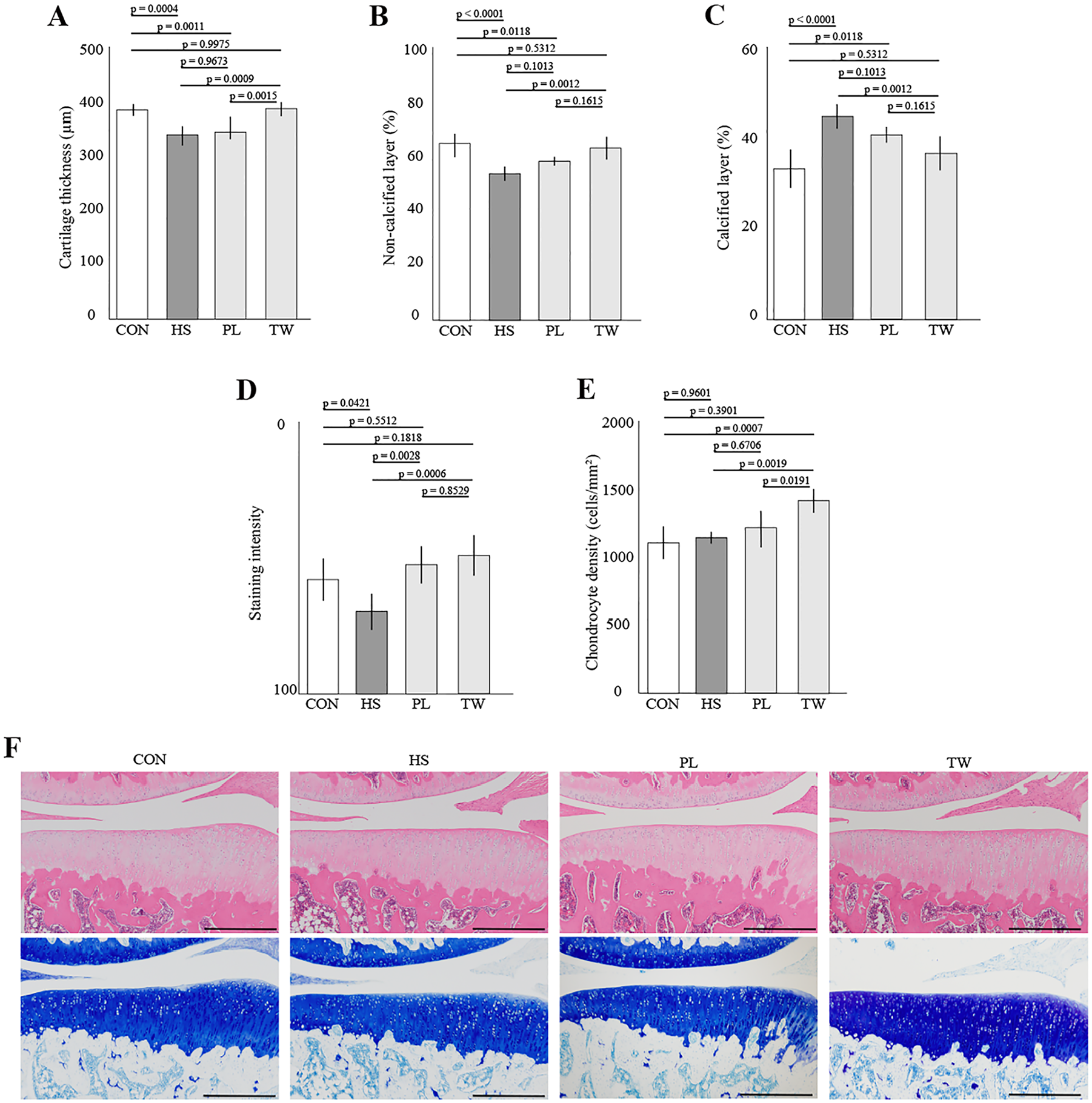
Four weeks of hindlimb suspension significantly decreased cartilage thickness (HS vs. CON, P = 0.0004) ( Fig. 2A ). Cartilage thickness in the PL group was not significantly different from that in the HS group and was significantly thinner than that in the CON group (PL vs. HS, P = 0.9673, and PL vs. CON, P = 0.0011). Cartilage thickness in the TW group was significantly thicker than that in the HS group and was not significantly different from that in the CON group (TW vs. HS, P = 0.0009, and TW vs. CON, P = 0.9975). The thickness in the TW group was significantly thicker than that in the PL group (P = 0.0015).
Histological effects of physiological loading and treadmill exercise on the articular cartilage during the unloading period. (
The proportion of noncalcified layers in the articular cartilage decreased significantly by hindlimb suspension ( Fig. 2B , CON vs. HS, P < 0.0001). The noncalcified proportion in the PL group was not significantly different from that in the HS group and was significantly thinner than that in the CON group (PL vs. HS, P = 0.1013, and PL vs. CON, P = 0.0118). However, the layer proportion in the TW group was significantly increased compared with that in the HS group and was not significantly different from that in the CON group (TW vs. HS, P = 0.0007, and TW vs. CON, P = 0.5312). Changes in the calcified layer showed the same trends as in the noncalcified layer ( Fig. 2C ).
The toluidine blue staining intensity decreased significantly after 4 weeks of hindlimb suspension (CON vs. HS, P = 0.0421) ( Fig. 2D ). Staining intensity in the PL and TW group was significantly higher than the HS group (PL vs. HS, P = 0.0028, and TW vs. HS, P = 0.0006).
No significant changes in chondrocyte density were observed by hindlimb suspension (CON vs. HS, P = 0.9601) ( Fig. 2E ). Similarly, no significant changes were observed by physiological loading (CON vs. PL, P = 0.3901). However, chondrocyte density was significantly higher in the TW group than in with the CON, HS, and PL groups (TW vs. CON, P = 0.0006; TW vs. HS, P = 0.0019; and TW vs. PL, P = 0.0191).
Overviews of the articular cartilage of the tibia are shown in
Figure 2F
. Detailed data on histomorphometric changes in the articular cartilage are shown in the supporting information (
Histomorphometric Analyses of the Bone
No significant differences in the area of the cancellous bone were detected among all groups ( Fig. 3A ). Four weeks of hindlimb suspension significantly reduced the area of the trabecular bone compared with the area in the CON group ( Fig. 3B , HS vs. CON, P = 0.0015). The trabecular bone area in the PL and TW groups was significantly smaller than that in the CON group ( Fig. 3B , PL vs. CON, P = 0.0142, and TW vs. CON, P = 0.0006). Similar trends were observed in bone volume ( Fig. 3C , CON vs. HS, P = 0.0015; PL vs. CON, P = 0.0029; and TW vs. CON, P = 0.0002). In addition, similar trends were observed for subchondral bone thickness ( Fig. 3D , CON vs. HS, P = 0.0002; PL vs. CON, P < 0.0001; and TW vs. CON, P = 0.0002).
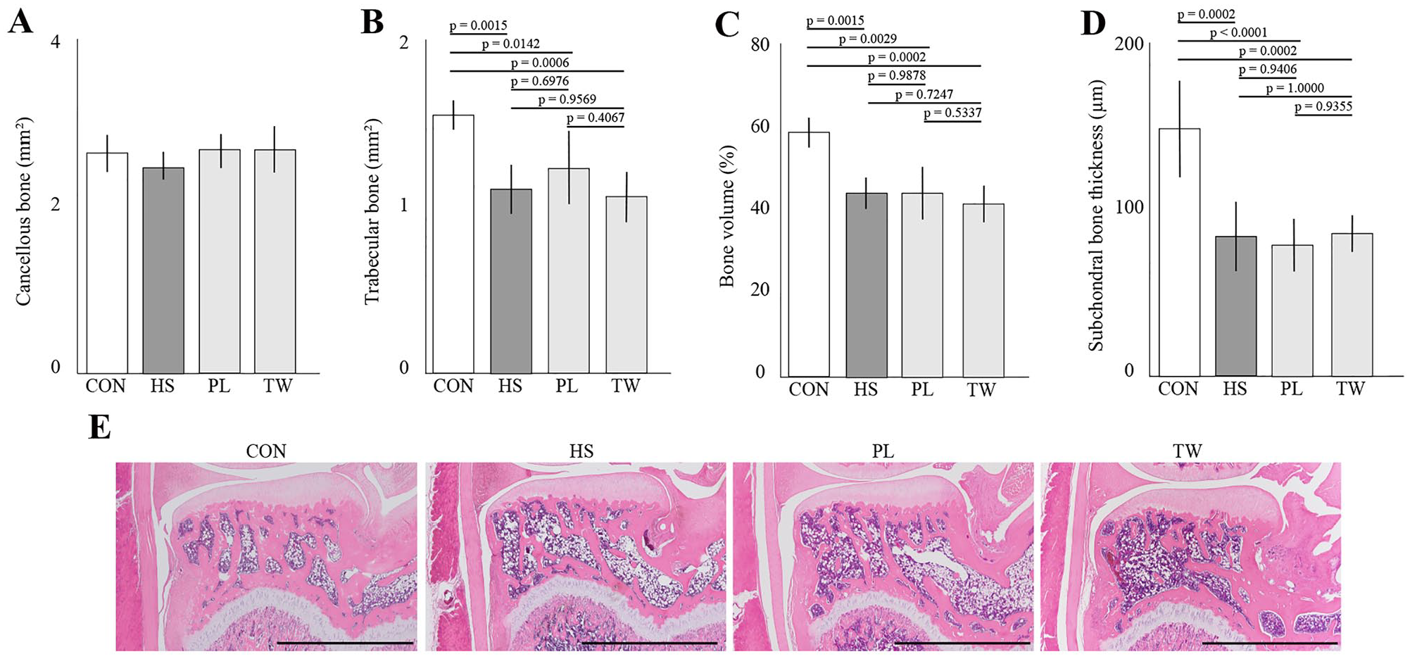
Histological effects of physiological loading and treadmill exercise on bone during the unloading period. (
Overviews of the bone and subchondral bone of the tibia are shown in
Figure 3E
. Detailed histomorphometric results for the bone and subchondral bone are shown in supporting information (
Immunohistochemical Analyses of the Articular Cartilage
The immunohistochemical results are shown in
Figure 4
and the supporting information (
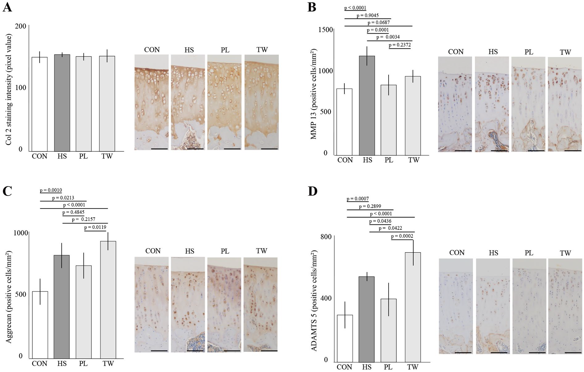
Immunohistochemical effects of reloading on cartilage atrophy in the medial tibia. (
Discussion
This study aimed to histologically examine the preventive effects of physiological loading and treadmill exercise on disuse atrophy of the articular cartilage in the rat knee joint during hindlimb suspension. The results showed that physiological loading did not prevent the progression of disuse atrophy of the articular cartilage, whereas treadmill walking had a significant preventive effect on the disuse atrophy in the thickness, staining intensity, and layer proportion. However, neither physiological loading nor treadmill walking prevented disuse changes in the subchondral bone and bone during hindlimb suspension.
The results of this study can contain novel and useful information from both basic and clinical perspectives. First, the inhibitory effect of mechanical stress on disuse atrophy during the unloading condition has been studied in skeletal muscles,27,28 but this is the first report of such a study conducted on the articular cartilage. In result, the histological findings of the preventive effects of exercise on the disuse atrophy of the articular cartilage revealed in the present study are novel. Second, although the ability of the articular cartilage to repair itself is poor, the results of this study and our previous studies suggest that disuse atrophy of the articular cartilage can be prevented and restored. 17 Thus, the pathology of disuse atrophy in the articular cartilage may be different from other cartilage diseases. Third, a significant number of patients can present with disuse atrophy of the articular cartilage in clinical practice. 11 The present study does not directly replicate clinical situations because of the prolonged period of unloading and the limited number of clinical situations in which walking is permitted during complete bed rest. However, from a rehabilitation perspective, unnecessary rest should be avoided, and standing and walking exercises are practiced from an early stage. Therefore, although this study is a preclinical model and the results do not directly relate to clinical practice, we believe that in addition to basic histological findings, the results of this study provide some useful information for clinical practice. Fourth, disuse atrophy of the articular cartilage contributes to the early onset and severity of OA, 18 so the histological findings of this study may be important for OA prevention. In light of the above four points, disuse atrophy of the articular cartilage may represent a new therapeutic target for orthopedics and rehabilitation.
Three interesting histological results were obtained in this study. First, treadmill walking, but not physiological loading, prevented disuse atrophy of the articular cartilage. Previous studies have shown that articular cartilage density influences cartilage volume, composition, and repair capacity, and articular cartilage volume correlates with exercise load.4,39 In the present study, a significant increase in chondrocyte density was observed only in the TW group. Therefore, chondrocyte density may have increased because the exercise load from treadmill walking was greater than the physiological loading in this study. Similar histological findings were observed in our previous study on the recovery of disuse atrophy of the articular cartilage, 17 suggesting that chondrocyte density is an important factor in the recovery and prevention of disuse atrophy of the articular cartilage.
The second result concerns the changes in ADAMTS5, which is an enzyme degrading Aggrecan.25,40 In this study, ADAMTS5-positive cell density significantly increased in the HS group and to an even greater extent in the TW group but did not increase in the PL group. A similar trend was observed in Aggrecan-positive cell density, and chondrocyte density also increased. These results suggest that exercise stimulates cartilage metabolism, resulting in a significant increase in both the production and degradation of cartilage matrix. However, our previous study on the recovery of disuse atrophy of the articular cartilage did not show an increase in ADAMTS5. 17 Significant increases in ADAMTS5 during the development of OA due to overload have also been reported.5,41 Furthermore, articular cartilage exposed to unloading conditions may have a lower threshold for mechanical loading and greater susceptibility to OA.8,42 Therefore, significant increases in ADAMTS5 in the TW group identified in this study may be related to the development of OA. Future studies should focus on safe exercise loads that prevent disuse atrophy of the articular cartilage and do not contribute to the onset of OA.
Third, bone loss and subchondral bone thinning were not prevented by physiological loading or treadmill exercise during the unloading period. In our previous study, disuse-atrophied bone and subchondral bone were restored after 2 and 4 weeks of reloading, respectively. 17 In addition, treatment with exercise therapy is effective against bone loss and osteoporosis. 43 In light of these facts, the absolute amount of mechanical stress defined by the amount of exercise, loading, and time may have been insufficient in this study. Another possible factor could be the difference that in the bone, multiple complex mechanisms may contribute to endochondral and membranous ossification, whereas in the cartilage, the mechanisms are dependent on matrix production.
This study has three major limitations. First, the animals used in this study were rodents. In general, metabolism, age at maturity, thickness, chondrocyte density, and layer composition ratios in small animals differ from those in humans.44 -47 Second, physiological loading and treadmill exercise conditions were single conditions, and the effects were observed at only one time point. Therefore, the optimal exercise conditions to prevent disuse atrophy of the articular cartilage are unknown. If the unloading period is short, physiological loading may also have a preventive effect. Third, mechanical stress is difficult to quantify. Although the histological results suggested that the mechanical stress applied to the TW group was greater than that of the PL group, we searched previous studies and found it difficult to quantify and compare the mechanical stress applied to the PL and TW groups.
In conclusion, treadmill walking during unloading conditions prevents the progression of disuse atrophy of the articular cartilage in rat knee joints. Combined with the results of our previous study, 17 our results suggest that exercise and loading can prevent or recover disuse atrophy of the articular cartilage. Future studies should focus on determining the optimal conditions for preventing and/or recovering disuse atrophy of the articular cartilage.
Supplemental Material
sj-docx-1-car-10.1177_19476035231154510 – Supplemental material for Treadmill Exercise Suppresses Histological Progression of Disuse Atrophy in Articular Cartilage in Rat Knee Joints during Hindlimb Suspension
Supplemental material, sj-docx-1-car-10.1177_19476035231154510 for Treadmill Exercise Suppresses Histological Progression of Disuse Atrophy in Articular Cartilage in Rat Knee Joints during Hindlimb Suspension by Ikufumi Takahashi, Taro Matsuzaki, Hiroshi Kuroki and Masahiro Hoso in CARTILAGE
Supplemental Material
sj-docx-2-car-10.1177_19476035231154510 – Supplemental material for Treadmill Exercise Suppresses Histological Progression of Disuse Atrophy in Articular Cartilage in Rat Knee Joints during Hindlimb Suspension
Supplemental material, sj-docx-2-car-10.1177_19476035231154510 for Treadmill Exercise Suppresses Histological Progression of Disuse Atrophy in Articular Cartilage in Rat Knee Joints during Hindlimb Suspension by Ikufumi Takahashi, Taro Matsuzaki, Hiroshi Kuroki and Masahiro Hoso in CARTILAGE
Supplemental Material
sj-docx-3-car-10.1177_19476035231154510 – Supplemental material for Treadmill Exercise Suppresses Histological Progression of Disuse Atrophy in Articular Cartilage in Rat Knee Joints during Hindlimb Suspension
Supplemental material, sj-docx-3-car-10.1177_19476035231154510 for Treadmill Exercise Suppresses Histological Progression of Disuse Atrophy in Articular Cartilage in Rat Knee Joints during Hindlimb Suspension by Ikufumi Takahashi, Taro Matsuzaki, Hiroshi Kuroki and Masahiro Hoso in CARTILAGE
Supplemental Material
sj-docx-4-car-10.1177_19476035231154510 – Supplemental material for Treadmill Exercise Suppresses Histological Progression of Disuse Atrophy in Articular Cartilage in Rat Knee Joints during Hindlimb Suspension
Supplemental material, sj-docx-4-car-10.1177_19476035231154510 for Treadmill Exercise Suppresses Histological Progression of Disuse Atrophy in Articular Cartilage in Rat Knee Joints during Hindlimb Suspension by Ikufumi Takahashi, Taro Matsuzaki, Hiroshi Kuroki and Masahiro Hoso in CARTILAGE
Footnotes
Author Contributions
All of the authors made substantial contributions to (1) the conception and design of the study, data acquisition, and analysis and interpretation of the data; (2) drafting the article or critically revising it for important intellectual content; and (3) providing final approval of the submitted version of the manuscript. The authors’ specific contributions were as follows:
1. Conception and design of the study: IT, TM, HK, and MH
2. Analysis and interpretation of the data: IT, TM, HK, and MH
3. Drafting of the article: IT, HK, and MH
4. Critical revision of the article for important intellectual content: IT, HK, and MH
5. Final approval of the article: IT, TM, HK, and MH
6. Obtaining funding: IT, TM, HK, and MH
7. Collection and assembly of the data: IT, TM, and MH
Ikufumi Takahashi (
Acknowledgments and Funding
The authors thank the members of the Department of Human Pathology at the Kanazawa University Graduate School of Medicine for offering advice regarding the histopathological techniques and performing the immunohistochemical staining. The author(s) disclosed receipt of the following financial support for the research, authorship, and/or publication of this article: This study was supported by a JSPS KAKENHI grant-in-aid for Early Career Scientists (number: 20K19444).
Declaration of Conflicting Interests
The author(s) declared no potential conflicts of interest with respect to the research, authorship, and/or publication of this article.
Ethical Approval
The study protocol was approved by the Animal Research Committee of the Graduate School of Medicine of Kanazawa University (Kanazawa, Japan; approval nos. 204125 and 214293) and Guidelines for the Care and Use of Laboratory Animals of Kanazawa University.
References
Supplementary Material
Please find the following supplemental material available below.
For Open Access articles published under a Creative Commons License, all supplemental material carries the same license as the article it is associated with.
For non-Open Access articles published, all supplemental material carries a non-exclusive license, and permission requests for re-use of supplemental material or any part of supplemental material shall be sent directly to the copyright owner as specified in the copyright notice associated with the article.