
Abstract
Objective
Surgical microfracture is considered a first-line treatment for talar osteochondral defects. However, current rigid awls and drills limit access to all locations in human joints and increase risk of heat necrosis of bone. Using a flexible water jet instrument to drill holes can improve the reachability of the defect without inducing thermal damage. The aim of this feasibility study is to determine whether water jet drilling is potentially safe compared with conventional microfracture awls by studying side effects and perioperative complications, as well as the quality of cartilage repair tissue.
Design
Talar chondral defects with 6-mm diameter were created bilaterally in 6 goats (12 samples). One defect in each goat was treated with microfracture created with conventional awls, the contralateral defect was treated with holes created with 5-second water jet bursts at a pressure of 50 MPa. Postoperative complications were recorded and after 24 weeks analyses were performed using the ICRS (International Cartilage Repair Society) macroscopic score and modified O’Driscoll histological score.
Results
Several practical issues using the water jet in the operating theatre were noted. Water jet drilling resulted in fibrocartilage repair tissue similar to the repair tissue from conventional awls.
Conclusions
These results suggest that water jet drilling gives adequate fibrocartilage repair tissue. Furthermore, the results highlight essential prerequisites for safe application of surgical water jet drilling: stable water pressure, water jet beam coherence, stable positioning of the nozzle head when jetting, and minimizing excessive fluid extravasation.
Introduction
(Osteo-)chondral defects of the talus (OCDT) are caused by damaged cartilage and underlying bone. Symptoms include prolonged deep ankle pain and swelling, while resulting in osteoarthritis and chronic disability over time. 1 Debridement and bone marrow stimulation, or microfracture, is a commonly used first-line treatment option for OCDT smaller than 15 mm.2,3 During this arthroscopic procedure, the defect is debrided and the calcified layer covering the defect is punctured, introducing blood with mesenchymal stem cells that will produce fibro-cartilaginous repair tissue. 4 Pain reduction, functional improvement, and patient satisfaction are described to be 61% to 86% in both primary and secondary OCDT.5,6 However, limited research is available on whether improvement of the surgical technique is possible. 7
The current technique mainly involves hammering microfracture awls or drilling K-wires. Both are rigid instruments that allow a limited approach of more posterior or lateral defects causing suboptimal distribution and nonperpendicular placement of the microfracture holes. This can cause the surgeon to wedge the instrument on the tibia when treating such a defect. Also, collateral damage to the surrounding bone structure can occur in terms of heat necrosis or impaction of bone when drilling with K-wires or using awls, respectively. 8
Water jet cutting is originally an industrial technique that uses a focused water beam to cut through material.
9
It is being used for medical treatment of soft tissue in fields such as oral implantology and the preparation of skin grafts, as well as in hepatobiliary, colorectal, and renal surgery.10-19 Experimental research has shown that water jets can also be used for cutting hard biological tissue such as bone,8,20 making it a feasible option for arthroplasty revision surgery21-24 and creating microfracture holes.8,25,26 This feasibility study in a live animal model is a necessary step in the development of a foreseen arthroscopic water jet surgical instrument (
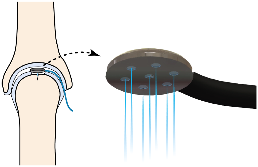
Graphic illustration of the proposed minimally invasive water jet surgical instrument. The flexible tubing that offers the water/saline supply facilitates the reachability in the tight joint spaces and the nozzle head can be designed such that it allows multiple water jets to be jetting simultaneously. As can be seen in the left picture, the opposing surface layer can act to offer the counterforce needed for the recoil and stable jetting.
The aim of this feasibility study is to determine whether water jet drilling can be safely applied as an alternative for conventional microfracture awls by studying potential side effects and perioperative complications, as well as the quality of cartilage repair tissue.
Materials and Methods
The study setup is similar to previous studies performed by our group using a caprine model. We use the caprine model, because of the reasonable similarity to a human situation in terms of cartilage thickness, weight, and healing potential28-31 and our extensive experience with this model.
Caprine Model
Six female Dutch milk goats (Capra hircus sana) were used in this study. The average weight was 77.7 kg (range 60.8-86.8 kg). Before entering the study, all goats were screened for absence of pregnancy and disease. Group housing was arranged starting at least 1 week before until 1 week after surgery to minimize stress on the animals.
Since no studies using pure water jets to drill in in vivo mammalian bone are available, no sample size calculation could be performed. Instead, the number of animals was based on earlier research on cartilage repair in large animal models.29-35 The study protocol was approved by the local Animal Welfare Committees (ORCA182).
Operative Technique
All surgical procedures were performed in a standardized manner by the first author (ACK) and an assistant using a protocol presented earlier.29,30 Through a posterolateral approach access was gained to the talus. Using a sharp surgical spoon, a chondral defect of approximately 6 mm diameter was created at the center of the talar dome. 36 The subchondral bone layer was kept intact to prevent spontaneous blood introduction and to allow the microfracture treatment by puncturing the subchondral bone layer, enabling the inflow of mesenchymal stem cells.
The chondral defects were treated with conventional microfracture or water jet drilling based on a computer-generated randomization scheme. The contralateral talus received the other treatment in the same session. This way, the goat served as its own control. Conventional microfracture was performed according to the technique originally described by Steadman et al. 4 A 1.1-mm K-wire was used to create at least 3 evenly distributed holes to a depth that resulted in adequate bleeding to produce a blood clot in the defect.
Water jet drilling was performed by using a custom-made setup. An air compressor connected to a 300-L accumulator was used to power the air-driven high-pressure pump (P160 Resato, Roden, Netherlands, www.resato.com;
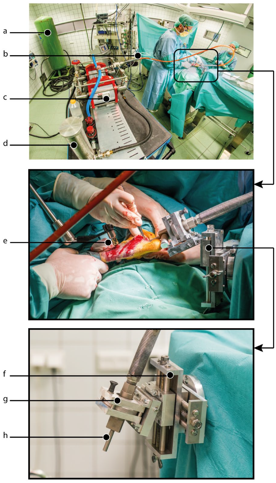
Overview of the complete water jet set up in the operating room. Top: overview of operating theatre. Center: water jet alignment of the water jet instrument with the defect in the talar bone and external fixator to fixate the goat’s leg to the operating table. Bottom: alignment tool and water jet nozzle head. (
All parts of the pump that would come into contact with saline were sterilized at 134°C and assembled under sterile conditions at the operating theatre. The pressurized saline was fed through a high-pressure hose (Holmatro, Raamsdonksveer, Netherlands) into a custom-made nozzle head consisting of a straight tube with a 0.4-mm diameter sapphire nozzle (Salomon Jetting Parts B.V., Maasdam, Netherlands). Using a low-pressure water stream, the nozzle head was aligned perpendicularly to the defect in the talar bone and fixed using a custom-made support structure that allowed omnidirectional movement (
After treatment, the surgical wound was rinsed and closed intracutaneously with absorbable sutures. Immediate postoperative weight bearing was encouraged. Anesthesia and pain medication protocols are presented in the appendix.
Follow-up
Follow-up was completed at an off-site farm under daily observation with neither food nor exercise restriction analogous to our previous published protocol.29,30 After 24 weeks, the goats were euthanized following the protocol presented in the appendix.
Analysis
To assess potential safety issues all wound and rehabilitation related adverse events were registered during surgery and daily onward until the end of the study. Also, the technical performance of the water jet and the effects of water jet drilling on the surrounding soft tissue were noted during the procedures. This provided descriptive data on the use of water jet drilling in an in vivo joint.
To determine if water jet drilling results in similar treatment execution as conventional microfracture, the visibility of holes and bleeding were noted during surgery. Also, resulting repair tissue quality was assessed by postmortem repair tissue analyses similar to those in our previous animal studies.30,31 In short, the tali were collected and photographed for macroscopic analysis using the ICRS (International Cartilage Repair Society) score by 2 independent and blinded observers (ACK, KTAL). 39 The ICRS score ranges from 0 to 12, with 0 being the worst possible outcome. Directly afterward 20 × 20 mm blocks were cut of the talus around the defect and these blocks were fixed in 4% formaldehyde in a 0.1 M phosphate buffer. A histological expert dehydrated and embedded these blocks in methylmetacrylate according to a standardized protocol. Representative histological slices at a quarter and halfway through of the defect cut, stained with hematoxylin and eosin and safranin-O and scored using the modified O’Driscoll score by 2 independent and blinded observers (ACK, KTAL). 40
Results
Complications
One postoperative complication was noted. In this case, the goat showed signs of a prolonged wound healing with swelling and reluctance to weightbearing starting 2 days after surgery on the water jet side. Antibiotics were administered which resolved the symptoms.
Water Jet Technique Performance
In 4 out of the 6 cases, the 4 to 6 water jet–drilled holes were visible after treatment in a comparable manner as the holes created with conventional microfracture (
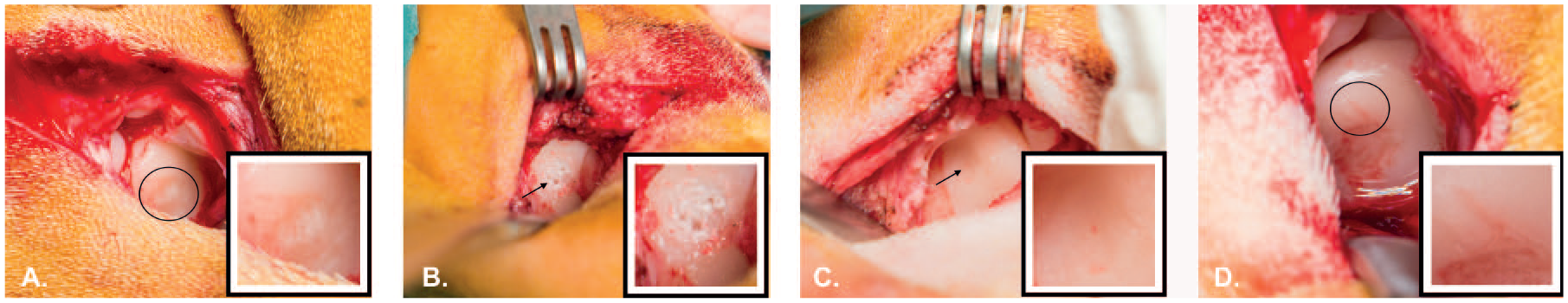
Perioperative photographs. (
We confirmed minor signs of bleeding from the water jet holes once created, but the blood took longer to appear than with conventional microfracture.
In addition, it was noted in several cases that the soft tissue around the surgical incision appeared swollen directly after treatment with the water jets due to extravasation of the saline. This phenomenon had a similar appearance as extravasation seen in prolonged arthroscopic procedures. The swelling subsided in the days after surgery.
Last, in 1 case the surgery had to be cancelled after anesthesia due to insufficient pressure build-up of the water jet pump to perform the treatment. The surgery was rescheduled and performed 1 week later after resolving the issue. Figure 4 shows representative macroscopic and histologic samples of the best and worst cases of each of the 2 surgical techniques.
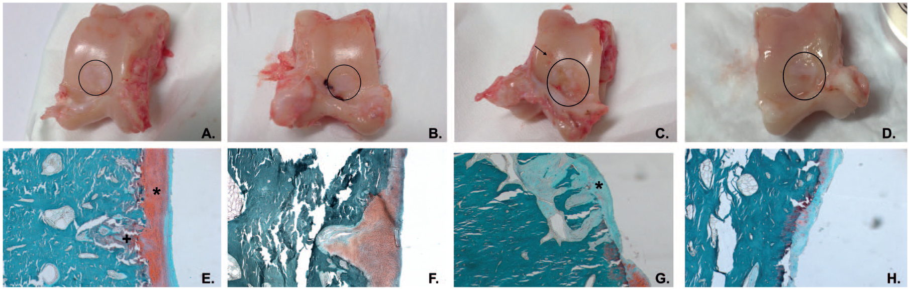
Representative examples of the macroscopic and microscopic analyses. (
Macroscopic ICRS Score
The median total ICRS score for the water jet tali was 9.5 (range: 6-12) for observer 1 and 9 (range 2-11) for observer 2 (
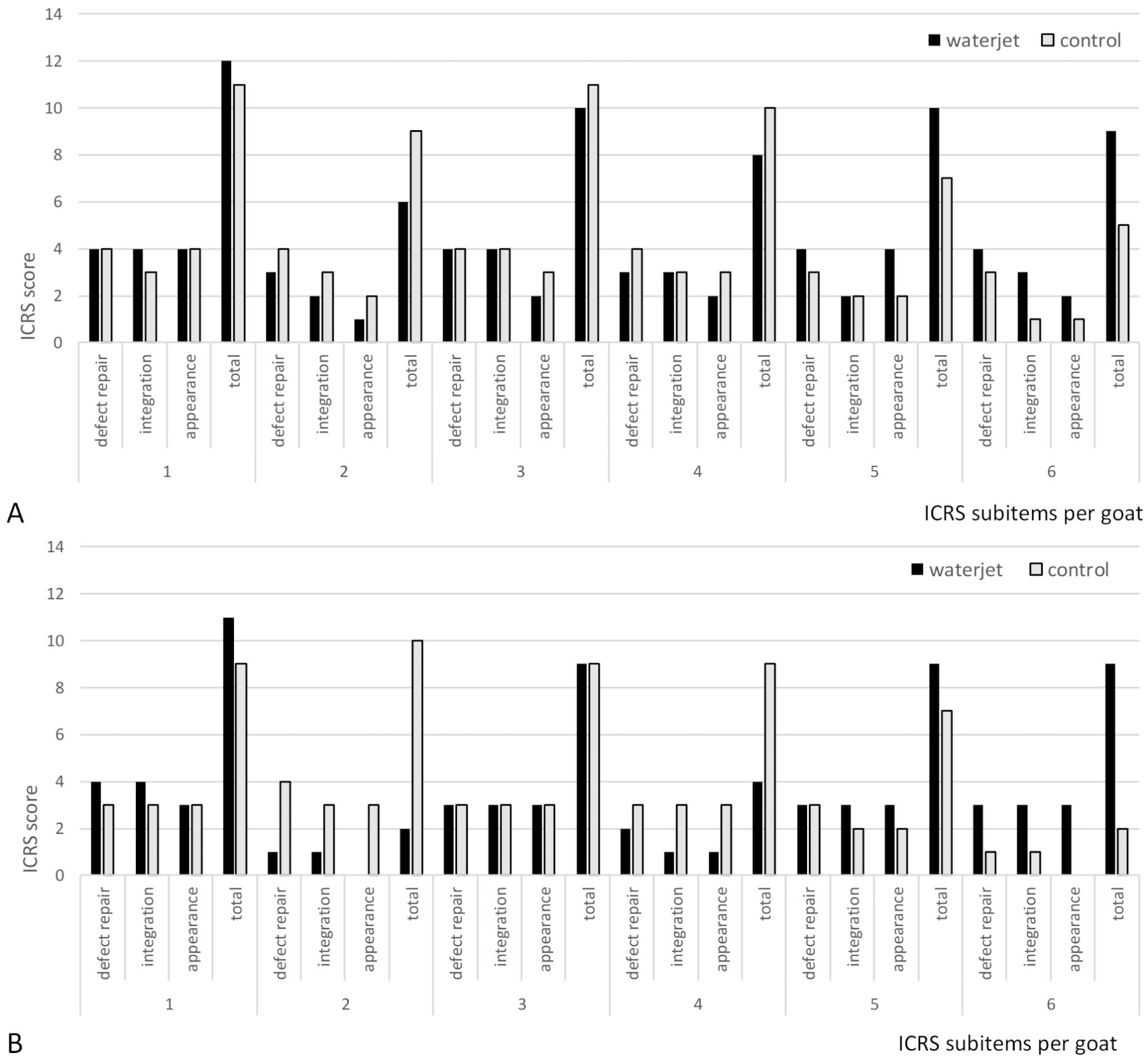
ICRS (International Cartilage Repair Society) subitems and total score for each individual goat. (
Histological Analysis
The median total modified O’Driscoll score for the water jet tali was 15 (range: 7-17) for observer 1 and 13 (range: 3-20) for observer 2 (
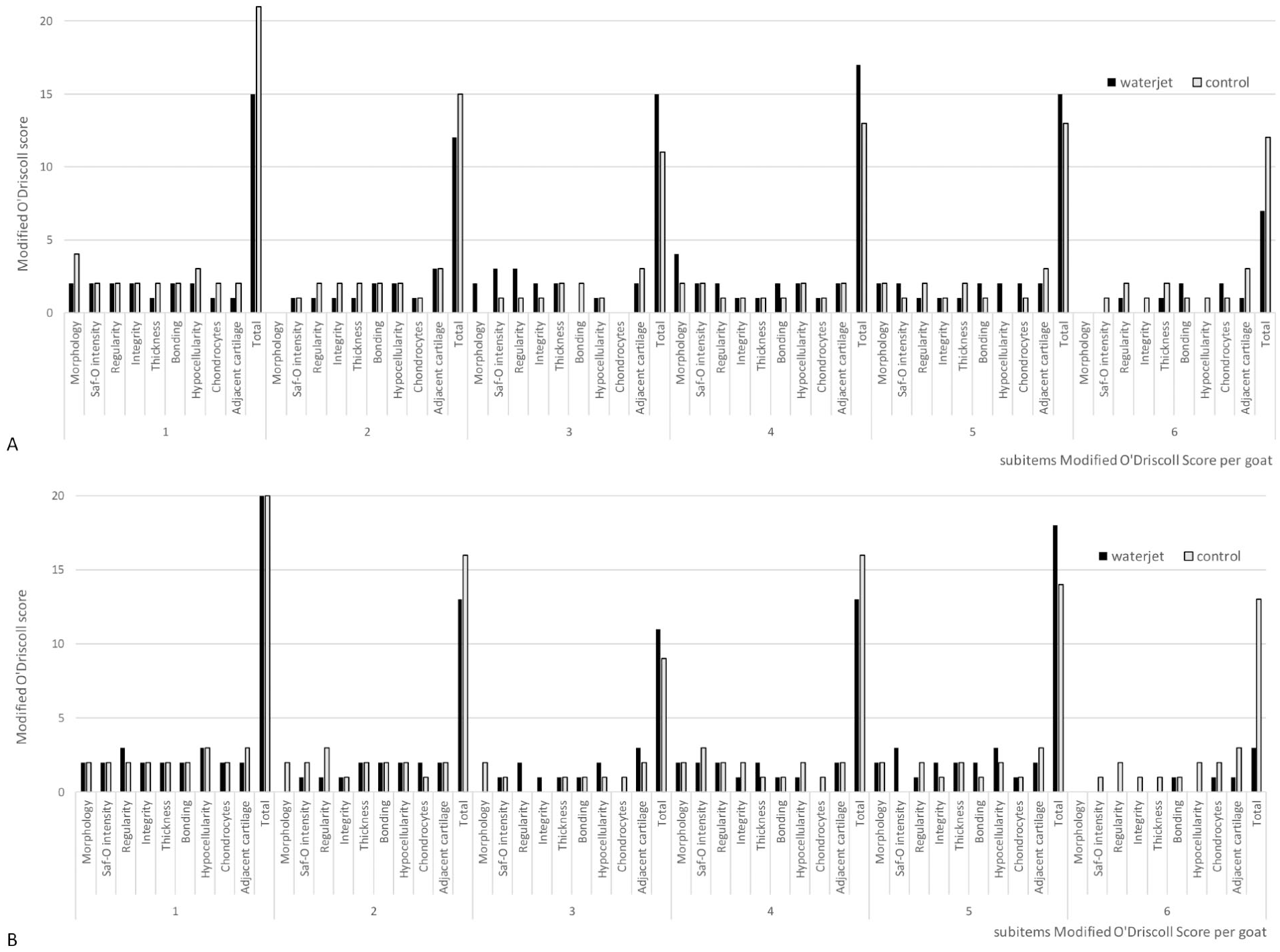
The modified O’Driscoll scores for each individual goat. (
Discussion
The aim of this study was to determine whether water jet is a safe alternative in comparison to conventional microfracture awls by studying potential side effects and perioperative complications, as well as the quality of cartilage repair tissue.
Concerning the safety and potential of water jet drilling for creating microfracture holes, we analyzed the actual effect of the drilling, the appearance of bleeding from the created holes and the postoperative complications.
There is no commercial medical water jet system available that could generate the desired water volume flow to drill into bone. To meet this high flow demand, a custom-made setup was built using all available knowledge. It was tested extensively during several pilot sessions using cadaveric caprine talar bones and total lower extremities. These pilot tests and previous studies.8,25,38 lead to the choice of conservative machine settings to guaranty a functional water jet without a potential overshoot in depth.
In the first 2 goats, we encountered difficulty with achieving a sufficiently stable fixation between the leg and the water jet nozzle because recoil from the drilling caused movement of the leg. Also, achieving the correct alignment of the nozzle over the defect to allow the desired geometry and perpendicular placement of the holes was hampered initially by relatively large nozzle head compared with the minimal exposure of the talus. This resulted in unsuccessful drilling in one case and undesired abrasion of cartilage outside the defect in another case (
The water jet setup was adjusted to fit a smaller and straight nozzle head, which increased maneuverability in alignment while producing a more coherent water jet beam and reducing potential pressure loss. Also, the caprine leg was more rigidly fixed to the operating table using an omni-tract during the procedure (
Finally, a last challenge with the custom-made set up was that in some cases the pressure during the water jet drilling would gradually drop from the initial 50 MPa at the beginning of the water jet burst to a critical low level of 35 MPa at the end. This was due to exhaustion of the compressed air in the 300-L accumulator. As it takes a couple hours for the accumulator to fill again, this effect impaired the drilling capacity of the water jet during the procedure so that not all desired holes could be drilled.
There was one superficial wound infection in our study. However, with the limited sample size in this study, we are unable to determine the actual risk of postoperative complications when using water jet drilling in talar defects compared with regular treatment at this time. Extravasation of irrigation fluid was observed into the soft tissue surrounding the caprine ankle. This was due to backsplash and to some extent this was expected. With the current settings, approximately 130 mL of water was used per water jet burst, thus in total around 0.8 L was used per talus. Extravasation of irrigation fluid has been a commonly known issue in shoulder and hip arthroscopy.43,44 It frequently leads to short-term discomfort for the patient, but true complications are rare. However, these can potentially have serious effects such as neuropraxia, increased muscle pressure leading to rhabdomyolysis, metabolic acidosis, or pulmonary edema and upper airway obstruction in the case of shoulder arthroscopy.43,45-47 Neither did we encounter such complications in our study, nor did we find a relationship between the postoperative swelling and an increased risk of wound related issues. However, the majority of the backsplash was able to leak out of the open surgical wound and when extrapolating this phenomenon to an arthroscopic setting where the water is introduced in a confined space, it could potentially become a complicating factor.
The one case in our study where the extravasation was particularly present also showed a low ICRS macroscopic score for both observers compared with the contralateral, conventionally treated, talus (6 vs. 9 for observer 1 and 2 vs. 10 for observer 2) with severe fissures in the macroscopic appearance and minimal integration into the adjacent healthy cartilage (
A previous in vivo water jet study on pigs indicated thromboembolic effects can arise due to abrasive water jet cutting, which uses a mixture of water, abrasives (hard solid cutting enhancing particles) and air (90 vol%). 24 In this study, a saline solution was used instead to avoid injecting the thromboembolic-inducing air, since previous research using only saline did not hinder the ability of water jet to drill bone25,26,38 and no respiratory anomalies were detected.
Analysis of quality of the repair tissue gave no clear difference in the macroscopic or microscopic quality of the repair tissue between groups apart from the subitem repair cartilage thickness of the modified O’Driscoll score for one observer in favor of the conventional treatment (
First, this is a feasibility animal model study with a limited sample size, artificially created chondral defects and an experimental setup of the water jet device, which makes the results sensitive to outliers and highlights limitations of the water jet setup. However, we believe the setup to be representative enough to have exposed major contraindications for water jet drilling in bone if present, such as severe impedance of the sterile surgical field or gross disruption of the cartilage repair process. Since no such undesired events occurred, using water jets seems to be a potentially safe alternative to the conventional rigid awls for microfracture in chondral defects. A follow-up study with the improved water jet set up and an ample sample size would shed light on the comparative results and could be designed based on the current results.
Second, visually confirming the created holes and assessing their geometry was more difficult than expected due to the small diameter of the holes and the longer time to visualize direct blood flow into the defect due to the supposedly high-pressure washout. We know from previous research that a smaller diameter of microfracture holes does not alter repair tissue quality.
30
Bleedings only occurred after treatment with either surgical technique, since the initial chondral defect with intact subchondral bone layer did not show any bleedings (
The next steps in the development of the water jet drilling technique will be the design of a medical pump that offers stable high pressures and a flexible arthroscopic water jet drill that is suitable for performing microfracture during an arthroscopic procedure (
In summary, this feasibility study indicates that water jet drilling in chondral defects in the caprine talus results in repair tissue quality that appears similar to repair tissue created using conventional microfracture awls. Additionally, the results highlight essential prerequisites for safe application of the water jet drilling technique: stable water pressure, water jet beam coherence, stable positioning of nozzle head when jetting, and minimizing excessive fluid extravasation.
Footnotes
Appendix
Authors’ Note
The customized set up was designed and developed at the Technical University Delft (TUD). Animal treatment was performed at the Centre for animal studies at the Vrije Universiteit Amsterdam. Histological samples were processed at the Academical Centre for Dentistry Amsterdam (ACTA).
Acknowledgments and Funding
We thank K. W. Meyer, P. Sinnige, G. Vink, K. Opdam, T. Pahlplatz, R. van Antwerpen, M. Stijntjes and T. Crijns for their practical assistance; A. Bakker and M. van Duin for their histological processing. R. van Antwerpen is also thanked for his assisting in designing the custom-made water jet head and frame. Finally, the company Resato is thanked for supplying the high-pressure pump. This work was supported by the Technology Foundation TTW, Applied Science Division of NWO, and the technology program of the Ministry of Economic Affairs, The Netherlands (Project No. 10851).
Ethical Approval
The study protocol was approved by the local Animal Welfare Committees (ORCA182).
Animal Welfare
The present study followed international, national, and/or institutional guidelines for humane animal treatment and complied with relevant legislation.