
Abstract
Objective
Excessive articular loading, for example, an ankle sprain, may result in focal osteochondral damage, initiating a vicious degenerative process resulting in posttraumatic osteoarthritis (PTOA). Better understanding of this degenerative process would allow improving posttraumatic care with the aim to prevent PTOA. The primary objective of this study was to establish a drop-weight impact testing model with controllable, reproducible and quantitative axial impact loads to induce osteochondral damage in caprine tibiotalar joints. We aimed to induce osteochondral damage on microscale level of the tibiotalar joint without gross intra-articular fractures of the tibial plafond.
Design
Fresh-frozen tibiotalar joints of mature goats were used as ex vivo articulating joint models. Specimens were axially impacted by a mass of 10.5 kg dropped from a height of 0.3 m, resulting in a speed of 2.4 m/s, an impact energy of 31.1 J and an impact impulse of 25.6 N·s. Potential osteochondral damage of the caprine tibiotalar joints was assessed using contrast-enhanced high-resolution micro-computed tomography (micro-CT). Subsequently, we performed quasi-static loading experiments to determine postimpact mechanical behavior of the tibiotalar joints.
Results
Single axial impact loads with a mass of 15.5 kg dropped from 0.3 m, resulted in intra-articular fractures of the tibial plafond, where a mass of 10.55 kg dropped from 0.3 m did not result in any macroscopic damage. In addition, contrast-enhanced high-resolution micro-CT imaging neither reveal any acute microdamage (i.e., microcracks) of the subchondral bone nor any (micro)structural changes in articular cartilage. The Hexabrix content or voxel density (i.e., proteoglycan content of the articular cartilage) on micro-CT did not show any differences between intact and impacted specimens. However, quasi-static whole-tibiotalar-joint loading showed an altered biomechanical behavior after application of a single axial impact (i.e., increased hysteresis when compared with the intact or nonimpacted specimens).
Conclusions
Single axial impact loads did not induce osteochondral damage visible with high-resolution contrast-enhanced micro-CT. However, despite the lack of damage on macro- and even microscale, the single axial impact loads resulted in “invisible injuries” because of the observed changes in the whole-joint biomechanics of the caprine tibiotalar joints. Future research must focus on diagnostic tools for the detection of early changes in articular cartilage after a traumatic impact (i.e., ankle sprains or ankle fractures), as it is well known that this could result in PTOA.
Keywords
Introduction
Osteoarthritis (OA) is the most common joint disorder, affecting over 250 million people worldwide.1,2 Although the cause of OA is considered multifactorial (genetic, inflammation, age), it is generally accepted that mechanical stress (e.g., overloading or trauma) is a key factor in the (onset and) degeneration of articular cartilage. 3 The development of posttraumatic osteoarthritis (PTOA) can remain silent for many years, since cartilage is not innervated and therefore degradation itself is not painful. Contrary to the hip and knee joints, ankle OA is predominantly of posttraumatic origin. The true prevalence of PTOA of the ankle joint is likely to be higher than the reported 78%,4,5 given the long interval between the actual trauma and the first symptoms. Ankle joint injuries are mainly caused by sports-related injuries, ranging from repetitive micro traumata (i.e., instability), sprains, and fractures. 6 As a consequence, relatively young and physically active people suffer from PTOA of the ankle joint.
Macroscopic damage occurring at the time of the actual trauma is considered to play an important role in the development of PTOA.6-8 It is known that an intra-articular fracture of the tibial plafond will result in PTOA of the ankle after decades.8,9
Unfortunately, despite ample clinical evidence that joint injuries initiate a slow process of progression towards PTOA, understanding of the etiology and pathogenesis is still limited. 10 This hampers early diagnosis following trauma and subsequent intervention to prevent development of PTOA. 11 Early osteochondral microdamage could initiate the process towards ankle PTOA 12 and therefore the current study aimed to induce osteochondral microdamage, such as fissures within the articular cartilage or microcracks in the subchondral bone. To visualize the osteochondral microarchitecture and potential damage at microscale level, contrast-enhanced micro-computed tomography (micro-CT) imaging is a commonly used imaging technique.13,14
The unique composition of hyaline cartilage enables load distribution and, together with synovial fluid, frictionless movement of articulating bones. 15 Water, the predominant component of articular cartilage (approximately 80%), provides stiffness and resilience to compression. Water is attracted to and bound by proteoglycans, which is composed of negatively charged glycosaminoglycans (GAGs) side chains attached to hyaluronic acid backbones. 16 The proteoglycans are constrained by a network of collagens, which also provides form and tensile strength. Because of the large amount of water, the chondrocytes within the articular cartilage experience mechanical stimuli as an all-sided, hydrostatic pressure, which has a chondrogenic effect.17,18 Mechanical loading thus is a key determinant of articular cartilage structure and functioning.
It is well established that ankle instability, ankle sprains, and (intra-articular) ankle fractures could result in PTOA of the ankle joint many years after the initial injury. However, it is unknown what changes occur to the articular cartilage and subchondral bone directly after the traumatic impact (i.e., ankle instability, sprains, and fractures). It is well-known that mechanical overloading will result in osteochondral damage and finally in PTOA. 19 However, from a clinical perspective we need to find out what loads are beneficial and what loads are devastating for both, intact and locally damaged articular cartilage of the ankle joint. Hence, the primary objective of this study was to establish a drop-weight impact testing model with controllable, reproducible, and quantitative axial impacts loads to induce osteochondral damage on microscale level in ex vivo caprine tibiotalar joints that allows to study the initiation of the process toward PTOA.
The secondary aims were to:
investigate what impact loads are needed to induce osteochondral damage on microscale level
visualize the osteochondral (micro)damage of the different axial impact loads, by using contrast-enhanced micro-CT
investigate the direct effects of a single axial impact load on the whole-tibiotalar-joint biomechanics.
We hypothesized that excessive articular loading in the form of a single axial impact will induce acute osteochondral damage, ranging from gross intra-articular fractures to osteochondral damage on microscale level (e.g., damaged collagen network), resulting in altered whole-tibiotalar-joint biomechanics.
Methods
Specimens
Eighteen fresh abattoir-derived tibiotalar joints of mature 3- to 5-year-old female Dutch milk goats with an estimated average weight of 80 kg (Firma Van der Horst, Maarssen, the Netherlands) were used in the current study. No further data were available concerning the exact age and weight of the specific goats. After collection, the tibiotalar joints were stored at −20°C until further preparation.
Before preparation, the specimens were thawed at room temperature for 12 hours in a bath containing phosphate buffered saline (PBS) to prevent dehydration. Thereafter, intact tibiotalar specimens were reduced to a longitudinal length of 7.5 cm in full extension through dissection of the specimens, in which the calcaneus and excessive soft tissue (muscles and tendons) had been removed but ligaments were left intact. Next, small unicortical screws were drilled into the distal talus and the proximal tibia for additional grip during the subsequent embedding process. The tibia and talus were potted within casting molds and partially embedded in a low-melting-point (48°C) bismuth alloy (Cerrolow-147). The joints were embedded in full extension with the talus directed upward. The casting mold on top of the talus was guided by a tube, which aligned the vertical axis of the tibiotalar joint in the direction of the applied loads and allowed for vertical translations and rotations of the tibiotalar joints.
Drop-Weight Impact Testing
To induce osteochondral damage in caprine tibiotalar joints, single impacts were applied using a self-developed drop-weight impact load testing system (
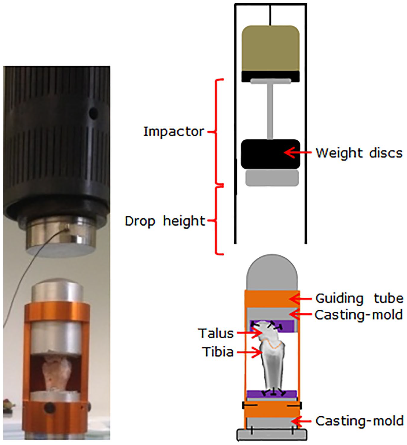
The drop-weight impact testing setup. A photographic and schematic image.
The drop-weight impact model allowed to control impacts, since the prescribed impactor properties, mass, and drop height, define both the impact energy and the impulse imposed. Except for the tibiotalar joint, all elements in the impact design were made of metal to limit their deformability, thereby assuring optimal transfer of energy and impulse to the tibiotalar joints.
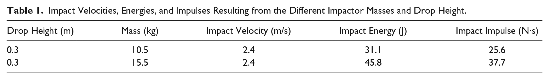
Impact testing was performed by dropping a weight of 10.5 kg or 15.5 kg onto the tibiotalar specimen from a height of 0.3 m. Three experimental groups were investigated: 2 groups with different impact magnitudes and a nonimpacted control group. Impact velocities, energies, and impulses corresponding to the impactor drop height and masses are shown in
Impact Velocities, Energies, and Impulses Resulting from the Different Impactor Masses and Drop Height.
Micro-CT Evaluation of Articular Cartilage and Subchondral Bone
The tibiotalar specimens were evaluated on the presence of impact-induced macroscopic damage, for example, intra-articular fracturing of the tibial plafond. Then, if no macroscopic damage was apparent, the osteochondral architecture of the tibiotalar specimens was assessed on microscale level with high-resolution micro-CT imaging. In order to raise the X-ray contrast (radiodensity) of microdamage in the articular cartilage, the anionic X-ray absorbing contrast agent Hexabrix 320 (Guerbet Nederland BV, Gorinchem, the Netherlands) was used.14,23,24
Prior to scanning, the specimens were immersed for 30 minutes in 200 mL of 40%/60% Hexabrix/PBS solution at 37°C. Thereafter, the specimens were fixed securely into the sample holder. To maintain a high humidity and prevent specimen dehydration, 4 mL PBS was poured into the sample holder and the top was sealed with Parafilm. Three-dimensional reconstructions of the tibiotalar articulations were obtained using a micro-CT 40 system (Scanco Medical AG, Brüttisellen, Switzerland). The specimens were scanned at room temperature, with a spatial resolution of 18 µm at 70 kVp, 114 µA, and 600 ms integration time. After the preimpact scan, the tibiotalar specimens were immersed in PBS for 3 hours to allow Hexabrix desorption prior to impact testing. Micro-CT data were evaluated on microarchitectural articular cartilage damage through a thorough examination of irregular patterns (such as fissures and grooves, dependent variable) within the homogeneous articular cartilage tissue. Contrast-enhanced micro-CT was used for a quantitative assessment of the morphology and composition of articular cartilage. We screened for structural changes in the articular cartilage by looking at cartilage thickness, volume, and micro-irregularities (i.e., fissures and grooves). Barium and sulfate ions diffused into and concentrated within void space and damaged articular cartilage. The micro-irregularities within the tibiotalar articular cartilage are quantified by counting their number and length. Then we looked at changes in proteoglycan (PG) content within the articular cartilage of the tibiotalar joint. Palmer et al. 25 showed a strong linear correlation between voxel attenuation and proteoglycan content in an EPIC-micro-CT assessment of bovine articular cartilage. The current study used Hexabrix as a negatively charged contrast agent, which equilibrates in cartilaginous tissues at concentrations inversely proportional to the local concentration of negatively charged proteoglycans or sulfated glycosaminoglycans (sGAGs). Regions of low PG content thus have relatively higher X-ray attenuation, whereas regions of higher PG content result in reduced contrast agent concentration and therefore reduced voxel density in the micro-CT images.
In order to visualize the microarchitecture of subchondral bone, a higher spatial resolution was required. Hence, the specimens were scanned for a second time using a SkyScan 1272 µCT system (Brüker micro-CT NV, Kontich, Belgium), with a spatial resolution of 3 µm at 100 kVp, 100 µA, and 800 ms integration time. The micro-CT cross sections were evaluated and visually inspected on microarchitectural damage (i.e., microcracks) in the subchondral bone or irregular patterns within the articular cartilage. Because the barium and sulfate ions from the contrast-agent Hexabrix precipitate in all void space, including the potential microcracks or fractures within the subchondral bone, the potential damage is stained by Hexabrix and appears as brighter voxels.26,27 Then the microcracks within the subchondral bone are quantified by counting their number and length.
Evaluation of Articular Mechanics
The quasi-static biomechanical properties of the tibiotalar joints were assessed at room temperature using a hydraulic mechanical testing device (Instron, model 8872, Instron and IST, Norwood, MA;
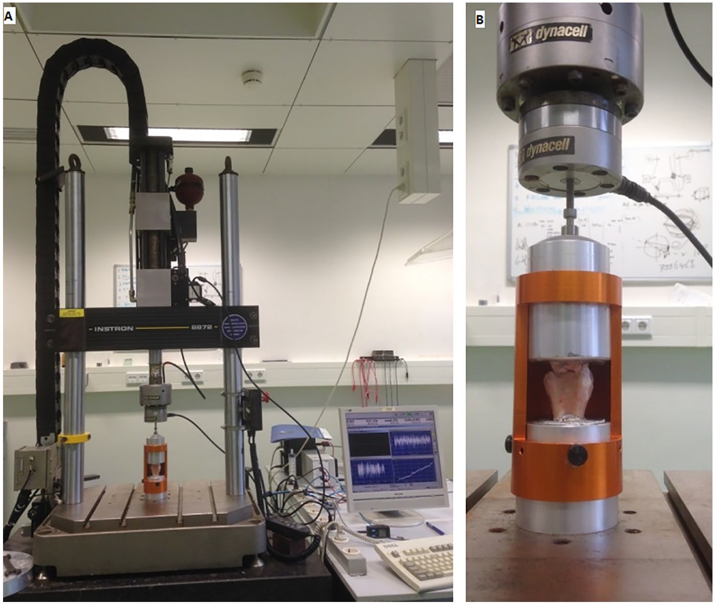
Biomechanical testing setup. (
A multi-axis FastTrack 8800 dynamic controller allowed controlling, recording, and digitizing of the position and force. The sampling frequency was set at 100 Hz. In quasi-static tests, the tibiotalar specimens were subjected to a compressive loading-unloading cycle up to a force of 200 N with a speed of 1 mm/min. A maximum compressive load of 200 N is considered well below the level of voluntary loading and was selected to avoid additional specimen damage. The time between two subsequent compression cycles was equal for both groups. A typical example of the data obtained during a quasi-static test is provided in
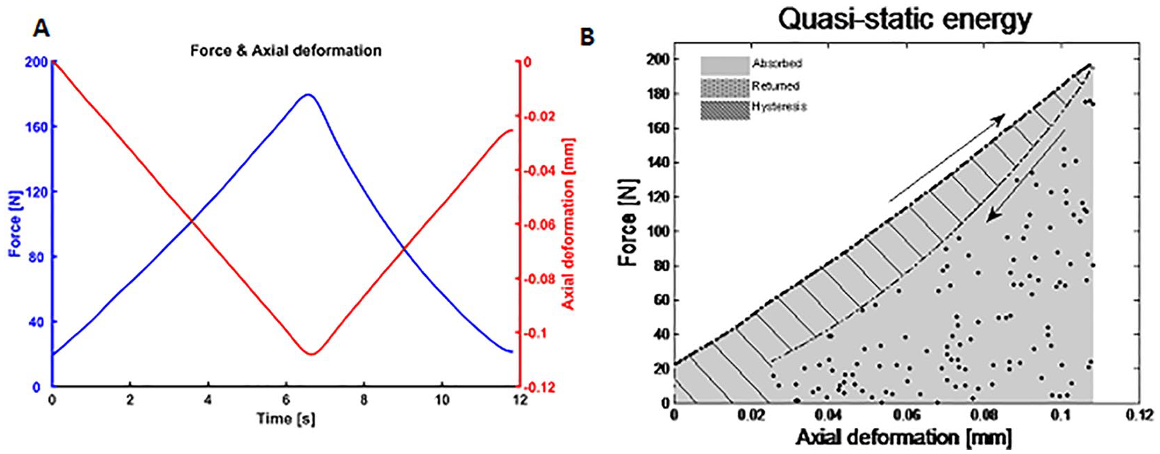
Force and axial deformation during quasi-static testing. (
Statistical Analysis
To study the acute effects of single impact loads on outcome parameters of both micro-CT imaging and biomechanical testing, three experimental groups were distinguished: (1) a group with an impactor mass of 10.5 kg, (2) a group with an impactor mass of 15.5 kg, and (3) a nonimpacted or control group. Statistical tests were performed using SPSS (version 20; IBM Corp, Armonk, NY, USA). P values <0.05 were considered as indicating statistical significance. Results were expressed as means and standard deviation (SD). Pre- versus postimpact within-subject changes in outcome parameters of both micro-CT imaging and biomechanical testing were investigated using paired t tests. For all outcome parameters of both micro-CT imaging and biomechanical testing, differences in pre- versus postimpact within-subject changes between groups were investigated using one-way independent analysis of variance (ANOVA). Thus, “time” is the within-subject factor, and “impact” is the between-subject factor. The independent variable was the condition (impacted or nonimpacted), the dependent variable was the outcome parameter. Significant effects were investigated by performing Bonferroni post hoc statistical tests. Finally, correlations between impact and biomechanical outcome parameters on the one hand and structural damage type and severity on the other hand were investigated using Pearson correlation coefficients. The assumption of normally distributed data was tested using histograms, QQ plots, values of skewness and kurtosis, and the Kolmogorov-Smirnov test. The assumption of homogeneity of variance was tested using the Levene’s test. If the assumptions of normality or homogeneity of variances were violated, the nonparametric Mann-Whitney test, Wilcoxon signed-rank test, Kruskall-Wallis test, and Kendall’s tau were carried out instead of the parametric independent t test, paired t test, one-way independent ANOVA, and Pearson correlation coefficient, respectively.
Results
All caprine tibiotalar joints that were subjected to an impactor with a mass of 15.5 kg dropped from a height of 0.3 m revealed intra-articular fractures of the tibial plafond (
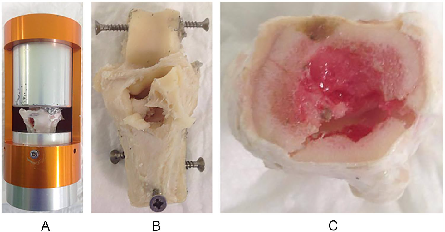
Intra-articular fractures of the impacted caprine tibiotalar joints. (
The articular cartilage and underlying subchondral bone of tibiotalar joints subjected to an impactor with a mass of 10.5 kg dropped from a height of 0.3 m, did not result in any rregular patterns or microcracks with micro-CT imaging (
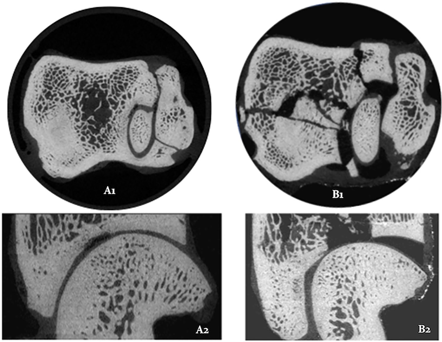
Micro-CT reconstructions from 2 impacted caprine tibiotalar joints. Typical examples of axial (1) and sagittal (2) reconstructions of caprine tibiotalar joints impacted with 10.5 kg from 0.3 m (
Because of the severe malalignment following intra-articular fractures of the tibial plafond, quasi-static testing of these joints was irrelevant. Hence, only impacted tibiotalar joints without intra-articular fractures (i.e., impacted with 10.5 kg dropped from 0.3 m) and nonimpacted tibiotalar joints were investigated in quasi-static testing.
The application of a single axial high impact load resulted in significant changes in the outcome parameters hysteresis and relative energy loss (
Hysteresis and Relative Energy Loss under Quasi-Static Loading of the Caprine Tibiotalar Joints. a
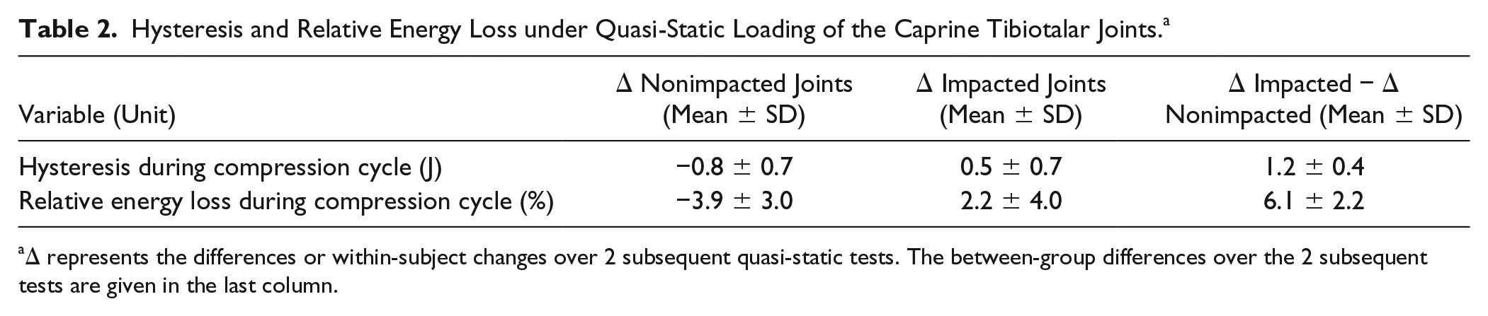
Δ represents the differences or within-subject changes over 2 subsequent quasi-static tests. The between-group differences over the 2 subsequent tests are given in the last column.
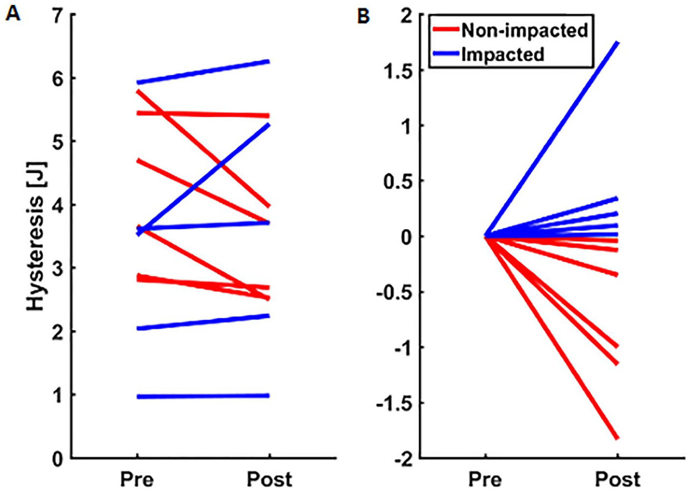
Changes in hysteresis during 2 subsequent compression cycles. Hysteresis during compression cycles in pre- and postimpact measurements for controls/nonimpacted (red) and impacted (blue) caprine tibiotalar joints, respectively.
Discussion
The primary objective of this study was to establish a drop-weight impact testing model with controllable, reproducible, and quantitative axial impacts loads to induce osteochondral damage on microscale level in caprine articulating tibiotalar joints that allows to study the initiation of the process toward PTOA. In general, single impact loads with an impactor mass of 15.5 kg dropped from a height of 0.3 m induced intra-articular fractures of the tibial plafond, whereas single impact loads with an impactor mass of 10.5 kg dropped from a height of 0.3 m did not yield any macroscopic osteochondral damage. While no macroscopic damage was observed when dropping an impactor with a mass of 10.5 kg from a height of 0.3 m, it was expected that these caprine tibiotalar joints would show osteochondral damage at the microscale level, since only 47% more impact energy and impulse resulted in intra-articular fractures in caprine tibiotalar joints. Furthermore, it is reported that a collision energy and an impulse of 50.1 J and 25.6 N·s, respectively, are required to induce intra-articular fractures of the tibial plafond in ex vivo human ankle joints. 9 This is comparable to the collision energy (45.8 J) and impulse (37.7 N·s) that was applied when dropping an impactor with a mass of 15.5 kg from a height of 0.3 m. Hence, the axial impact loads induced in these ex vivo caprine tibiotalar joints are well comparable to real-life human ankle traumas. Despite relatively high-impact loads (10.5 kg from 0.3 m), contrast-enhanced high-resolution micro-CT did not reveal impact-induced osteochondral damage, neither in the microarchitecture of the tibiotalar articular cartilage nor in the underlying subchondral bone. During contrast-enhanced high-resolution micro-CT, the anionic X-ray absorbing contrast agent Hexabrix 320 was used in order to raise the X-ray contrast (radiodensity) of microdamage in the tibiotalar articular cartilage.14,23,31 The mechanism of its action is that the negative charge of Hexabrix and sGAGs repulse each other. As sGAGs form the main component of the negative fixed charge density (FCD) in cartilage, 25 the influx of the negatively charged Hexabrix is inversely related to the sGAG content. We hypothesized that a high axial impact load will induce damage of the collagen fibers within the extracellular matrix that results in an outflow and loss of proteoglycans (sGAGs). Despite the use of postmortem samples, the proteoglycans (sGAGs) are still able to flow in and out of the articular cartilage because the specimens are immersed in PBS and Hexabrix, respectively. This loss of sGAGs could be visualized by changes in the Hexabrix density or voxel density on micro-CT. However, no differences were seen between the controls/nonimpacted and impacted caprine tibiotalar joints with contrast-enhanced high-resolution micro-CT. Perhaps osteochondral damage occurred on lower microscale or even submicron (nanoscale) levels, which could not be visualized with our currently used micro-CT scanners.
The rationale for whole-joint biomechanical testing was the study of Fazaeli et al., 32 who showed that even mild disruption or subtle damage of the collagen fibers lead to mechanical softening of articular cartilage. This affected its mechanical stability and changed the total joint metrics. Fazaeli et al. 32 induced collagen damage by digestion with collagenase, where we hypothesized that the application of a single axial high impact load will result in collagen damage that could result in mechanical changes of the whole-joint mechanics. The biomechanical tests consisting of loading-unloading cycles indeed revealed a change in mechanical behavior at macroscopic level. The control group, consisting of nonimpacted tibiotalar joints showed a decrease in hysteresis and relative energy loss over 2 subsequent quasi-static tests. This decrease is probably attributable to the extrusion of interstitial fluid in the first loading-unloading cycle, making the articular cartilage more elastic in the second loading-unloading cycle. An analogous phenomenon was observed in a study with ex vivo caprine intervertebral discs, in which a few days of diurnal physiological loading was required to achieve a dynamic equilibrium. 33 In contrast, we observed a significant increase in hysteresis and relative energy loss in the tibiotalar joint that were subjected to a single axial impact load.
Interestingly, as shown in Figure 6, the delta hysteresis (i.e., the change in hysteresis over 2 subsequent quasi-static loading cycles before and after application of a single axial impact load) is negative for all nonimpacted specimens and positive for all impacted specimens.
This is in line with the study of Malekipour et al., 29 where they applied single impact loads on equine tibiotalar joints and found a significantly higher relative energy loss in single axially impacted tibiotalar joints compared to the intact joints. Edelsten et al. 34 also described an increase in hysteresis after a controlled impact on articular cartilage. A possible explanation could be the impact-induced damage of the collagen network, allowing proteoglycans and bound water to escape. 35
While impacted tibiotalar joints showed different quasi-static behavior compared with nonimpacted tibiotalar joints, between-subject variability was still present. This variability may be attributable to differences in response to the applied impact loads. The severity of architectural damage following impact may have been affected by genetically inherited and acquired properties such as the size, previous weight, and joint surface characteristics of the particular goat.
Caprine tibiotalar joints were employed for several reasons. First, we chose an ex vivo model because this allows better quantification of both impact and damage of the articular cartilage and osteochondral bone. Second, the osteochondral properties of caprine joints translate well to humans.36-38 Third, an animal model was used because it is essential to compare the impacted cartilage with healthy cartilage. Healthy human cadaveric ankle joints vary strongly in age, size, health condition and are often derived from elderly people in whom articular cartilage may already be in a more degenerated state; this would preclude studying the initiation of PTOA.
The outcomes of the current study should be interpreted in the light of several limitations. The relatively strong curvature of the caprine tibiotalar joint surface made it difficult to pinpoint the exact location of the impact-induced damage. A second limitation of the model was the inevitable disruption of the articular capsule and subsequent loss of synovial fluid during specimen preparation. As a consequence, synovial fluid could not contribute to shock absorption during impact loading. 39 Altogether, we consider caprine tibiotalar joints to be a representative, but suboptimal ex vivo joint model to study the effect of relatively high-impact loads on articular cartilage.
The vast majority of previously conducted impact testing studies investigated the osteochondral impact response of “open-joint” models, either with in vitro explants21,40-48 or in vivo.22,49-51 However, the mechanical environment deviates considerably from actual blunt impact loads, since articular cartilage is manipulated directly in open-joint models. Besides, testing of isolated tissues, often performed in in vitro studies, does not represent the mechanical response of these tissues in situ, in that the response of articular cartilage in isolation is different29,42,44 than when firmly attached to underlying subchondral bone. 52 Thus, “articulating joint” models better mimic live trauma or impact loading conditions. Furthermore, contrary to the few previously conducted closed-joint studies,9,53 the current study attempted to induce and investigate osteochondral damage on microscale level instead of macroscopic damage or gross intra-articular fractures. 54
The clinical relevance of the current study is that the single high-impact loads affected the quasi-static biomechanical behavior of caprine tibiotalar joints, but no microscale damage of the articular cartilage or subchondral bone was visible with high-resolution contrast-enhanced micro-CT. This finding emphasizes the difficulties with visualization and quantification of trauma-induced damage at the microscale level. Moreover, it may explain why the current clinically applied imaging techniques are inadequate for detecting acute trauma-induced articular cartilage damage. Currently, osteoarthritic characteristics such as reductions in the thickness of articular cartilage, osteophyte formation, presence of subchondral sclerosis and subchondral cysts are only diagnosed after longer periods of articular cartilage degeneration (i.e., osteoarthritis) and not in an early stage directly after the actual joint trauma.
The observed biomechanical changes in quasi-static behavior may be the first step in the process toward PTOA of the ankle joint. A local bruise within the articular cartilage could initiate a subclinical process of articular cartilage degeneration that eventually results in a painful ankle joint due to posttraumatic osteoarthritis. We believe that the vicious circle of cartilage degeneration, described for knees 55 as well as intervertebral discs, 19 explains how impact-induced osteochondral damage potentially initiates the onset of trauma-induced OA. Although the initiating events of OA are not fully understood, there is growing consensus that a disturbed biomechanical environment of the articular cartilage or its underlying subchondral bone triggers a pathomechanical cascade, which eventually leads to articular cartilage degeneration. 56
In order to elucidate how impacts may initiate a process toward PTOA, future research must focus on the understanding of its etiology and pathogenesis. From a clinical perspective, the long-term goal is to find out what loads are beneficial and what loads are devastating for articular cartilage. Our trauma impact model could be used in loaded-ankle bioreactor studies in which we preserve the tibiotalar joint models and allow for a well-controlled environment with dynamic mechanical loading. Since articular cartilage is neither innervated nor vascularized, we know that fluid flow is necessary for the transport of nutrients and waste products. But mechanical overloading (e.g., a single high-impact load) is devastating for articular cartilage and results in cartilage damage, degeneration and finally PTOA.
The current study established a drop-weight impact testing model with 2 different single axial impact loads that induced (1) macroscopic damage (i.e., intra-articular fractures of the tibial plafond) and (2) significant changes in the whole-tibiotalar-joint biomechanics. The key message is that despite the lack of any osteochondral damage on microscale level, as assessed with high-resolution contrast-enhanced micro-CT, the whole-joint biomechanics altered after the application of a single axial impact load. Diagnostic tools must be developed that allow detection of early trauma-induced changes in articular cartilage and subchondral bone characteristics to prevent degeneration and progression toward PTOA of the ankle joint.
Footnotes
Acknowledgments and Funding
The author(s) received no financial support for the research, authorship, and/or publication of this article.
Declaration of Conflicting Interests
The author(s) declared no potential conflicts of interest with respect to the research, authorship, and/or publication of this article.