
Abstract
Purpose
An important feature of biomaterials used in cartilage regeneration is their influence on the establishment and stabilization of a chondrocytic phenotype of embedded cells. The purpose of this study was to examine the effects of a porous 3-dimensional scaffold made of cross-linked hyaluronic acid on the expression and synthesis performance of human articular chondrocytes.
Materials and Methods
Osteoarthritic chondrocytes from 5 patients with a mean age of 74 years were passaged twice and cultured within the cross-linked hyaluronic acid scaffolds for 2 weeks. Analyses were performed at 3 different time points. For estimation of cell content within the scaffold, DNA-content (CyQuant cell proliferation assay) was determined. The expression of chondrocyte-specific genes by embedded cells as well as the total amount of sulfated glycosaminoglycans produced during the culture period was analyzed in order to characterize the synthesis performance and differentiation status of the cells.
Results
Cells showed a homogenous distribution within the scaffold. DNA quantification revealed a reduction of the cell number. This might be attributed to loss of cells from the scaffold during media exchange connected with a stop in cell proliferation. Indeed, the expression of cartilage-specific genes and the production of sulfated glycosaminoglycans were increased and the differentiation index was clearly improved.
Conclusions
These results suggest that the attachment of osteoarthritic P2 chondrocytes to the investigated material enhanced the chondrogenic phenotype as well as promoted the retention.
Introduction
Articular cartilage has a limited potential for self-regeneration, whereby minor traumas are often the initial event in the onset of osteoarthritis. Because of synovial joint injuries, the risk of osteoarthritis is increased. The results are localized and whole joint responses which lead to progressive damage and degeneration. Osteoarthritis that develops after joint injuries, posttraumatic joint abnormalities or primarily instability causes lifelong pain for millions of people. As the number of patients increase it becomes more and more a social economic burden.
Strategies for the treatment of cartilage defects (reviewed in Farr et al. 1 and Ahmed and Hincke 2 ) range from marrow stimulating techniques (abrasion arthroplasty, 3 microfracture, 4 drilling, and spongialization) over osteochondral allograft or autograft transplantation (mosaicplasty) to cell-based therapies. 5 The latter are either based on the transplantation of autologous chondrocytes directly into the defect (autologous chondrocyte implantation [ACI]) or in combination with biomaterials (matrix-associated autologous chondrocyte transplantation [MACT])6-9 or on the implantation of an “empty” scaffold combined with marrow-stimulating techniques (e.g., autologous matrix-induced chondrogenesis [AMIC]).10-13 In the latter case, bone marrow–derived cells (blood-derived cells and mesenchymal progenitor cells) and growth factors infiltrating into the implanted scaffold might contribute to the regeneration process. 13
While on the application of marrow-stimulating techniques mainly fibrocartilage3,4 is generated, approaches combining these or cell-based techniques with biomaterials (tissue engineering) allow the formation of hyaline or hyaline-like cartilage exhibiting improved biomechanical properties and prolonged durability and thus resembling better the native, healthy tissue,6,13-15 and showing better clinical outcome. 10 Thereby, the employed biomaterial has to serve as a cell carrier matrix (scaffold) featuring good biocompatibility and biodegradability and providing optimal physical and biochemical conditions for proper cell adhesion and the establishment and/or stabilization of a chondrogenic phenotype of cells, which is crucial for the quality of the repair tissue. Currently, a series of different biomaterials composed of natural polymers (collagen,10,11,16,17 fibrin, 18 gelatin, agarose, alginate,19,20 chitosan, 21 or hyaluronic acid [HA]7,9,22-24), synthetic polymers (polyglycolic acid [PGA],12,14 polylactic acid [PLA], poly(lactic-co-glycolic acid) [PLGA],6,13 poly(ethylene glycol) [PEG]) or composites thereof8,25-27 are under experimental investigation or already clinically applied. 2
Of these, HA, an ubiquitous macromolecular polysaccharide of repeating disaccharide units of N-acetyl-
Already there is a huge variety of HA scaffolds available, but only a few are clinically used. 37 Most of these HA scaffolds like Hyalograft C or Hyalofast use an esterified derivative of hyaluronic acid to compose a biodegradable scaffold for regenerative medicine. The advantage of these nonwoven membranes, fibers, or sponges is their thermal stability that leads to best mechanical properties and promising results in both in vitro and clinical studies.38,39 Alternatives for building HA biomaterials is by auto-cross-linking, where HA self-aggregates over hydrophobic interactions. These interactions are weak, and so different conditions and temperatures can affect aggregate formation and dissociation. ACP (auto-cross-linked polymer) a white lyophilized powder, is hydrated to a transparent gel and commonly used to repair tissue defects. 38
Cross-linking with polyfunctional groups can produce scaffolds that offer the advantages of controlled degradation rates, best mechanical properties and good biological responses. 40
When chondrocytes are liberated from cartilage tissue and expanded in 2-dimensional monolayer culture, cells lose properties associated with the chondrocyte phenotype. This is characterized by the expression of cartilage-specific genes (e.g., collagen II and aggrecan) and increased synthesis of collagen I and versican, which is also reflected by the deterioration of a differentiation index (ratio of type II collagen to type I collagen). Embedding chondrocytes into an appropriate 3-dimensional environment, dedifferentiation can be halted and the occurring redifferentiation process can be used as a model system for the determination of the impact of a biomaterial on the chondrocyte phenotype. 39 The aim of this study was to examine the suitability of a porous 3-dimensional matrix of cross-linked HA as a scaffold for cartilage regeneration. Therefore, dedifferentiated, human chondrocytes were cultured within the HA scaffold and the effect of the material on the expression of cartilage specific genes, the differentiation status and sulfated glycosaminoglycans (sGAG) synthesis was evaluated.
Materials and Methods
Isolation of Cells
Human articular cartilage was received from the County Hospital Krems from osteoarthritis patients (grade 2 or 3 in the Kellgren and Lawrence system for classification of osteoarthritis of the knee) undergoing total knee arthroplasty. In all cases (5 patients [4 female, 1 male] aged between 68 and 84 years), informed consent was obtained and the study was approved by the regional Ethical Committee (GS4-EK-4/064-2009).
For chondrocyte isolation, articular cartilage was minced into 2 mm3 pieces prior to enzymatic digestion with Liberase TM (0.2 WU/mL, Roche Diagnostics GmbH, Mannheim, Germany) in medium (GIBCO DMEM/F12 GlutaMAX-I, Invitrogen, LifeTech Austria, Vienna, Austria) with antibiotics (penicillin 200 U/mL; streptomycin 0.2 mg/mL and Amphotericin B 2.5 µg/mL [Sigma-Aldrich Chemie GmbH, Steinheim, Germany]) under permanent agitation for 18 to 22 hours at 37°C. The resulting chondrocyte suspension was passed through a Cell Strainer with 40 µm pores (BD, Franklin Lakes, NJ) to remove undigested debris, washed with phosphate-buffered saline, centrifuged (10 minutes, 500g, room temperature) and resuspended in growth medium (i.e., medium supplemented with antibiotics (see above), 10% fetal calf serum (PAA Laboratories GmbH, Linz, Austria) and 0.05 mg/mL ascorbic acid (Sigma-Aldrich Chemie GmbH, Steinheim, Germany)). Viability was determined via trypan blue (Sigma-Aldrich Chemie GmbH, Steinheim, Germany) staining and cells were counted using a hemocytometer.
The isolated cells were seeded in growth medium in 75 cm2 culture flasks (Nunc, Rochester, NY) at a density of 1 × 10⁴ cells/cm2 and cultivated at 37°C in a humidified environment with 5% CO2. Medium was changed every 2 to 3 days till 80% confluency. For passaging, cells were harvested by use of accutase (1.5 mL/flask; PAA Laboratories GmbH, Linz, Austria), counted, and seeded again (P1 cells). After this second round of expansion, cells were harvested again and used for seeding onto HA scaffolds (P2 cells).
Hyaluronic Acid Scaffolds
Scaffolds made of cross-linked HA were produced and provided by Croma Pharma GmbH. HA was derivatized with a linker containing a thiol group. Thiol-modified HA was dissolved in deionized water at a concentration of 1.5% (w/v) and the pH adjusted to 7.1 to 7.3 using NaOH. The solution was poured into 12-well plates (0.9-2 g per well for different hydrogel heights) and kept at room temperature for 5 hours. Under these conditions, thiol groups slowly form disulfide bridges. The resulting hydrogel was lyophilized using the following parameters: The plates were frozen overnight in a −80°C freezer, followed by the primary drying step for 24 hours. Temperature of product footprint was −10°C to −15°C, while the condenser temperature was −50°C to −55°C. The vacuum applied was 0.05 to 0.1 mbar. Secondary drying step was for 2 hours using the same parameters. The resulting HA scaffolds were punched to obtain sizes suitable for in vitro experiments. Scaffolds from 3 different batches exhibiting similar structural organization and pore sizes were used for 3 independent experiments. For determination of pore structure, the scaffold was observed by using a Hitachi Tabletop Microscope TM-1000.
Morphometric Analysis
For determination of the pore size the cross-linked HA scaffolds were rehydrated and then dehydrated by using the Tissue-Tek VIP 5 Jr. (Sakura, Torrance, CA) before they were embedded in paraffin. Slices of 8 µm thickness were cut from the paraffin blocks by using the CUT 5062 microtome (SLEE, Mainz, Germany) and colored by hematoxylin and eosin staining. The following analyzation was done by Leica DM 1000 light microscope and the Leica QW analysis software (Leica, Wetzlar, Germany).
Seeding and Cultivation of Scaffolds
Circular scaffolds with a diameter of 22 mm were cut into 4 equally sized pieces of area = 0.95 cm2. Each quarter was placed in an Eppendorf reaction tube and seeded with 2 × 10⁵ human, osteoarthritic chondrocytes (P2) in 100 µL growth medium. After a 4-hour incubation period at 37°C allowing for cell attachment, 900 µL growth medium was added per tube and incubation at 37°C was continued for 1, 7, or 14 days, respectively. Half of the medium was exchanged every 3 to 4 days.
Electron Microscopy of Seeded Scaffolds
Cell morphology within the scaffold was studied by scanning electron microscopy. Samples were fixed with 3% glutaraldehyde in 0.2 M sucrose in phosphate-buffered saline for 24 hours at 4°C, dehydrated in a graded ethanol series, dried with hexamethyldisilazane, coated with gold in a Q150R rotary-pumped sputter coater (Quorum Technologies Ltd, East Grinstead, UK) and observed on a Hitachi Tabletop Microscope TM-1000.
Quantification of DNA Content
DNA content within seeded scaffolds was determined in order to estimate alterations in cell numbers during the period of cultivation. At the time points indicated (1, 7, and 14 days after seeding), scaffolds were centrifuged (5 minutes, 3.890g, room temperature) and frozen at −80°C after removal of the supernatants. DNA quantification was performed using the CyQUANT DNA kit (Molecular Probes, LifeTech Austria, Vienna, Austria) according to the manufacturer’s instructions. In brief, frozen scaffolds were thawed, dissolved by addition of 1 µL β-mercaptoethanol (Sigma-Aldrich Chemie GmbH, Steinheim, Germany) and supplemented with lysis buffer containing the CyQuant dye. After a 5-minute incubation period, fluorescence was measured using a Synergy 2 microplate reader (BioTek Instruments, Inc., Winooski, VT) with excitation at 480 nm and emission detection at 520 nm. A cell number standard curve was used to convert sample fluorescent units into cell number per scaffold. λ-DNA standard was used to prepare a standard curve for DNA content. The experiment was performed twice (with cells derived from 2 different patients) with scaffold seeding in triplicate for each time point (n = 6).
Gene Expression Analysis
To monitor the expression of chondrocyte-specific genes, the amounts of mRNA of collagen II (COL2A1), aggrecan (ACAN), SRY (sex determining region-Y)-box 9 (SOX9) as well as glyceraldehyde-3-phosphate dehydrogenase (GAPDH; for normalization) and collagen I (COL1A1; marker for dedifferentiation) were determined by quantitative reverse transcriptase-polymerase chain reaction (RTqPCR) from chondrocytes grown in monolayer or in scaffolds at 1, 7, and 14 days after cell seeding. Therefore, using the RNeasy plant mini kit (Qiagen, Hilden, Germany), the centrifuged scaffolds were dissolved with β-mercaptoethanol and transferred to a QIAshredder spin column placed on a 2 mL collection tube. The further procedure for isolating total RNA was executed as described in the RNeasy Mini Handbook (Qiagen, Hilden, Germany, 06/2001). RNA was stored at −80°C until it was used for reverse transcription. Complementary DNA (cDNA) was synthesized with 1st Strand cDNA Synthesis Kit for reverse transcriptase PCR (Roche Diagnostics GmbH, Mannheim, Germany) and Random Primer p(dN)6 according to the supplier’s instruction. cDNA was stored at −20°C until it was used for real-time PCR.
A dual-labeled probe–based real-time PCR was performed with FastStart TaqMan Probe Master (Roche Diagnostics GmbH, Mannheim, Germany) and with gene-specific primers (Eurofins MWG Synthesis GmbH, Ebersberg, Germany) in triplicate on the iCycler iQ (Bio-Rad Laboratories, Hercules, CA). Probes and primers were selected by use of Universal Probe Library System and by applying in silico PCR (Roche). The primer-dependent optimal annealing temperature was determined experimentally. qPCR was carried out as follows: initial denaturation step at 95°C for 10 minutes, further denaturation at 95°C for 30 seconds, an annealing step at 55°C to 62°C optimized for the respective primers ( Table 1 ) for 30 seconds, a polymerization step at 72°C for 15 seconds.
Sequences of Primers and Conditions Used in Real-Time Polymerase Chain Reaction.
The data resulting from the fluorescence measurement were relatively quantified without efficiency correction with R = 2−∆Ct [MEAN target − MEAN reference] method. 41
Gene expression analysis was performed for three independent experiments with cells derived from 3 different patients in triplicate for each time point (n = 9). For control purpose and in order to demonstrate further dedifferentiation of chondrocytes in 2-dimensional monolayer culture, cells grown in 6-well plates (data from 3 experiments in triplicate each for day 1 [n = 9]; from 2 experiments in triplicate for day 7 and day 14 [n= 6]) in parallel were analyzed additionally.
Measurement of Sulfated Glycosaminoglycans (sGAG)
The quantification of sGAG was conducted according to Barbosa et al. 42 In brief, scaffold culture supernatants collected during media exchange and the scaffolds were treated overnight with 25 U/mL proteinase K (Sigma, St. Louis, MO) at 56°C. After inactivation of the enzyme (90°C, 10 minutes) the scaffold was destroyed with a pestle and pooled with the respective supernatants from media exchange before the whole sample was centrifuged. The supernatant was collected in ultrafree filter reaction tubes of 0.1 µm pore size (Millipore, Billerica, MA) and centrifuged (12,000g, 4 minutes, room temperature). One milliliter of a 1.9-dimethyl-methylene blue solution (DMMB) was added to 100 µL filtrate and vigorously mixed to allow the formation of complexes of DMMB and sGAG in the sample. The complexes were pelleted via centrifugation (12,000g, 10 minutes, room temperature) and subsequently dissolved in decomplexation solution. After 30 minutes of shaking, the absorbance at 656 nm was measured photometrically using an Ultrospec 3300 pro photometer (Amersham Bioscience plc, Amersham, UK). The sGAG amount was calculated from a standard curve with shark chondroitin sulphate (Sigma, St. Louis, MO). The experiment was performed twice (with cells from 2 different patients) with scaffold seeding in triplicate for each time point (n = 6). sGAG synthesis rate was calculated using mean values.
Statistical Analysis
All data are expressed as means ± standard deviations. Statistical analysis was performed using the independent-samples T test using SPSS software (SPSS, Inc, Armonk, NY). Statistical significance was set at P ≤ 0.05.
Results
Scaffold Structure
The pore structure and pore size distribution of the HA scaffold was monitored by electron microscopic and morphometric analysis. Figure 1 shows the surface structure of the material at the corner of a quartered scaffold (A) and the inner pore structure of a sliced scaffold (B). Measurement of the pore sizes showed that more than 95% of the pores were in a range between 50 and 200 µm. Pore walls were perforated with small, circular ports interconnecting the pores. The surface was partially closed with enough pores remaining open to allow for a good seeding efficiency of the material.
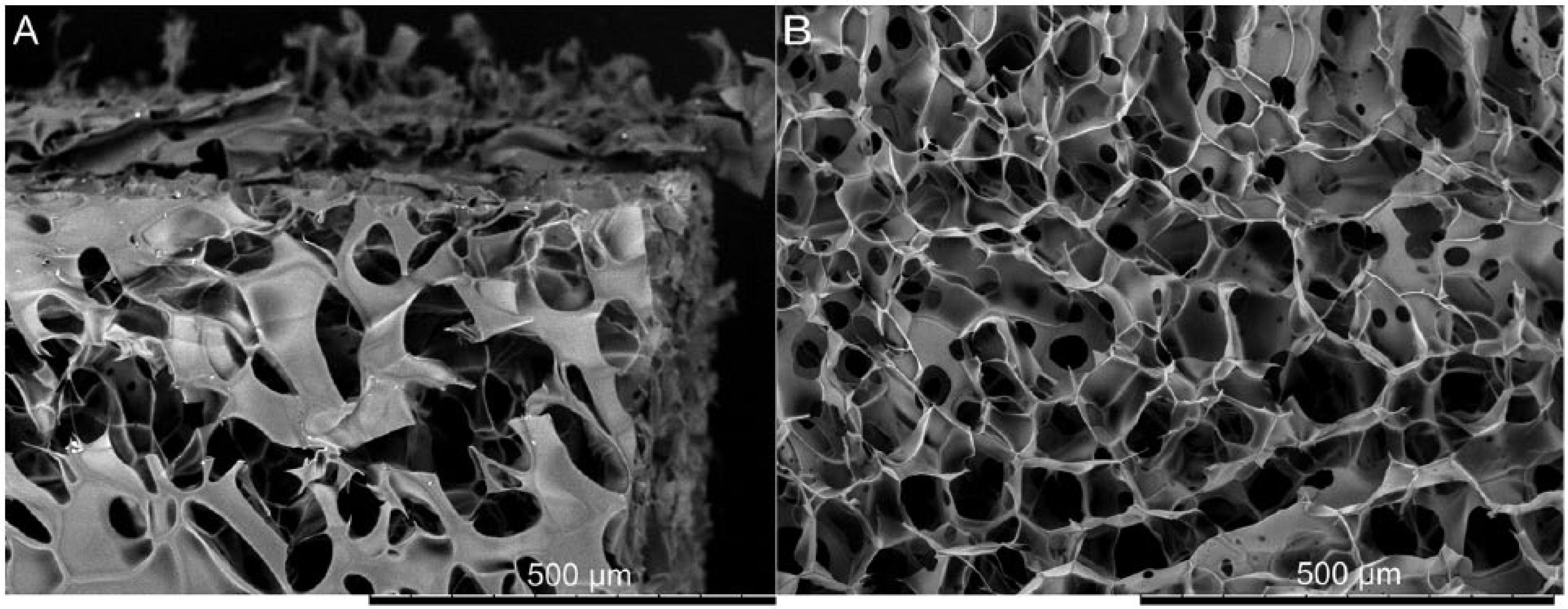
Pore structure of the hyaluronic acid (HA) scaffold. (
Scaffold Seeding and DNA Quantification
Dedifferentiated human, osteoarthritic chondrocytes were cultured in HA scaffolds (2 × 10⁵ cells per scaffold) for up to 14 days. Because of the open surface structure, the cell suspension was taken up quickly and cells were well distributed ( Fig. 2A ). Single as well as clusters of cells of spherical morphology were attached to the scaffold ( Fig. 2B ); while in the monolayer the cells showed their typical elongated morphology.
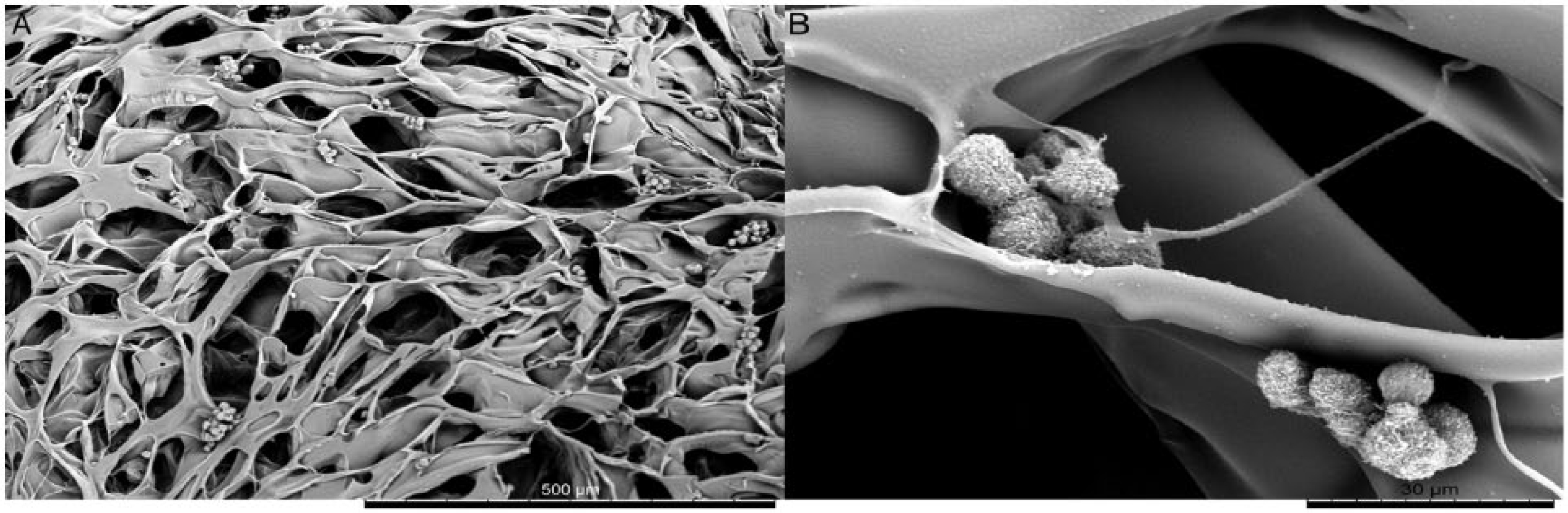
Scanning electron microscopy images of cell-seeded scaffolds 1 day after seeding with magnifications of 200 (
The cell number per scaffold at different time points after seeding was determined by measurement of DNA content and using a cell number standard curve in order to assess the seeding efficiency and potential cell proliferation. The seeding efficiency determined on day 1 after seeding was 85.5%. However, during the 14 day cultivation period, cell numbers continuously decreased significantly from about 1.7 × 10⁵ to about 1.2 × 10⁵ cells per scaffold ( Fig. 3 ).
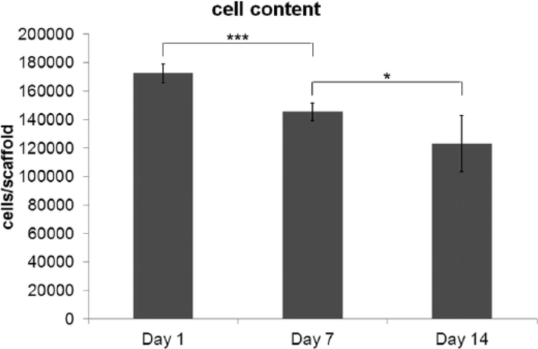
Cell content of hyaluronic acid (HA) scaffolds at different time points after seeding (day 1, day 7, and day 14).
Expression of Cartilage-Specific Genes and Cell Redifferentiation
In control cells grown in monolayer, the mRNA level of Col I ( Fig. 4A ) stayed constant during the 14-day cultivation period. However, Col I expression by cells cultured in HA scaffolds immediately decreased to half of the level of control cells on day 1, continued to decline until day 7 and remained at this low level until the end of the cultivation period. In contrast to Col I, Col II expression ( Fig. 4B ) rapidly dropped from day 1 to day 7 and even further until day 14 in cells grown in monolayer reflecting the continuing dedifferentiation process. On the contrary, cells cultivated within the 3-dimensional HA scaffold, though initially exhibiting a lower Col II expression than control cells, kept their Col II mRNA level constant throughout the whole cultivation period. The next cartilage-specific gene tested, ACAN ( Fig. 4C ), did not show such dramatic changes like Col I or Col II. But its mRNA level rose at least slightly within cells grown in HA scaffolds until day 14, while it stayed constant in control cells. The transcription factor SOX9 playing a crucial role in skeletal development and chondrocyte differentiation and driving the expression of cartilage specific genes as Col II and ACAN. SOX9 was expressed to significantly lower levels in control cells than in cells from HA scaffolds throughout the whole test period ( Fig. 4D ).
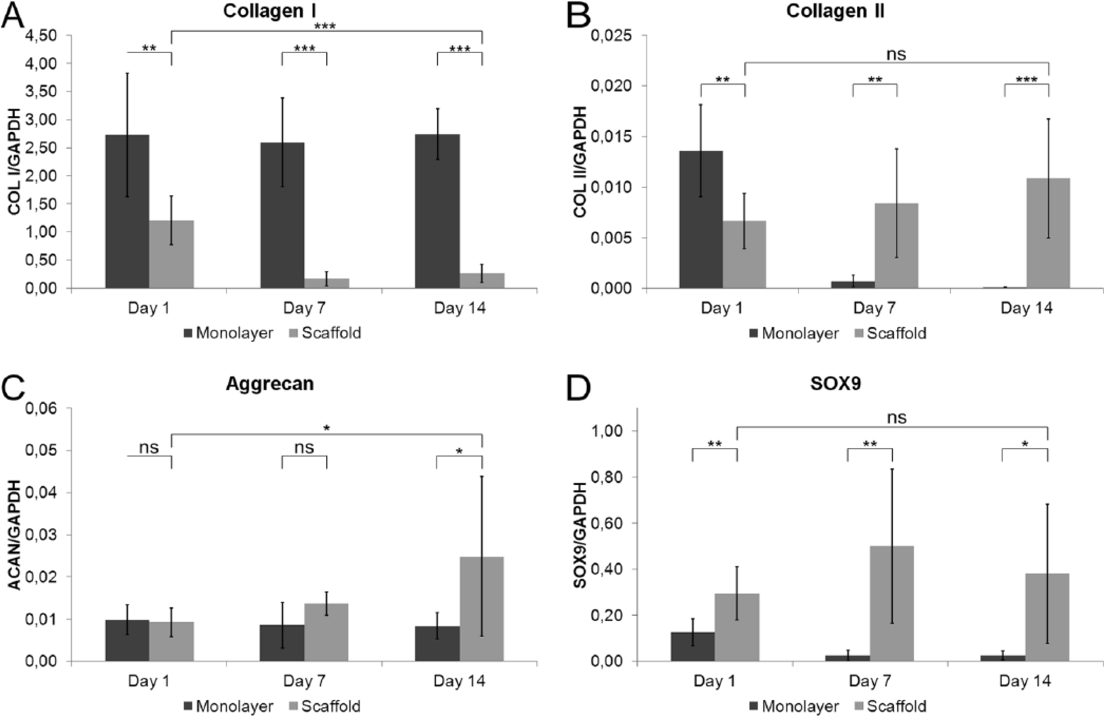
Semiquantitative analysis of Col I (
To evaluate the influence of the 3-dimensional HA scaffold on the differentiation status of chondrocytes, a differentiation index (ratio of type II collagen to type I collagen) of control cells and cells cultured in scaffolds were calculated for each time-point ( Fig. 5 ). Control cells showed a continuous decline of the differentiation index from day 1 to day 14 which was caused by an overall decrease of Col II with constant levels of Col I expression by these cells. Cells from HA scaffolds showed a similar initial differentiation status as control cells (day 1). However, it was increased by an order of magnitude within the first week of cultivation and stayed the same during the rest of the test period demonstrating clearly the maintenance of the differentiated phenotype of the cells. This improvement of differentiation index is based on the reduction of Col I with simultaneous constantly high Col II expression.
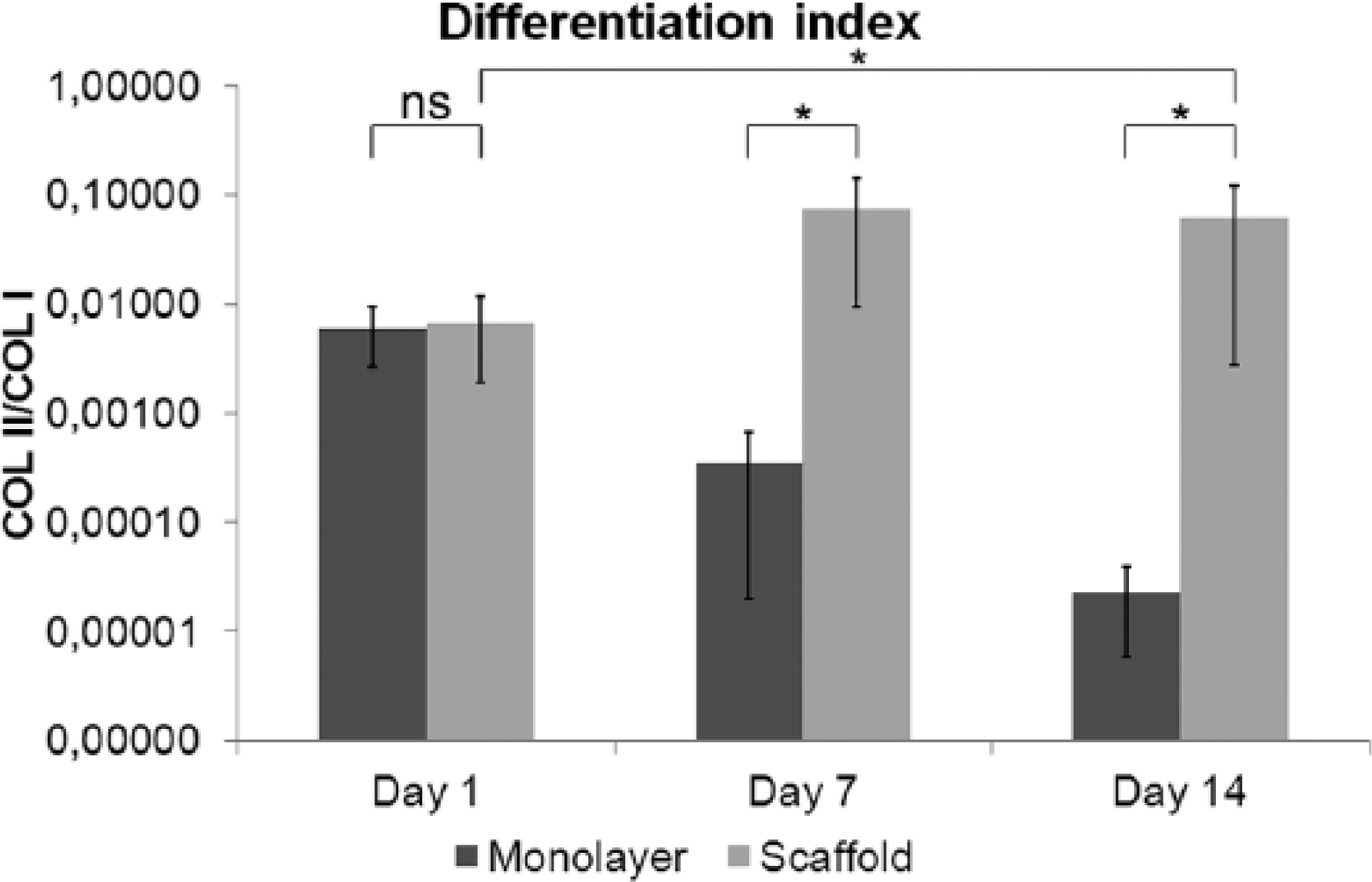
Differentiation index (Col II/Col I ratio). Human, osteoarthritic chondrocytes grown in monolayer (dark gray bars) or cultured in hyaluronic acid (HA) scaffolds (light gray bars) at different time points (1, 7, and 14 days). Results of 2 or 3 different experiments performed in triplicate each (n = 9 for HA-cultured cells at all time points and monolayer cells on day 1 and n = 6 for monolayer cells on day 7 and day 14) are shown.
Synthesis of sGAG
To correct the sGAG to the cell numbers, sGAG contents were normalized to total DNA. The outcome was an augmented sGAG content as the extent of the net increase went up significantly during the first and second week of incubation ( Fig. 6 ).
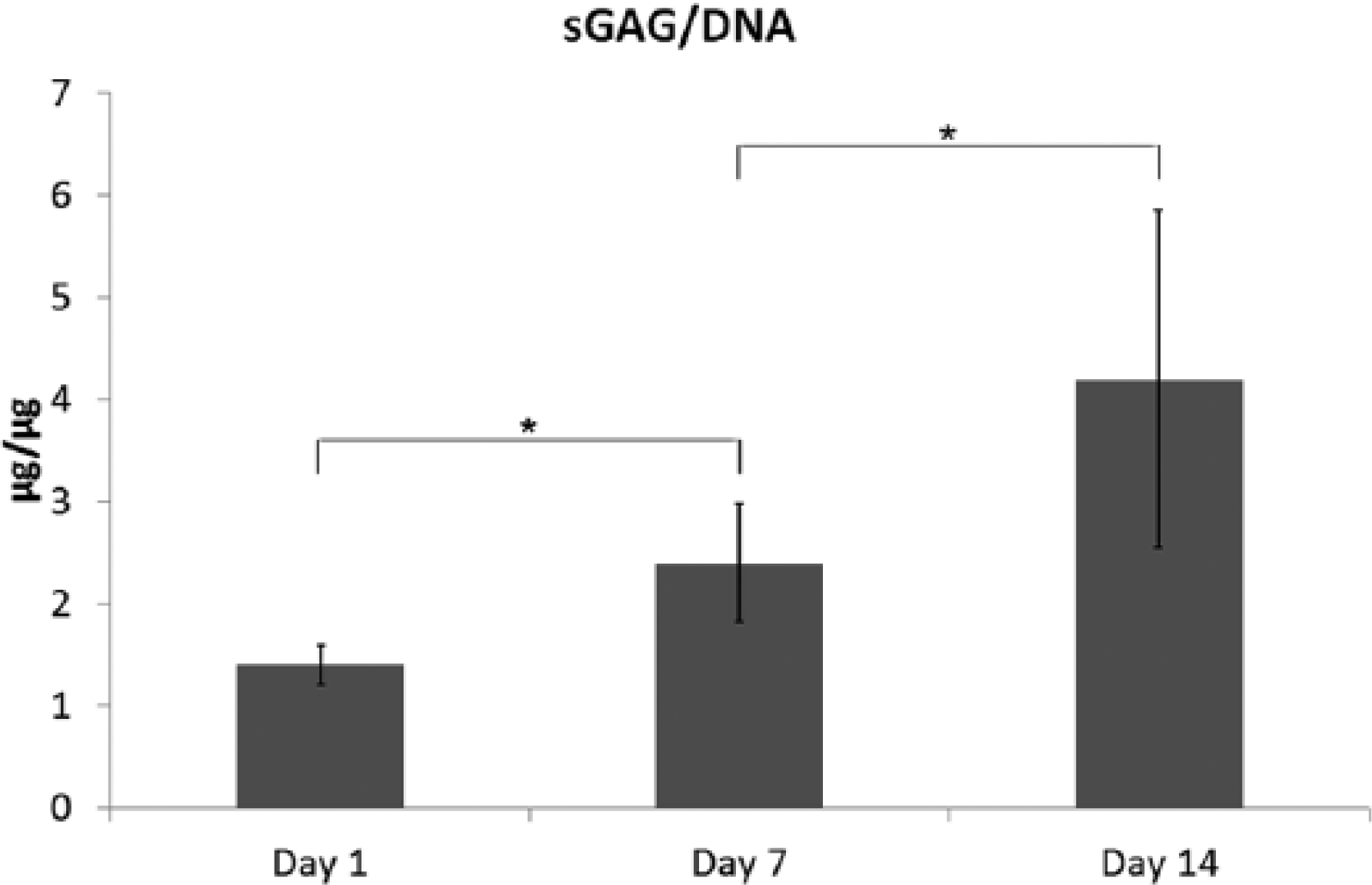
Analysis of sulfated glycosaminoglycans (sGAG) production. Hyaluronic acid (HA) scaffolds seeded with human, osteoarthritic chondrocytes were analyzed up to the different time points (1, 7, and 14 days) after cell seeding. Results of 2 different experiments performed in triplicate each (n = 6) are shown.
Discussion
Restorative techniques for cartilage defect treatment, including tissue engineering approaches, have been found to be superior to strict reparative techniques that are based on bone marrow stimulation alone with regard to the quality of the newly synthesized tissue. 2 The latter lead to the production of collagen type I–rich fibrocartilage, which allows filling of the defect and covering the exposed bone, but has inferior biomechanical properties than the native tissue and, thus exhibits only limited durability. In contrast, tissue engineering based strategies that employ biodegradable scaffolds in combination with autologous cells have the potential to generate hyaline or hyaline-like cartilage tissue with its composition and properties resembling better the native tissue.13,14 Thereby, the composition of the scaffold has a major impact on the performance of embedded cells. 43 Cell-matrix interactions and the effects of the selected biomaterial on the cellular phenotype lay the foundation of a proper healing process. Thus, the ability of a biomaterial to modulate the cellular phenotype and synthesis performance is a crucial criterion for its applicability in tissue engineering.
Today there are always discussions if osteoarthritic chondrocytes fulfill the prerequisites for the use in tissue engineering. 7 It might be able to regenerate cartilage, if cells are taken from macroscopically intact cartilage regions and are applied under suitable conditions. Ex vivo, osteoarthritic chondrocytes show similar expression patterns for collagen type I and II and aggrecan to that of healthy cells. Differences were shown in the gene expression of inflammatory factors like interleukin-1β or tumor necrosis factor-α. These factors could cause problems for the regeneration process with transplants, as they reduce type II collagen expression and activate catabolic factors. 44 Also important is the upregulation of the hypertrophic marker collagen type X in osteoarthritic chondrocytes, which might limit their use for tissue engineering as well.45,46 However, in a study using 3-dimensional HA scaffolds (Hyaff-11), differences in gene expression between osteoarthritic and healthy chondrocytes were diminished as osteoarthritic chondrocytes show a comparable chondrogenic differentiation. Also, the risk of differentiation into the hypertrophic cartilage lineage was not increased. It is suggested that chondrocytes from human osteoarthritic cartilage can fulfill the requirements for use in cartilage regeneration, 47 if they are expanded in vitro in a quality suitable for scaffold-augmented autologous chondrocyte transplantation. 44
The aim of this study was to evaluate the suitability of a new type of scaffold made of cross-linked HA for employment in cartilage regeneration. Therefore, we determined its impact on the redifferentiation as well as the synthesis performance of dedifferentiated human osteoarthritic chondrocytes in an in vitro cell culture system. The pore size of the used scaffold ranged between 50 and 200 µm and it showed good pore interconnectivity, which was reflected by quick rehydration and homogenous distribution of cells.
Within the 2-week cultivation period, the cell number decreased significantly. This indicates that embedded cells did not proliferate or were lost during media exchange or through degradation of the scaffold. Indeed, data gained from gene expression analyses and the measurement of sGAG synthesis fitted perfectly into this scenario. The quick elevation of the SOX9 mRNA level immediately after the introduction of the cells into the 3-dimensional environment of the HA scaffold compared with that of control cells grown in monolayer indicated the immediate onset of genetic reprogramming within the cells. As a consequence of dedifferentiation, collagen II expression decreased dramatically within control cells, while aggrecan and collagen I mRNA levels stayed constant. In contrast, chondrocytes cultured within HA scaffolds, exhibited constant expression of the cartilage specific collagen II and an increase of the aggrecan mRNA synthesis. The lower collagen II mRNA level in cells cultured in HA scaffold on day 1 might be reasoned by adaptation process occurring within cells on delivery into the 3-dimensional environment or through matrix effects. The slight increase of aggrecan expression and the significantly higher expression of SOX9 compared with control cells fitted perfectly to the observations of collagen II expression. It showed that genetic reprogramming within these cells directing chondrogenic re-differentiation started in the first week on the delivery of the cells into the 3-dimensional environment. The decline of SOX9 expression from day 7 to day 14 after initial boost occurring between day 1 and day 7 might be a result of a negative feedback regulation of gene expression. Taking these data together, the significant recession of collagen I of embedded cells in a 3-dimensional environment is more evident than cells cultured in 2 dimensions. This was also validated by the clear improvement of the differentiation index in cells grown in HA scaffolds, while it was continuously lowered in control cells. The chondrogenic performance of cells within the HA scaffold observed on RNA level could be confirmed by analyses of the synthetic behavior with respect to sGAG. The synthesis of sGAG is another indicator for the establishment of a chondrocytic phenotype of the cells and allows the evaluation of the cellular synthesis performance. The decreased cell number from day 1 to day 14 and the rising amount of sGAG per µg DNA clearly point to an increasing synthetic activity of those cells remaining within the scaffold.
Thus, our data rule out that the investigated material, a new type of scaffold made of cross-linked HA, has the potential to modulate the chondrocyte phenotype in a way favoring a more differentiated status. It has a positive impact on the synthetic performance of embedded chondrocytes with respect to the production of cartilage-specific components like collagen II, aggrecan and sulfated glycosaminoglycans. It would have been very interesting and not a weakness of this study if the novel HA scaffold would have been compared with already existing and clinically used HA scaffolds like Hyaff-11 or Hyalofast. However, the in vivo performance of the material has to be tested in animal studies that are currently being carried out.
Footnotes
Acknowledgments and Funding
The authors would like to acknowledge the Austrian Research Promotion Agency (FFG, grant 820129) and Croma Pharma GmbH Austria for supporting this study.
Declaration of Conflicting Interests
The author(s) declared the following potential conflicts of interest with respect to the research, authorship, and/or publication of this article: Renate R. Baumgartner and Sonja Höller are employees of Croma Pharma.
Ethical Approval
Ethical approval for this study was obtained from the regional Ethical Committee (GS4-EK-4/064-2009).
Informed Consent
Written informed consent was obtained from all subjects before the study.