
Abstract
Objectives
Schefflera leucantha leaves have traditionally been used to relieve respiratory symptoms. This study aimed to develop and evaluate a stable herbal lozenge containing S. leucantha extract, emphasizing physicochemical properties, standardization of bioactive markers, and cytotoxicity assessment to ensure safety and quality.
Methods
Four solvent systems were explored to extract phytoconstituents from S. leucantha leaves. The aqueous extract (M1), obtained by decoction, was selected based on extraction yield, moisture content, and phytochemical profile. M1 was standardized for betulinic acid using validated HPLC-DAD and evaluated for antioxidant, anti-inflammatory, and antimicrobial activities. Safety assessments included cytotoxicity, heavy metal, and microbial contamination tests. Lozenges formulated with M1 were examined for pharmacopoeial quality and stability over six heating–cooling cycles.
Results
M1 exhibited the highest extraction yield (31.29 ± 0.87%), lowest moisture content (0.03 ± 0.01%), and highest betulinic acid content. It demonstrated strong antioxidant and anti-inflammatory activities (IC50 = 1.41 ± 0.02 mg/ml) and mild antimicrobial activity against Staphylococcus aureus, while remaining non-cytotoxic. Lozenges met USP 41–NF 36 standards for hardness, friability, and content uniformity, with >98% betulinic acid release within 30 min and maintained stability (98.3 ± 1.8% to 98.2 ± 2.0%) after testing.
Conclusion
The aqueous extract of S. leucantha leaves was rich in betulinic acid and phenolic compounds, showing antioxidant, anti-inflammatory, and antimicrobial effects without cytotoxicity, as confirmed by the MTT assay. Compressed lozenges containing the extract, Aerosil® 200, magnesium stearate, and peppermint powder showed rapid betulinic acid release within 30 min, remained non-cytotoxic, and met microbial and heavy metal safety standards. All quality parameters before and after stability testing complied with USP 41–NF 36 requirements. These findings support the potential of S. leucantha lozenges as a safe, stable, and effective natural product for respiratory health.
Introduction
Respiratory conditions are commonly associated with bronchial inflammation and microbial infections, often characterized by coughing accompanied by mucus production. 1 Commercial treatments for these conditions typically contain synthetic ingredients, which may cause adverse side effects. In contrast, herbal-based formulations offer a promising alternative, leveraging natural compounds with potential therapeutic properties and a lower risk of toxicity. For instance, leaves of Schefflera leucantha, known in Thai as “Hanuman Prasankai,” belongs to the Araliaceae family, have traditionally been used in Thai herbal medicine to treat cough, respiratory inflammation, and bacterial infections.2–4 One of its key phytoconstituents, betulinic acid—a pentacyclic triterpenoid—exhibits multiple pharmacological effects, including antioxidant, anti-inflammatory, bronchodilatory, and anti-allergic activities, with no reported toxicity following oral administration. 5 Notably, betulinic acid has been shown to mitigate particulate matter (PM2.5)-induced lung inflammation and tissue damage by downregulating pro-inflammatory mediators and enhancing endogenous antioxidant defenses. 6 Furthermore, this compound has demonstrated antiviral activity by inhibiting the 3CL protease, thereby impairing the replication of severe acute respiratory syndrome coronavirus 2 (SARS-CoV-2). 7 In light of these findings, this study aimed to develop standardized herbal lozenges containing S. leucantha leaf extract, using betulinic acid as a marker compound. The objective was to provide a novel, natural remedy for supporting respiratory health, particularly for the relief of chronic cough and throat inflammation.
The evolution of herbal lozenges is rooted in a long-standing tradition of local medicine, which employs combinations of medicinal herbs to manage respiratory symptoms. To formulate lozenges containing the bitter-tasting S. leucantha extract, additional herbal powders were incorporated to mask the bitterness and enhance the overall therapeutic efficacy. Specifically, herbal ingredients such as Glycyrrhiza glabra roots, Phyllanthus emblica fruits, Solanum indicum fruits, and Zingiber officinale rhizomes were selected based on their well-documented bronchodilatory, antioxidant, antimicrobial, and anti-inflammatory properties. 8 Licorice, derived from the root of G. glabra (Leguminosae family), is widely used in Ayurvedic medicine to manage respiratory ailments such as sore throat, cough, and bronchitis. Glycyrrhizin, the main sweet-tasting compound in licorice, functions as both an expectorant and a demulcent. 9 Indian gooseberry (P. emblica), recognized for its sour flavor, is rich in bioactive compounds such as ascorbic acid, emblicanins, phyllaemblicin B, and punigluconin. These constituents exhibit antioxidant, anti-inflammatory, antimicrobial, and immunomodulatory properties, 10 which may support respiratory health and relieve inflammation-related symptoms. S. indicum (Solanaceae family), also known as poison berry, is traditionally used in Ayurvedic formulations to treat respiratory issues including cough and dyspnea. Its slightly sour fruits contain bioactive compounds such as steroidal alkaloids, coumarins, and flavonoids, 11 which contribute to its therapeutic effects. Ginger (Z. officinale), a well-known medicinal plant from the Zingiberaceae family, is rich in bioactive constituents such as gingerols, shogaols, and paradols. These compounds provide its characteristic pungency and are responsible for its antioxidant, anti-inflammatory, and immunostimulant activities. 9 Collectively, the inclusion of these four adjunct herbal ingredients not only improves the sensory properties of the lozenges but also enhances their biological activity in managing respiratory conditions. Therefore, this study aims to develop and evaluate standardized herbal lozenges containing S. leucantha extract and synergistic herbal components for the effective relief of cough, throat irritation, and inflammation.
Lozenges are solid, single-dose preparations designed to dissolve slowly in the mouth, providing localized effects in the oral cavity and throat. This makes them particularly suitable for relieving cough and soothing throat irritation. As the lozenge gradually erodes, the active ingredients are released and dissolved in saliva, enabling sustained local action. The formulation of lozenges is critically influenced by the selection of active ingredients and excipients, especially binders, which affect both the dissolution rate and patient acceptability, including taste and mouthfeel. 12
In light of these findings, this study aimed to develop standardized herbal lozenges containing S. leucantha leaf extract, using betulinic acid as a marker compound. The objective was to provide a novel natural remedy to support respiratory health, particularly in relieving chronic cough and throat inflammation. To achieve this, a potent leaf extract was selected and incorporated into a standardized lozenge formulation, which was evaluated for quality control parameters, bioactive marker content, cytotoxicity, and stability to ensure the safety, efficacy, and robustness of the final product.
Materials and Methods
Study Location and Duration
All experiments, including extraction, phytochemical analysis, biological activity evaluation, and formulation development, were conducted at the Faculty of Pharmacy, Silpakorn University, Nakhon Pathom, Thailand, from September 2023 to December 2024.
Materials and Reagents
Dried leaves of S. leucantha were purchased from CPS Pharma, a Thai herbal shop in Nakhon Pathom, Thailand, in September 2023. The plant material was identified by our research group and authenticated by Assistant Professor Dr Arissarakorn Sirinamarattana. A herbarium specimen (No. VS002) was deposited at the Faculty of Pharmacy, Silpakorn University, Thailand. Herbal excipients were obtained from the Ban-Hua-Tung Herb Shop in Phitsanulok, Thailand. Citric acid, mannitol, sodium benzoate, magnesium stearate, peppermint oil, and menthol were purchased from Chemical Express Co., Ltd, Bangkok, Thailand. Polyvinylpyrrolidone (PVP) K30 and colloidal silicon dioxide (Aerosil® 200) were sourced from Gibthai Co., Ltd, Bangkok, Thailand, and Wacker Chemie AG, Munich, Germany, respectively.
Folin-Ciocalteu reagent and inductively coupled plasma-mass spectrometry (ICP-MS) multi-element standard solution XIII were purchased from Loba Chemie, Mumbai, India, and Agilent Technologies, Santa Clara, USA, respectively. 2, 2-Diphenyl-1-picrylhydrazyl (DPPH), ferric reducing antioxidant power (FRAP) reagent, ascorbic acid, gallic acid, chloramphenicol, diclofenac diethylamine, nitric acid, potassium bromide (KBr), silica gel 60 F254 aluminium plates, silica gel 60 (0.2-0.5 mm), Iscove's Modified Dulbecco's Media (IMDM), 3-(4,5-dimethylthiazol-2-yl)-2,5-diphenyltetrazolium bromide (MTT), microbiological culture media, and reagents for phytochemical screening were acquired from Merck KGaA, Darmstadt, Germany. Betulinic acid (HPLC grade ≥ 98%) was obtained from Sigma-Aldrich, Missouri, USA. The solvents and other chemicals used in this study were of analytical grade and obtained from Merck KGaA, Darmstadt, Germany.
Preparation of Extracts
Approximately 500 g of S. leucantha leaves were dried in the shade with frequent turning to reduce moisture content and prevent fungal growth. The dried leaves were then ground using an electric herbal powder grinder (Powder Grinder, ECO model, Spring Green Evolution, Thailand) and sieved through a 40-mesh sieve to obtain a fine powder with particle size smaller than 425 µm. Approximately 500 g of S. leucantha dried leaf powder (average particle size: 345.85 ± 2.23 µm) was boiled in 5 L of distilled water for 30 min. Additional water was added as needed to maintain the volume during decoction. The powder was also macerated three times in 5 L of 40%, 70%, and 95% v/v ethanol solutions for three days each, with periodic stirring at room temperature (25 ± 1 °C), resulting in four dark brown extracts labeled M1, M2, M3, and M4, respectively. The extraction methods were adapted and modified from Potduang et al (2007). 4 After extraction, each filtrate was individually concentrated to dryness at 45 °C using a rotary evaporator (CH-9230, Flawil, Switzerland). All extracts were stored in the dark at 4 °C prior to analysis and formulation development. The moisture content of each extract was determined using the hot-air oven drying method.
Analysis of Phytochemical Constituents
Standard phytochemical screening procedures,13,14 with slight modifications, were employed to detect the presence of tannins, reducing sugars, alkaloids, saponins, terpenoids, flavonoids, steroids, and glycosides in the four S. leucantha extracts.
Thin-layer chromatography (TLC) was performed according to our previously established protocol, 14 with modifications to the mobile phase systems. The solvent systems used were ethyl acetate:methanol:acetic acid (19:0.8:0.2, v/v/v) and methanol:ethyl acetate:dichloromethane:cyclohexane (1:6:3:3, v/v/v). Spraying reagents included tetrazolium blue, anisaldehyde–sulfuric acid, vanillin–sulfuric acid, and Dragendorff's reagent.
Fourier-transform infrared (FTIR) spectra of the extracts were recorded using a spectrometer (Nicolet Magna 4700, Thermo Fisher Scientific, Santa Clara, CA, USA) with the potassium bromide (KBr) pellet method.
Determination of Total Phenolic Contents (TPC)
The total phenolic content (TPC) was determined in triplicate using the Folin-Ciocalteu method, as described previously. 14 Methanolic solutions of each extract were mixed with Folin-Ciocalteu reagent and sodium carbonate solution, then incubated in the dark for 90 min. The absorbance was measured at 765 nm using a UV-visible spectrophotometer (U-2990, Hitachi, Tokyo, Japan). TPC values were calculated from the standard calibration curve of gallic acid (y = 0.0296x + 0.0796, r² = 0.9997) and expressed as milligrams of gallic acid equivalents per gram of dried extract (mg GAE/g extract).
Validation of HPLC Method and Determination of Betulinic Acid Content
A high-performance liquid chromatography method with a diode-array detector (HPLC-DAD; HPLC 1100 series, Agilent Technologies, Santa Clara, USA) was developed and validated for the separation and quantification of betulinic acid in S. leucantha extracts and in samples obtained from dissolution studies. Chromatographic separation was performed using an Inertsil® ODS-3 reversed-phase analytical column (4.6 × 250 mm, 5 µm; GL Sciences, Inc., Ibaraki, Japan) with isocratic elution. The mobile phase consisted of acetonitrile, methanol, and acidified water (pH 3.0, adjusted with formic acid) in the ratio of 70:20:10 (v/v/v), at a flow rate of 1.0 ml/min. Detection was carried out at a wavelength of 210 nm. The procedures for separation, quantification, and method validation were adapted from the general approaches described by Patel and Trivedi, 15 who reported the analysis of betulinic acid using a Kromasil 100 RP-C18 column (5 µm, 250 × 4.6 mm i.d.) with an isocratic mobile phase of methanol–acetonitrile–water (90:5:5, v/v/v; pH 2.5, adjusted with orthophosphoric acid), a flow rate of 1.3 ml/min, and detection at 270 nm.
Method validation was performed according to the International Conference on Harmonization (ICH) guidelines for the validation of analytical procedures (ICH Q2[R1]), ensuring the reliability and reproducibility of the developed HPLC method. 16 For this purpose, five concentrations of standard betulinic acid solution (0.025-0.800 mg/ml) were analyzed to assess linearity, accuracy, and precision. Intraday accuracy and precision were determined by analyzing five replicates of each concentration within a single day, while interday assessments were carried out once daily over six consecutive days.
To evaluate extraction recovery, the extracts were spiked with known concentrations of betulinic acid, and the spiked samples were analyzed in comparison to unspiked controls. Recovery values were expressed as a percentage of the measured concentration relative to the theoretical spiked amount.
The limits of detection (LOD) and quantitation (LOQ) were determined based on the linear regression data using the standard deviation of the response and the slope of the calibration curve.
To prepare the sample solutions, each dried extract was dissolved in methanol to achieve a working concentration of approximately 1.0 mg/ml. The solutions were filtered through a 0.45 µm membrane filter, and 20 µl aliquots of the clear filtrate were injected into the HPLC system. All experiments were conducted in triplicate.
Evaluation of Biological Activities
The antioxidant activities of the extracts (n = 3) were evaluated using the DPPH and FRAP assays, based on previously established protocols.14,17 For the DPPH assay, each extract was mixed with DPPH solution and incubated in the dark at ambient temperature for 30 min. The decrease in absorbance was measured at 515 nm using a UV-visible spectrophotometer (U-2990, Hitachi, Tokyo, Japan). A calibration curve was prepared using L-ascorbic acid (y = 9.9438x + 7.2724, r² = 0.9997). The DPPH radical scavenging activity was expressed as SC50 values (µg/ml). In the FRAP assay, each sample was incubated with the FRAP reagent in the dark at 37 °C for 30 min, and the absorbance was measured at 593 nm. A calibration curve was generated using L-ascorbic acid (y = 0.2028x + 0.1694, r² = 0.9994). FRAP results were expressed as milligrams of ascorbic acid equivalents per gram of dried extract (mg AAE/g extract).
In vitro anti-inflammatory activity was evaluated using the albumin denaturation inhibition method, as previously described. 18 Fresh egg albumin, phosphate-buffered saline (pH 7.4), and sample solutions were mixed and incubated at 37 °C for 15 min, followed by heating at 70 °C for 5 min. After cooling to room temperature, the absorbance of the reaction mixtures was measured at 660 nm using a UV-visible spectrophotometer. A calibration curve was constructed using diclofenac diethylamine as a standard (y = 14.717x + 39.953, r² = 0.9993). The anti-inflammatory activity was expressed as the IC50 value (mg/ml), representing the concentration required to inhibit 50% of albumin denaturation.
A modified paper disc diffusion method was employed to assess the antimicrobial activity of the extracts. Sterile filter paper discs (6 mm in diameter) were individually loaded with 2.5 mg of extract and placed on the surface of Mueller-Hinton agar plates that had been inoculated with a 5-h culture of Staphylococcus aureus ATCC 19381, containing approximately 1.5 × 10⁸ colony-forming units (CFU)/ml. Discs impregnated with the corresponding solvents served as negative controls, while chloramphenicol (30 µg/disc) was used as a positive control. After incubation at 37 °C for 18 h, the diameters of the inhibition zones were measured. The minimum inhibitory concentrations (MICs) against S. aureus were determined using the micro-broth dilution method according to the Clinical and Laboratory Standards Institute (CLSI) guidelines (M07, 11th edition). 19 The initial bacterial concentration in Mueller-Hinton broth was adjusted to 1.0 × 10⁶ CFU/ml. Chloramphenicol and inoculated broth without extract were used as positive and negative controls, respectively. Following incubation at 37 °C for 24 h, the MIC was recorded as the lowest concentration at which no visible turbidity was observed. All experiments were conducted in triplicate.
Cytotoxicity
The cytotoxicity of the selected S. leucantha extract and the final formulated lozenges was evaluated using the MTT colorimetric assay 20 on human dermal fibroblasts (ATCC CRL-2076; Manassas, Virginia, USA). Each sample was diluted in Iscove's Modified Dulbecco's Medium (IMDM) to achieve the desired concentration range. Cells were seeded at a density of 1 × 10⁴ cells/well in a 96-well microplate and cultured until reaching 80%–90% confluency. The cells were then treated with 100 μl of media containing the samples and incubated for 24 h at 37 °C in a 5% CO2 incubator. Following this, 10 μl of 5 mg/ml MTT solution was added to each well and incubated for an additional 2 h. After the supernatant was removed, 100 μl of dimethyl sulfoxide (DMSO) was added to dissolve the formazan crystals formed by metabolically active cells. Absorbance was measured at 550 nm using a Fusion universal microplate analyzer (Model A153601, Packard BioScience Company, Connecticut, USA). Cell viability was calculated as a percentage relative to untreated control cells.
Preparation of S. leucantha-Based Herbal Lozenges
The granule ingredients—including S. leucantha aqueous extract (M1), various herbal powders, and excipients—are listed in Table 1. Lozenges were prepared in 500 g laboratory-scale batches using the wet granulation method. Medicinal powders (40-80 mesh), such as M1, G. glabra roots, P. emblica fruits, S. indicum fruits, and Z. officinale rhizomes, were thoroughly blended with pulverized citric acid, plum powder, and mannitol using geometric dilution in a planetary mixer for 10 min. PVP K30 and sodium benzoate were dissolved in two-thirds of the prescribed binder (distilled water or 70% v/v ethanolic solution), according to the formulation in Table 1. The resulting solution was gradually added to the herbal mixture under continuous stirring for 10 min.
Extraction Methods, Extracting Solvents, Yields, Water Contents, Retention Factors (Rf) on Thin-Layer Chromatography (TLC) Plates, and Wavenumbers (cm−1) on Fourier-Transform Infrared (FTIR) Spectra of S. leucantha Extracts.

*Each value is represented as mean ± SD (n = 3); means with different superscript letters (a–d) within the same column are significantly different (p < .05), as determined by one-way ANOVA followed by Tukey's post hoc test using SPSS Statistics version 16.0.
**The mobile phase consists of ethylacetate: methanol: acetic acid (19:0.8:0.2).
***The mobile phase consists of methanol: ethylacetate: dichloromethane: cyclohexane (1:6:3:3).
The resulting mass was further granulated using the remaining binder and passed through a mesh-12 sieve to obtain wet granules. These were dried at 60 °C until a moisture content of 3%–5% was achieved, then passed through a No. 16 sieve. The dried granules were blended with Aerosil® 200, magnesium stearate, and peppermint powder for 20 min. The composition of S. leucantha lozenges is presented in Table 2. Each lozenge, with a total weight of 1 g, contained 100 mg of dried S. leucantha extract and was compressed using a biconvex single-punch tablet press machine. 21
Total Phenolic Contents (TPC), Antioxidant Activities (DPPH and FRAP Assays), Anti-Inflammatory Activities, Antimicrobial Activities Against S. aureus ATCC 19381 of S. leucantha Extracts.

Each value is represented as mean ± SD (n = 3); means with different superscript letters (a–e) within the same column are significantly different (p < .05), as determined by one-way ANOVA followed by Tukey's post-hoc test using SPSS Statistics version 16.0; N/A: not applicable.
Evaluation of Lozenges
The specifications for the final formulated S. leucantha lozenges were established based on three production batches. Five in-process tests—weight variation, thickness, diameter, hardness, friability, and disintegration time—were conducted in accordance with the United States Pharmacopeia 41 - National Formulary 36 (USP 41 - NF 36). 22 These tests were selected to monitor process consistency, which is critical to ensuring product quality.
For the weight variation test, ten lozenges were randomly selected from each production lot and individually weighed using an electronic balance (BP210S, Sartorius, Göttingen, Germany). The results were reported as the mean weight ± standard deviation (SD). Additionally, thirty lozenges were randomly sampled to evaluate physical parameters. The thickness and diameter were measured using a Vernier caliper, while hardness and friability were assessed using a tablet hardness tester (Monsanto Type, BEXCO, Busan, South Korea) and a tablet friability tester (TA-10, Erweka GmbH, Langen, Germany), respectively. The percentage of friability was calculated using the following equation:
The disintegration time of the lozenges was tested using a basket-rack assembly disintegration apparatus (Sotax DT3, Allschwil, Switzerland) operating at a constant frequency of 29–32 cycles/min. Distilled water maintained at 37 ± 0.5 °C was used as the disintegration medium. The mean disintegration time and standard deviation were calculated from three experiments (n = 6).
For the in vitro dissolution study, the release of betulinic acid (the bioactive compound) from the lozenges was evaluated using a USP Type II (paddle) dissolution apparatus (PWS3C, Pharma Test, Hainburg, Germany). The dissolution was carried out in 900 ml of simulated salivary fluid (pH 6.8) at 37 ± 0.5 °C with a paddle rotation speed of 50 rpm. Aliquots of 10 ml were withdrawn at 10, 15, 20, 25, and 30 min and immediately replaced with an equal volume of fresh medium. All samples were filtered through a 0.45 µm nylon syringe filter before being analyzed using an HPLC-DAD system (HPLC 1100 series, Agilent Technologies, Santa Clara, USA). The cumulative percentage of betulinic acid released was calculated using an equation derived from a standard calibration curve.
Stability Study
Prior to testing, 30 lozenges from each of the three production batches were individually packed in glass bottles with screw caps and protected from light. Stability testing was performed using heating–cooling cycles, in which the S. leucantha lozenges were subjected alternately to 4 °C for 24 h and 45 °C with 75% relative humidity for 24 h. This cycle was repeated for a total of six cycles (12 days) to simulate stress conditions, as described in standard stability testing procedures for solid oral dosage forms.21,23 At the end of the study, the lozenges were evaluated for their physical characteristics and betulinic acid content to assess their stability under these accelerated conditions.
Heavy Metal Contamination
The concentrations of heavy metals in S. leucantha extracts and the formulated lozenges were determined using an ICP-MS instrument (7500CE Series, Agilent Technologies, Santa Clara, USA). Sample preparation involved microwave-assisted digestion with 60% v/v nitric acid, following the method described in our previous study. 14 Calibration curves for each heavy metal were established using a range of concentrations from a multi-element standard solution.
Microbial Limit Testing
Microbial limit testing was performed in accordance with the specifications outlined in USP 41 – NF 36. 22 Plate count methods were used to assess the presence of viable microorganisms, including the total aerobic microbial count (TAMC), total combined yeast and mold count (TYMC), Enterobacteriaceae, Clostridium spp., Salmonella spp., Escherichia coli, and S. aureus.
Statistical Analysis
All quantitative data are presented as mean ± standard deviation (SD). Statistical differences between groups were analyzed using one-way analysis of variance (ANOVA) performed with SPSS software, version 16.0. 24 The analysis was based on triplicate measurements. A p-value of less than .05 was considered statistically significant.
Results
Extraction Yields and Moisture Content
Dried leaves of S. leucantha (Figure 1) were pulverized and extracted under varying conditions, yielding four dark-brown extracts: aqueous (M1), 40% v/v ethanol (M2), 70% v/v ethanol (M3), and 95% v/v ethanol (M4) (Figure 2). The extraction yields ranged from 12.21 ± 0.75% to 31.29 ± 0.87%, while the water contents varied between 0.03 ± 0.01% and 1.85 ± 0.01%, as summarized in Table 1. Among these, M1 (prepared by decoction) exhibited the highest yield and the lowest water content. The extraction yields from maceration using ethanol at different concentrations decreased in the following order: 0% (distilled water) > 40% > 70% > 95% v/v ethanol. These results suggest that extraction efficiency increases with the polarity of the solvent used.

Schefflera leucantha dried leaves.

Schefflera leucantha extracts including aqueous (M1), 40% v/v EtOH (M2), 70% v/v EtOH (M3), and 95% v/v EtOH (M4) extracts.
Phytochemical Screening and Identification of Betulinic Acid
The findings of this study were consistent with those obtained from phytochemical screening, TLC, and FTIR analyses, as summarized in Table 1. Various phytochemicals—including tannins, reducing sugars, alkaloids, saponins, terpenoids, flavonoids, and glycosides—were detected in all extracts, whereas steroids were found only in M2, M3, and M4. TLC plates sprayed with specific reagents revealed compounds of varying colors and distinct R f values across different mobile phase systems. When using a mobile phase consisting of ethyl acetate : methanol : acetic acid (19:0.8:0.2, v/v/v), extracts M1, M2, and M4 exhibited four compounds with R f values of 0.54, 0.63, 0.69, and 0.79. In contrast, M3 showed eight compounds with R f values of 0.28, 0.35, 0.45, 0.54, 0.63, 0.69, 0.79, and 0.87. Compared to the reference standard, betulinic acid (a triterpene) was observed in all extracts as a bluish-purple spot after spraying with anisaldehyde–sulfuric acid and heating at 100 °C. This spot had an R f value of 0.72 on a silica gel TLC plate developed with methanol : ethyl acetate : dichloromethane : cyclohexane (1:6:3:3, v/v/v/v) as the mobile phase. Moreover, the FTIR spectra of all extracts exhibited characteristic peaks consistent with those of betulinic acid, as detailed in Table 1. These findings indicate that S. leucantha leaves contain bioactive constituents—particularly betulinic acid—that can serve as markers for the identification, standardization, and quality control of lozenges formulated with S. leucantha extract.
Method Validation and Quantification of Betulinic Acid
For the HPLC analysis of betulinic acid, various mobile phase systems were initially evaluated to achieve optimal separation and resolution. The most suitable system was an isocratic elution consisting of acetonitrile: methanol: acidified water (pH 3.0, adjusted with formic acid; 70:20:10, v/v/v), which provided clear separation of betulinic acid using an Inertsil® ODS-3 reversed-phase analytical column, as illustrated in Figure 3. The retention time of betulinic acid was 12.574 ± 0.1 min.

HPLC chromatograms of the standard betulinic acid (A) and M1 (B) were obtained under the following chromatographic conditions: Inertsil® ODS-3 (4.6 x 250 mm, 5 μm) column, acetonitrile: methanol: acidified water (pH 3.0 with formic acid, 70:20:10 v/v/v), detection wavelength at 210 nm, and a flow rate of 1.0 ml/min.
Quantification was performed using a calibration curve constructed over the concentration range of 0.025–0.800 mg/ml. The curve showed excellent linearity with an r² value of 0.9999 and was described by the equation y = 1998.8x + 20.709, where y is the peak area and x is the concentration. Betulinic acid concentrations in the S. leucantha extracts (M1–M4) were calculated from this calibration curve and are summarized in Table 2. Among the four extracts, M1 contained the highest concentration of betulinic acid, followed by M2, M3, and M4.
Precision was evaluated through both intraday and interday analyses, with relative standard deviation (RSD) values of 1.54% and 1.36%, respectively—both within the acceptable limit of ≤ 2%, indicating good reproducibility. Accuracy was assessed via recovery studies, which showed betulinic acid recoveries ranging from 98.65% to 99.28%, falling within the acceptable range of 98%–102%.
The LOD and LOQ were calculated using the standard deviation (SD) of the response and the slope of the calibration curve, based on linear regression analysis. The equations used were LOD = (3.3 × SD) / slope and LOQ = (10 × SD) / slope. The LOD and LOQ of betulinic acid were determined to be 0.30 µg/ml and 0.91 µg/ml, respectively.
Total Phenolic Content, Antioxidant, and Anti-Inflammatory Activities
Table 2 summarizes the TPC and various biological activities of S. leucantha extracts. The DPPH radical scavenging activity (SC50) of the extracts ranked from highest to lowest as follows: M2, M1, M3, and M4. In contrast, the FRAP values followed the order: M3, M1, M4, and M2. Notably, M2 demonstrated the strongest DPPH radical scavenging activity, with an SC50 value of 169.18 ± 2.63 µg/ml, whereas M3 exhibited the highest FRAP activity, with a value of 11.53 ± 0.07 µg AAE/g extract.
In addition, M3 exhibited the highest TPC (72.36 ± 0.14 mg GAE/g extract), which correlated with its strong FRAP activity. Interestingly, M4 demonstrated the most potent anti-inflammatory activity, with an albumin denaturation IC50 value of 0.47 ± 0.03 mg/ml, followed by M1 (IC50 = 1.41 ± 0.02 mg/ml), as shown in Table 2.
Antimicrobial Activity Against S. aureus
The antimicrobial activity of S. leucantha extracts against S. aureus ATCC 19381 was evaluated using the disc diffusion method. The diameters of the inhibition zones for extracts M1–M4 were 10.33 ± 0.47 mm, 9.23 ± 0.17 mm, 8.83 ± 0.19 mm, and 8.95 ± 0.32 mm, respectively. In comparison, the positive control, chloramphenicol, exhibited a significantly larger inhibition zone of 23.00 ± 1.30 mm. According to common interpretive standards, inhibition zones below 12 mm are generally considered to indicate weak antimicrobial activity, reflecting limited bacterial growth suppression.
To further assess the antimicrobial efficacy, MICs were determined using the microbroth dilution method, as shown in Table 2. All S. leucantha extracts showed MIC values exceeding 20 mg/ml, indicating no significant inhibitory effect against S. aureus. In contrast, chloramphenicol demonstrated potent antimicrobial activity, with a MIC value of 0.010 mg/ml. These findings are consistent with the small inhibition zones observed in the disc diffusion assay, indicating that while the extracts possess minimal inhibitory potential at high concentrations, their overall antibacterial effect is weak. Such agreement between diffusion and dilution methods supports their complementary use in confirming the antimicrobial properties of herbal extracts in future studies.
Extract Selection for Lozenges Formulation
Among the four extracts obtained from S. leucantha leaves (M1–M4), M1—extracted by decoction with distilled water—demonstrated the most promising characteristics for further development into a lozenge formulation. It exhibited the highest extraction yield (31.29 ± 0.87%), which is desirable for large-scale production. In addition, it had the lowest water content (0.03 ± 0.01%), which is beneficial for storage stability and minimizing microbial contamination.
Phytochemical screening, TLC, and FTIR analyses confirmed the presence of several bioactive constituents in all extracts, including tannins, reducing sugars, flavonoids, terpenoids, and particularly betulinic acid, a compound known for its anti-inflammatory and antimicrobial activities. Among all extracts, M1 contained the highest concentration of betulinic acid, reinforcing its suitability for therapeutic applications.
Regarding anti-inflammatory activity, M1 demonstrated moderate inhibition of albumin denaturation with an IC50 of 1.41 ± 0.02 mg/ml. In the antimicrobial assays, M1 produced the largest inhibition zone against S. aureus ATCC 19381 (10.33 ± 0.47 mm); however, its antibacterial effect was limited, as reflected by the relatively high MIC value (>20 mg/ml) compared with chloramphenicol.
Taken together, these results strongly support the selection of M1 as the optimal extract for lozenge formulation. Its combination of high yield, favorable physicochemical properties, high betulinic acid content, antioxidant and anti-inflammatory effects, and moderate antibacterial activity makes it a suitable candidate for an herbal throat lozenge. Furthermore, its preparation method (decoction) aligns with traditional practices, enhancing its acceptability and relevance to ethnomedicinal knowledge. These findings suggest that lozenges containing M1 extract could potentially offer symptom relief for conditions such as sore throat, cough, and minor throat infections.
Safety Evaluation: Cytotoxicity, Heavy Metals, and Microbial Contaminants
This study primarily evaluated the cytotoxicity, toxic heavy metal content, and microbiological safety of S. leucantha leaf extracts. The cytotoxicity of M1 was assessed using human dermal fibroblasts (CRL-2076) at six different concentrations through the MTT assay. As shown in Figure 4A, after 24 h of treatment, M1 at concentrations ranging from 0.08 mg/ml to 2.50 mg/ml did not exhibit cytotoxic effects compared to the untreated control.

Percentages of CRL-2076 viability after 24-hr incubation with S. leucantha extract M1 (A) and lozenge formulation F4 (B).
In terms of heavy metal safety, the concentrations of mercury (Hg), lead (Pb), cadmium (Cd), and arsenic (As) in all extracts were found to be below the permissible limits specified by the World Health Organization (WHO) and the European Medicines Agency (EMEA), which are 1 ppm for Hg, 10 ppm for Pb, 0.3 ppm for Cd, and 10 ppm for As (Table 3).
Mean Concentrations (ppm) of Heavy Metals in S. leucantha Extracts and Lozenge Formulation F4 Along with Permissible Limits Regulated by WHO and EMEA Guidelines.

Each value is expressed as mean ± SD (n = 3). ND: not detectable; detection limits were 0.889, 0.697, 1.012, and 0.863 ppb for Hg, Pb, Cd, and As, respectively. Means within the same column with different superscript letters (a, b) are significantly different (p < .05), as determined by one-way ANOVA followed by Tukey's post-hoc test using SPSS Statistics version 16.0.
Microbiological analysis revealed that all extracts had TAMC below 104 CFU/g and TYMC below 103 CFU/g. No specific pathogens—including Enterobacteriaceae, Clostridium spp., Salmonella spp., E. coli, or S. aureus—were detected in any of the extracts. These findings comply with the microbiological standards outlined in USP 41–NF 36.
Overall, M1 demonstrated a favorable safety profile, with no cytotoxic effects, low levels of toxic heavy metals, and an absence of harmful microbial contaminants, supporting its potential for safe therapeutic use in lozenge formulations.
Development and Evaluation of Lozenges
The compositions of formulations F1–F4, which were preliminary trial formulations for granule production, are detailed in Table 4. A feasibility study on the wet granulation of various formulations containing S. leucantha extract (M1) and different quantities of bioactive herbs demonstrated that F1–F4 could be successfully processed into lozenges. The quantities of herbal ingredients were adjusted to improve flavor and ensure an optimal content of M1. During the granule development phase, formulations F2 and F3 contained 10.34% w/w M1, with differing amounts of mannitol (11.63% w/w and 9.56% w/w, respectively) and PVP K30 (2.06% w/w and 4.13% w/w, respectively). F1, which did not contain M1, was prepared as a control formulation to evaluate the physicochemical performance of the excipient base. F2 and F3 were designed to investigate the effect of increasing M1 content and varying binder and diluent levels on granule cohesiveness. Sodium benzoate was intentionally omitted from F1–F3, as these formulations were intended for preliminary evaluation without the need for long-term microbial stability.
Ingredients and Quantities for Preparation of S. leucantha Granules.

*Percentage of binder was calculated from the total weight of the solid mixture.
As illustrated in Figure 5A, the optimized formulation F4 contained a higher concentration of M1 (11.50% w/w), along with G. glabra root powder (29.00% w/w), P. emblica fruit powder (22.00% w/w), citric acid (1.80% w/w), plum powder (22.00% w/w), and PVP K30 (4.60% w/w). In contrast, it contained lower amounts of S. indicum fruit powder (5.00% w/w), Z. officinale rhizome powder (2.00% w/w), and mannitol (2.00% w/w) compared to F2 and F3. The addition of 0.10% w/w sodium benzoate in F4 and the use of 70% v/v ethanol instead of distilled water were aimed at enhancing microbial stability and mechanical properties, ensuring robust lozenges suitable for further evaluation and storage. To inhibit microbial growth in F4, 0.10% w/w sodium benzoate was added as a preservative, and 60 ml of 70% v/v ethanol was used in place of 30 ml of distilled water. The final composition of lozenges containing S. leucantha granules is shown in Table 5. Among the four formulations developed, F4 lozenges were the most favorably received due to their desirable organoleptic properties, including a light brown color, pleasant odor, sour flavor, smooth texture, and suitable physical characteristics, as depicted in Figure 5B.

Granules (A) and lozenges (B) of F4.
Composition of S. leucantha Lozenges.

*Peppermint powder was prepared by dissolving 50 g of menthol in 6 g of peppermint oil, followed by triturating the resulting liquid with 44 g of Aerosil® 200, and then grinding the mixture with a mortar and pestle until well blended.
The evaluation results of S. leucantha lozenges are summarized in Table 6, covering parameters such as weight variation, thickness, diameter, hardness, friability, and disintegration time. The average weights of all formulations were within the acceptable ±5% range. However, a statistically significant difference (p < .05) was observed, with formulation F1 exhibiting a slightly higher mean weight than the others. The thickness and diameter were generally consistent across all formulations, with no significant difference observed in diameter. Nonetheless, formulation F4 displayed a significantly lower thickness compared to the other groups (p < .05).
Weight Variation, Thickness, Diameter, Hardness, Friability, and Disintegration Time of S. leucantha Lozenges.

Values are presented as mean ± standard deviation (SD) (n = 3). Statistical analysis was performed using one-way ANOVA followed by Tukey's post hoc test (SPSS software, version 16.0). Different superscript letters (a–c) within the same column indicate statistically significant differences (p < .05).
The results of the dissolution study, presented in Table 7, illustrate the release profiles of the lozenges. Formulations F2, F3, and F4 demonstrated effective release of betulinic acid, with each achieving at least 80% dissolution within 30 min. Notably, formulation F4 exhibited the highest release rate, reaching 98% dissolution within the same period, outperforming the other formulations. Based on these findings, F4 was identified as the most promising formulation containing S. leucantha leaf extract and was selected for subsequent stability studies.
Percentages of Betulinic Acid Release from in Vitro Dissolution Testing.
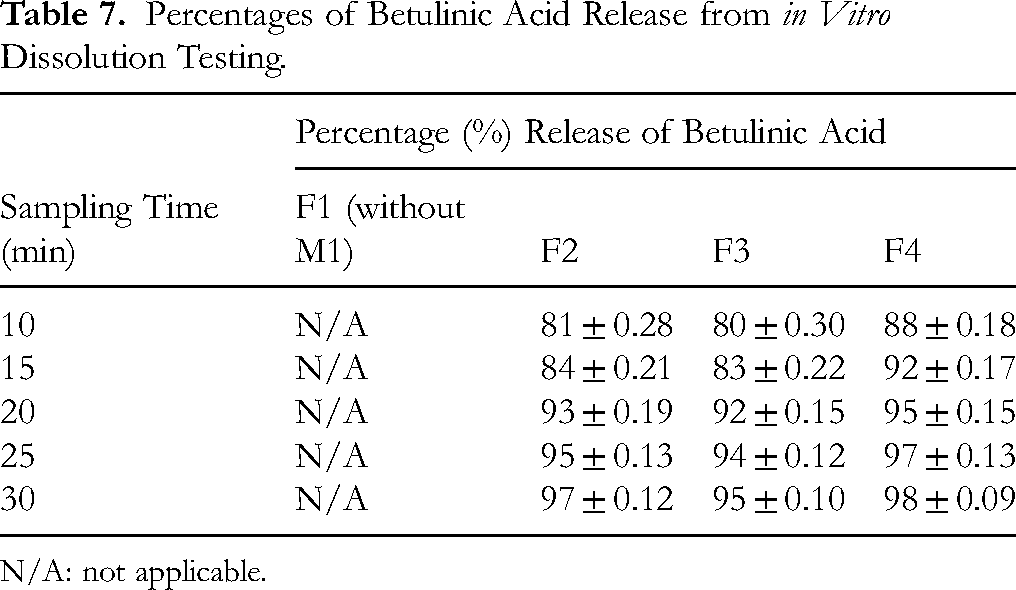
N/A: not applicable.
In the cytotoxicity assay, CRL-2076 human dermal fibroblasts treated with formulation F4 for up to 24 h at concentrations below 3.1 mg/ml showed no statistically significant reduction in cell viability compared to the control group (Figure 4B). Additionally, microbial and heavy metal contamination levels in F4 were within acceptable limits. These safety data confirm that the optimized F4 formulation is suitable for human use and that the rationale for adding sodium benzoate in F4, but not in F1–F3, appropriately balances preservative use with experimental objectives.
Stability of Lozenges
The optimized formulation F4 of S. leucantha lozenges was selected for accelerated stability testing. After six cycles, the physical characteristics—including appearance, average weight, thickness, diameter, friability, hardness, and disintegration time—remained largely unchanged across all test groups. However, the moisture content increased slightly from 1.64 ± 0.21% to 1.78 ± 0.14%, likely due to the hygroscopic nature of the excipients. Throughout the stability study, the lozenges consistently met microbiological safety criteria. Nonetheless, the slight increase in moisture content may elevate the risk of microbial growth during prolonged storage. To address this, it is recommended to replace glass bottle packaging with aluminum foil packaging to better protect against moisture uptake. In addition, herbal raw materials should be subjected to gamma irradiation at appropriate doses to reduce microbial contamination prior to formulation.
HPLC-DAD analysis showed no statistically significant difference (p > .05) in the betulinic acid content between the start (98.3 ± 1.8%) and the end (98.2 ± 2.0%) of the stability testing period. Dissolution rates remained consistently high, exceeding 98 ± 0.23% within 30 min. Overall, after six cycles of accelerated testing, all observed parameters across different batches exhibited minimal changes, and the betulinic acid content remained within acceptable limits.
Discussion
The aqueous decoction (M1) exhibited the highest extraction yield and the lowest moisture content compared with the ethanol-based extracts (M2–M4). This aligns with previous findings that polar solvents, particularly water, are more effective for extracting hydrophilic phytochemicals. 25 The low moisture content of M1 is advantageous for storage stability and microbial safety, as it minimizes the risk of contamination in herbal products.21,26 Decoction also proved effective for extracting thermally stable, water-soluble phytochemicals such as tannins, reducing sugars, and certain glycosides. 27 Phytochemical screening confirmed the presence of tannins, flavonoids, saponins, terpenoids, alkaloids, and glycosides in all extracts, while steroids were detected only in M2–M4. Betulinic acid, a bioactive triterpene with anti-inflammatory and antimicrobial activities, was identified in all extracts, with the highest concentration in M1.28,29 These findings are consistent with previous reports.2–4 Moreover, FTIR spectra of all extracts displayed characteristic peaks corresponding to betulinic acid. 28 Collectively, these results highlight M1 as the most suitable extract for standardization and quality control in lozenge formulation.
The HPLC-DAD method developed in this study allowed precise and reproducible quantification of betulinic acid, validated according to ICH Q2(R1) guidelines. 16 The low LOD and LOQ values confirmed its reliability for quality control of both extracts and formulated lozenges. In terms of biological activities, the differences observed between DPPH and FRAP assays reflected their distinct mechanisms. 30 While the DPPH assay primarily detected phenolics and flavonoids with radical scavenging ability, FRAP assessed overall reducing capacity. M1 showed strong antioxidant activity, supporting its role in protecting throat tissues from oxidative stress. Anti-inflammatory activity was most pronounced in M4, with M1 also showing moderate inhibition of albumin denaturation (IC50 = 1.41 ± 0.02 mg/ml), suggesting potential benefits for soothing throat inflammation. 31
Although S. leucantha extracts produced measurable inhibition zones against S. aureus (10.33-8.83 mm), these were 2.2–2.5 times smaller than the positive control, chloramphenicol (23.00 ± 1.30 mm). Consistently, MIC values for all extracts exceeded 20 mg/ml, confirming limited antibacterial activity. The mild effects may be attributed to the presence of bioactive compounds such as betulinic acid, which has been reported to exert antibacterial activity through multiple mechanisms, including disruption of bacterial membranes and inhibition of key enzymes such as DNA gyrase and beta-lactamase. 32 Additionally, betulinic acid demonstrated antibiofilm activity against interkingdom S. aureus–Candida albicans biofilms by altering cell membrane properties, reducing early adhesion and biofilm formation, and modulating biofilm-related gene expression without direct microbicidal effects. 33 Minor differences between disc diffusion and MIC results may reflect factors such as compound solubility, diffusion in agar, and interactions with media components.
While M1 showed limited antimicrobial activity, it did not exhibit cytotoxicity toward human dermal fibroblasts (CRL-2076) at concentrations up to 2.50 mg/ml, suggesting that the combination of phytochemicals could provide supportive or synergistic effects in lozenge formulations without compromising safety. Taken together, based on extraction yield, physicochemical properties, bioactive content, antioxidant and anti-inflammatory activities, and safety profile, M1 was selected as the optimal extract for lozenge development. Its preparation via traditional decoction further supports its ethnomedicinal relevance and suitability for respiratory health applications.
The four S. leucantha granule formulations (F1–F4) were developed to systematically evaluate the effects of extract concentration, excipient composition, and granulation solvent on lozenge properties. F1, which contained no S. leucantha extract, served as the control to assess the physicochemical performance of the excipient base alone. F2 and F3 were preliminary trial formulations containing 10.34% w/w M1 extract, with varying amounts of mannitol and PVP K30, designed to optimize granule cohesiveness and organoleptic characteristics. Sodium benzoate was intentionally omitted from F1–F3 to evaluate the intrinsic stability and safety of the granules without preservative interference. The optimized formulation, F4, contained a higher extract concentration (11.50% w/w M1) along with selected herbal powders and increased PVP K30, using 70% ethanol as the granulating solvent to enhance mechanical strength, reduce moisture content, and improve microbial stability. In F4, 0.10% w/w sodium benzoate was included to provide preservative protection suitable for storage and human use.
In terms of mechanical properties, F4 demonstrated the greatest hardness (6.17 ± 0.20 kg/cm²), which was significantly higher than that of the other formulations (p < .05). This enhancement in hardness corresponds with the higher PVP K30 concentration and ethanol binder, confirming the rationale that increasing binder levels and using a more effective granulating solvent improve tablet strength. 34 Correspondingly, F4 also exhibited the lowest friability (0.05 ± 0.04%, p < .05), indicating superior mechanical durability. All formulations passed the friability test, with values below the acceptable limit of 1%. 35 Disintegration times among the formulations ranged from 5.46 ± 0.29 to 9.05 ± 0.24 min, all of which met the pharmacopeial requirements. 36 A statistically significant difference (p < .05) was observed, with F4 showing the longest disintegration time (9.05 ± 0.24 min), potentially due to its increased hardness and decreased friability. 37 Overall, these results support the rationale that adjustments in extract content, binder concentration, and solvent type can modulate the mechanical strength and disintegration behavior of lozenges, while the use of 70% v/v ethanol in F4 contributed to the lowest moisture content (1.64 ± 0.21%), further supporting its superior mechanical stability. The improvements observed in hardness, friability, disintegration time, and betulinic acid dissolution confirm the effectiveness of adjusting binder concentration, solvent type, and preservative inclusion in achieving an optimal lozenge formulation.
Among the four trial formulations, F4 exhibited the most favorable organoleptic and physical properties. Accelerated stability testing showed minimal changes in mechanical characteristics, microbial safety, and betulinic acid content after six heating–cooling cycles, although a slight increase in moisture content indicated the potential benefit of protective packaging and pre-treatment of herbal materials to ensure long-term stability.34–37
This study successfully developed a standardized herbal lozenge containing S. leucantha aqueous extract (M1) using a modern pharmaceutical approach. The extract, selected for its high yield, low moisture content, abundant phytoconstituents—particularly betulinic acid—and promising biological activities, validated the traditional use of S. leucantha leaves in managing respiratory ailments such as cough and sore throat. The optimized lozenge met pharmacopeial quality standards for weight variation, friability, hardness, content uniformity, and disintegration time, while also maintaining stability and betulinic acid content under accelerated storage conditions.
Some limitations should be acknowledged. First, antimicrobial activity was assessed only against a limited pathogen panel, with relatively mild results. Broader testing, including antiviral assays, would strengthen the evidence for therapeutic potential. Second, although in vitro cytotoxicity confirmed extract safety, further in vivo safety and pharmacokinetic studies are required. Third, clinical efficacy was not evaluated; thus, future trials are needed to confirm therapeutic benefits in patients with respiratory symptoms. Additionally, antibacterial testing was not performed on granule formulations (F1–F4), as the presence of multiple herbal ingredients could interfere with the activity of M1; therefore, antimicrobial evaluation was limited to the extract itself.
Despite these limitations, this research provides a strong foundation for the development of S. leucantha-based lozenges and supports their potential as a novel phytotherapeutic product for respiratory health.
Conclusion
Based on the findings of this study, the aqueous extract of S. leucantha leaves was selected as the active ingredient in lozenges due to its rich phytochemical composition—including betulinic acid and phenolic compounds—and its beneficial biological activities, such as antioxidant, anti-inflammatory, and antimicrobial effects. The extract demonstrated no toxicity at the tested concentrations, as confirmed by the MTT assay. Granules containing the S. leucantha aqueous extract were successfully prepared using the wet granulation method, with PVP K30 and 70% v/v ethanol serving as binders. These granules, combined with Aerosil® 200, magnesium stearate, and peppermint powder, were compressed into lozenges using a biconvex single-punch tablet press. The resulting lozenges exhibited rapid release of betulinic acid within 30 min, showed no cytotoxic effects, and complied with stringent microbial and heavy metal contamination limits. All quality parameters—both before and after stability testing—were consistent with USP 41–NF 36 standards. These results support the potential of S. leucantha leaf-derived lozenges as a safe, stable, and effective therapeutic product for respiratory health.
Footnotes
Abbreviations
Acknowledgements
We gratefully acknowledge the support of the Faculty of Pharmacy, Silpakorn University, and Dontum Hospital in Nakhon Pathom, Thailand.
Ethical Considerations
This study did not involve human participants or animal subjects; therefore, ethical approval was not required.
Author Contributions
All authors have made meaningful and direct contributions to the research and approved the final manuscript for publication.
Ponphaiboon, J., Limmatvapirat, S., Ingsurarak, M., Auparigtatipong, W., and Limmatvapirat, C. contributed to the formulation of research objectives, design of the methodology, data visualization and interpretation, statistical analysis, manuscript drafting, and critical revision.
Mahadlek, J., Tuntarawongsa, S., Siangjong, L., Meetam, P., Krongrawa, W., Ekachampaka, J., Sarunyakasitrin, K., and Limmatvapirat, C. were involved in data collection, data handling and analysis, and participated in the critical evaluation and editing of the manuscript.
Limmatvapirat, C. led the acquisition of research funding, provided overall supervision, approved the final version of the work, and coordinated the research direction, planning, and data management.
Funding
The authors disclosed receipt of the following financial support for the research, authorship, and/or publication of this article: This research was supported by the Postdoctoral Fellowship Program, Silpakorn University, Thailand, fiscal year 2025 [grant number: SURDI Postdoctoral/68].
Declaration of Conflicting Interests
The authors declared no potential conflicts of interest with respect to the research, authorship, and/or publication of this article.