
Abstract
Introduction
Coronary artery disease (CAD) is characterized by a reduction in or cessation of blood flow to the myocardium due to fully or partially blocked coronary arteries, which increases the risk of myocardial infarction (MI) in the heart muscle.1,2 Generally, MI is linked to a number of pathophysiological indicators, such as the electrocardiogram (ECG), troponin-I blood levels, cardiac enzyme markers, and histological changes in cardiac tissues. 3 Unrestrained inflammation and an imbalance of myocardial oxidants and antioxidants lead to a reduction in cell viability, which has a major impact on heart function. 4 Different external effectors like exposure to gamma radiation and receiving doses of certain drugs or certain diseases like cancer might increase the oxidative burden on the biological system and provoke the condition of oxidative stress. 5
The myocardial infraction might result from the imbalance between blood supply and demand. 6 MI, an acute condition of necrosis of the myocardium, is the most lethal manifestation of CAD that usually results when blood flow stops to a part of the heart, causing damage to the heart muscles.2,7
Through the development of modern science, ionizing radiation (γ-rays) technology has been used extensively in several fields, including military, medicine, agriculture, and industry, making human exposure to increased levels of ionizing radiation inevitable. 8 Exposure to unruly levels of ionizing radiation causes a copious production of free radicals that altar cell constituents of macromolecules through enforcing lipids peroxidation (LPO), protein oxidation, and DNA strand breaks. 9 Heart cells are affected by radiation exposure through the development of RIHD (radiation-induced heart disease) syndrome, which comprises a spectrum of heart diseases, including pericarditis, cardiomyopathy, coronary artery disease, valvular heart disease, and cardiac conduction abnormality. 10
The usage of Isoproterenol (ISO), a synthetic nonselective β-adrenergic agonist, induces many cardiac disorders, such as calcium overload, myocardial oxidative stress, coronary hypotension, and energy depletion. 11 A high dose of ISO might encourage oxidative stress by releasing abundant free radicals and suppressing antioxidants. 12 The formation of free radicals as well as accumulation of lipid peroxides has been recognized as one of the possible biochemical mechanism for the myocardial damage caused by ISO. 12 It has been reported that ISO-induced myocardial necrosis show membrane permeability alterations which bring about the loss of function and integrity of myocardial membranes. 13 Of all the macromolecules to leak from damaged tissues, enzymes, because of their tissue specificity and catalytic activity, are the best markers of tissue damage. 14
Many shreds of evidence suggest that compounds containing powerful antioxidants may significantly protect against MI. 15 Van der Pole et al 16 stated that some experimental and clinical studies have shown that myocardial infarct size can be limited by increasing endogenous antioxidants and suppressing free radical generation. The current research examines nattokinase, a serine protease discovered in 1980 and extracted from natto, a traditional Japanese food, made by boiling or steaming soybeans and fermenting them with Bacillus subtilis natto. 17 Do Prado et al 18 reported that N.K., a soybean-derived enzyme, may treat certain cardiovascular diseases (CVD) by possessing a variety of favorable cardiovascular effects, such as fibrinolytic activity, anti-hypertensive, antiatherosclerotic, and lipid-lowering, antiplatelet, and neuroprotective effects. 19
Its anti-hypertensive, 20 anti-atherosclerotic, 21 anti-platelet/anticoagulant, 22 and neuroprotective effects 23 were extensively documented. With the biotechnological advent NK was studied for its sensorial, pharmacokinetic and toxicology aspects as well.24,25 These pharmacologic actions of NK have relevance to the prevention and treatment of CVD. In fact, patients with chronic kidney disease, exhibits a high risk for developing an occlusive vascular thrombus, which may lead to deleterious cardiovasicular diseases, such as myocardial infarction and stroke. 26
Based on the above, The purpose of this study is to examine the antioxidant and cardio and reno-protective properties of N.K. against myocardial damage brought on by radiation exposure or ISO injection, as well as the effect these properties have on kidney function. This aim was achieved through the measurement of ANP (Atrial natriuretic peptide)), ANG-II(AngiotensinII), NGAL (neutrophil gelatinase-associated lipocalin) and cTnI (Troponin I) in heart tissue along with ALD (Aldosterone), KIM-1 (kidney injury molecule-1), in the kidney as well as LDH (lactate dehydrogenase) and Serum IL-18 besides heart and kidney eNOS, in rats exposed to gamma radiation and/or supplemented with ISO and treated with N.K.
Materials and Methods
Materials
Nattokinase was procured from Doctor's Best (USA). Meanwhile, Isoproterenol, along with other chemicals and reagents used in the experimental examination, were purchased from Sigma-Aldrich Chemical Co., USA.
Animals
In the present study, Male albino rats (Sprague Dawley substrain derivation of the strain (Norway rat, Rattus norvegicus)) weighing 120-150 g used in this study were obtained from the experimental animal house lab of the National Center for Radiation Research and Technology, Egyptian Atomic Energy Authority. Upon receiving the experimental animals, they are left for ac climatize for a period of 1 week concurrent with the presence of standard commercial pellet diet and water ad libitum. Rats were housed in standard conditions of temperature (22-24 °C), humidity (60 ± 10%), and a 12 h light/dark cycle. Animals were fed a commercial standard pellet diet (containing necessary nutritive elements 23% protein, 4.68% fats, 2.6% fibers, and soya free to minimize natural phytoestrogen supplementation) and water ad libitum during this period. The care and use of experimental animals of this study were done complied with the recommendations of National Institute of Health (NIH no 85:23, revised 1996) and in the obedience of guidelines adopted by the NCRRT ethic committee, which approved all experimental procedures (41 A/21).
Ethics Statement
This experiment was carried out according to recommendations in the Guide for the Care and Use of Laboratory Animals of the National Institutes of Health (NIH no. 85:23, revised 1996) and in compliance with ethical regulations of the National Centre for Radiation Research and Technology-NCRRT). With approval by the Central Scientific Publishing Committee of the Egyptian Atomic Energy Authority (Ref No: 188/10/2019) (Approval No 40A/21). All efforts were made to minimize the suffering of animals.
Radiation Facility
Throughout the experiment procedures, whole-body gamma irradiation of male albino rats was performed using Canadian γ cell-40 (Cs137) (housed at NCRRT, Cairo, Egypt) at an accumulative dose of 5Gy delivered in fractions of 0.5Gy/ day for ten consecutive days (dose rate 0.401 Gy/min).
Experimental Design
80 Rats will be divided into eight groups (10 rats each) as follows:
(1) Control group, C: Normal rats have not received any treatment or been exposed to gamma radiation. (2) Irradiated Group, R: rats were exposed to γ-irradiation (0.5Gy/ day ten times till reached 5 Gy accumulative dose delivered day by day for ten consecutive days. (3) Nattokinase Group, N.K.: rats were treated with oral water suspension of N.K. alone, at a dose of 720 FU/kg bw /day for ten consecutive days according to Fadl et al 27 The N.K. doses are often indicated in fibrin units (F.U.). The 2000 FU translates to roughly 100 milligrams (mg), and 3000 FU translates to 150 mg). (4) R + NK Group: rats were exposed to γ-irradiation as in group 2 and treated with N.K. as in group 3. (5) Isoproterenol group (ISO): rats were subcutaneously injected with ISO 150 mg/kg body weight/day,28,29 for two successive days starting on day one of the experiment. (6) ISO + R Group: rats were subcutaneously injected with ISO as in Group 5 and exposed to radiation as in Group 2. (7) Isoproterenol and Nattokinase group (ISO + NK): rats were subcutaneously injected with Isoproterenol as in Group 5 and treated with N.K. as in group 3. (8) ISO + R + NK Group: rats were subcutaneously injected with ISO as in group 5, exposed to γ-radiation as in group 2, and treated with N.K. as in group 3, respectively. The zero time of the experiment is the injection of ISO starting on day one of the experiment and the end of the experiment after the last dose of N.K.
Fasting overnight was applied to rats in all experimental groups before undergoing anesthesia by urethane after that immolation. The tissue (heart and kidney) samples and Serum were collected and prepared for the implementation of biochemical and histopathological investigations.
Biochemical Assay
In heart tissue, the levels of (ANP (Cat No: MBS726614) and cTnI (Cat. No: MBS727624)) were measured. In kidney, the levels of (Ang-II (Cat. No.: MBS705139), KIM-1 (Cat. No: MBS2702467), ALD (Cat. No.: MB287151), NGAL (Cat. No.: MBS564123)) and heart & kidney (TNF-α (Cat. No: MBS2507393); MDA (Cat. No: MBS738685); GSH (Cat. No: MBS265966); eNOS (Cat. No: MBS721860)) were measured. In Serum, (LDH (Cat. No: MBS043166) and IL-18 (Cat. No: MBS260091) were measured. All parameters were measured by enzyme-linked immunosorbent assays (My BioSource ELISA KIT- According to MyBioSource elisa kit, ∼300 µL extraction buffer was added per ∼5 mg tissue), according to the manufacturer's instructions via an ELISA microplate reader (DV990 BV 416; Gio.DE VITA and CO., Rome, Italy).
Histopathological Examination
Autopsy from heart and kidney tissues of rats in different groups were collected and fixed in formalin saline (at 10%) for 24 h, then washed with tap water. After that, serial dilutions of alcohol (methyl, ethyl, and absolute ethyl) will be used for different dehydration samples. The tissue samples were cleared in xylene and then preserved at 56 °C in a hot air oven for 24 h before embedding in paraffin. The paraffin beeswax of tissue blocks was prepared and sectioned at 4 mm thickness by slide microtome. Finally, the obtained tissue sections were collected on glass slides and de-paraffinized, then stained by hematoxylin and eosin (H&E) stain for routine examination via the Olympus BX50 (Japan) light electric microscope. 30
Grading of Histopathological Alterations
Histopathological alterations of the heart and kidneys were scored or graded as (0), which indicated no changes; (+), (++), and (+++) indicated mild, moderate, and severe changes, respectively, according to the methods mentioned by Sandamali et al 31 & Arsad et al 32 Also, the pathological changes in the myocardium were scored following the grading system of Acikel et al, 33 in which the grading of pathological aberrations reporting specific grading criteria, as follows: grade (0) refers to normal histology, while grade (I) stands for mild focal myocyte damage or small multifocal degeneration with slight degree of inflammation, whereas grade (II) denotes moderate-extensive myofbrillar degeneration and/or difuse inflammatory process, meanwhile grade (III) describes severe necrosis with difuse inflammatory infiltration.
Statistical Analysis
Statistics were performed using ANOVA (one-way analysis of variance), followed by Duncan's Multiple Range test using the statistical package of social science (SPSS) version 17.0 for Windows. The level of significance had been considered at P ˂ .05.
Results
Effect of Nattokinase on Heart Muscle of γ-Irradiated Rats Pretreated by Isoproterenol
Figure 1 (a&b) showed that the heart levels of ANP and Troponin increased significantly by (564.4%,458.6%, 8.65%) and (284.8%, 216.9%, 25.89%) (P < .05) in a rat of the Iso, R, and N.K. groups compared to control group, respectively. The concentration of GSH in the myocardium of the same groups decreased significantly (by 20.8%, 14.6%, 0.669%) (P < .05), while the concentration of MDA increased significantly (by 5129.7%, 5089.1%, 55.07%) (P < .05) compared to Control (Figure 1 c&d). Moreover, the concentration of TNF-α and eNOS in the myocardium increased meaningly (by (303.4%, 234.09%, 43.18%) and (74.46%, 67.98%, 2.33%) (P < .05) when comparing the values of these parameters to their equivalent values in control rats (Figure 1 e&f). The severity of changes for all the previously mentioned parameters was highest in the Iso + R group. While, the changes in all these parameters were significantly lessened by (ANP 42.68%, TROPONIN 39.43%, GSH 5.83%, MDA 28.05%, TNF-α 14.96% and eNOS 91.57%) in R + NK group compared to R group. However, the changes in all these parameters were significantly decreased in Iso + R, Iso + NK by (ANP 70.76%, TROPONIN 65.89%, GSH 10.62%, MDA12.54%, TNF-α 30.42% and eNOS 30.98%) and (ANP 39.36%, TROPONIN 35.96%, GSH 5.39%, MDA 20.35%, TNF-α 12.95% and eNOS 107.7%), respectively compared to their corresponding values in Iso group and in Iso + R + NK compared to Iso + R group by (ANP 29.49%, TROPONIN 29.37%, GSH 35.63%, MDA 23.5%, TNF-α 28.29% and eNOS 51.02%). That to say, the N.K. administration might preserve the myocardium against threats due to exposure to ISO and/or radiation.

Impact of N.K. on Myocardial Indicators (a: ANP and b: Troponin in Serum, Redox Status: c, GSH, and d, MDA and Inflammatory Status: e, TNF, and f, eNOS in Heart Muscles) of Different Animal Groups: Each Column Represents the Mean of 6 Values ± SEM. Different Letters Overhead the Columns Mean That the Differences are Significant Statistically Between the Inter-Compared Groups (P < .05), Whereas Similar Letters Mean the Differences are not Significant (P > .05). C: Normal Rats, R: Rats Exposed to Fractionated Dose γ-Irradiation, N.K.: Rats Receiving Treatment Drug (N.K.) Alone, R + NK: Rats will be Exposed to Fractionated Dose γ-Irradiation (0.5Gy/ Day ten Times till Reach 5 Gy). They will be Treated with N.K. as in a Group, ISO: Rat will be Subcutaneously Injected with ISO 150 mg/kg Body Weight, ISO + R: Rat will be Subcutaneously Injected with ISO as in Group 5 and Exposed to γ-Radiation as in Group 5, ISO + NK: Rat will be Subcutaneously Injected with ISO as in Group 5 and will be Treated with N.K. as in Group 3), ISO + R + NK: Rat will be Subcutaneously Injected with ISO at the First two Days of the Experiment) as in Group 5 and will be Exposed to Radiation as in Group5 and will be Treated with N.K. as in Group 3.
Effect of Nattokinase on NGAL and Aldosterone Activity in Kidney of γ-Irradiated Rats Pretreated by Isoproterenol
The data illustrated in (Figure 2a) showed that the levels of NGAL were significantly increased in the R and ISO groups by (138.67%, 246.11%) and decreased substantially in the N.K. group by (14.11%) in kidney of γ-Irradiated Rats Pretreated by ISO. In the meantime, levels of NGAL were significantly increased in NK + R, ISO + NK, ISO + R, and ISO + NK + R rats groups by (24.77%, 37.02%, 55.25%, 35.58%), respectively when compared to their equivalent values in R, Iso and Iso + R groups respectively (Figure 2a). The severity of changes for NGAL is highest in the Iso + R group. On the other hand, no significant changes were observed in the N.K. rats’ group compared to the control group. However, the changes in NGAL are significantly lessened in R + NK, Iso + NK, and Iso + R + NK compared to their corresponding values in R, Iso, and Iso + R groups, respectively.

Impact of N.K. on the NGAL Concentration and Aldosterone Activity in Different Rat Groups’ Kidney (Pg/g Tissue). Each Column Represents the Mean of 6 Values ± SEM. Different Letters Overhead the Columns Mean That the Differences are Significant Statistically Between the Inter-Compared Groups (P < .05). In Contrast, Similar Letters Mean the Differences are Insignificant (P > .05). Legend as in Figure 1.
According to the data presented in Figure 2b, the kidney of rats treated with γ-irradiation before receiving ISO had significantly higher levels of Aldosterone in the R and ISO groups by (162.5% and 232.8%) and considerably lower levels in the N.K. group by 16.78% ompared to control group. Comparing the values of these parameters to their equivalent values in rats of R, Iso, Iso and Iso + R groups respectively, the levels of Aldosterone were markedly higher in the NK + R, ISO + NK, ISO + R, and ISO + NK + R rat groups by (26.12%, 57.14%, 63.09% and 43.93%), respectively (Figure 2). For every trait listed above, the alterations are most pronounced in the Iso + R group.
Effect of Nattokinase on Kidney of γ-Irradiated Rats Pretreated by Isoproterenol
Figure 3 (a&b) shows that the kidney levels of TNF-α and KIM-1 increased significantly (P < .05) in the rat of the Iso, R, and N.K. groups by (243%, 182%, 19%) and (282.8%, 176.9%, 11.76%), respectively. The concentration of GSH in the myocardium decreased significantly (P < .05), while the concentration of MDA increased significantly (P < .05) in Iso, R, and N.K. groups by ((2.26%, 2.27%, 2.77%) and (1122.6%, 971.6%, 49.8%)), compared to Control group (Figure 3 c&d). Moreover, the concentration of ANG-II and eNOS in the myocardium increased meaningly by (300%, 227.2%, 27.27%) and (218.1%, 150.5%, 16.16%) (P < .05), respectively when comparing these parameters’ values to their equivalent in the control rat (Figure 3 e&f).
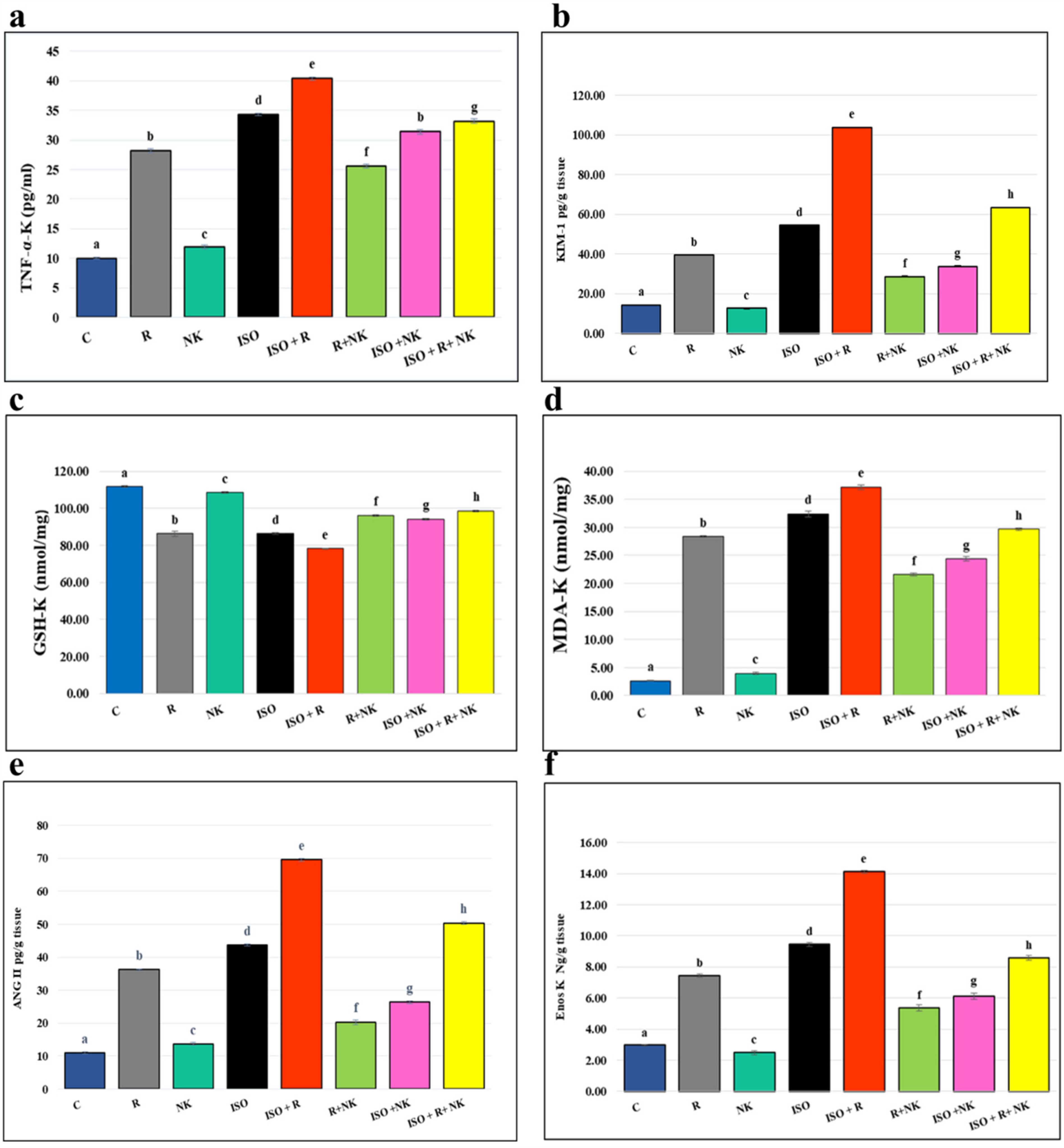
Impact of N.K. on Kidney; e (ANG-II), Redox Status: c(GSH) and d (MDA) and Inflammatory Status: a (TNF-α), b (KIM-1), f (eNOS), of Different Animal Groups. Each Column Represents the Mean of 6 Values ± SEM. Different Letters Overhead the Columns Mean That the Differences are Significant Statistically Between the Inter-Compared Groups (P < .05), Whereas Similar Letters Mean the Differences are not Significant (P > .05). Legends, as in the Figure 1.
The changes in the Iso + R group are the most severe for all the previously indicated characteristics by (17.78% TNF-α, 90.85% KIM-1, 9.51% GSH, 14.9% MDA, 59.09% ANG-II, 49.52% eNOS), compared to Iso group. However, compared to their comparable values in the R, Iso, and Iso + R groups, the changes in all of these parameters are dramatically lessened in the R + NK, Iso + NK, and Iso + R + NK groups by ((9.21% TNF-α, 27.21% KIM-1, 11.43% GSH, 23.9% MDA, 44.4% ANG-II, 27.82% eNOS), (8.45% TNF-α, 37.9% KIM-1, 9.004% GSH, 24.75% MDA, 40.9% ANG-II, 35.23% eNOS) and (17.82% TNF-α, 38.97% KIM-1, 26.09% GSH, 59.49% MDA, 28.57% ANG-II, 39.27% eNOS)) respectively. Stated differently, the N.K. administration may protect the kidney from harm caused by exposure to radiation or ISO.
Effect of Nattokinase on Serum IL-18 & LDH of γ-Irradiated Rats Pretreated by Isoproterenol
The serum levels of LDH and IL-18 in rats in the Iso, R, and N.K. groups increased significantly by ((292.5%, 356.2%, 1166%) and (494.01%, 394.5%, 4.36%)) (P < .05), as shown in Figure 4 (a&b). Comparing R + NK, Iso + NK, and Iso + R + NK to their corresponding values in R, Iso, and Iso + R groups shows a considerable amelioration of the alterations in LDH and IL-18, by ((21.09%, 17.39%, 69.4%) and (41.67%, 69.23%, 29.45%)), respectively. The Iso + R group has the most significant degree of change in these two metrics by (36.3%and 69.23%), respectively compared to Iso group.

Effect of Nattokinase on Serum IL-18 & LDH of γ-Irradiated Rats Pretreated by Isoproterenol. Each Column Represents the Mean of 6 Values ± SEM. Different Letters Overhead the Columns Mean That the Differences are Significant Statistically Between the Inter-Compared Groups (P < .05), Whereas Similar Letters Mean the Differences are not Significant (P > .05). Legend as in the Figure 1.
Histopathological Findings
Heart tissue from the Control negative group showed normal cardiac parenchyma with standard myocardial muscle bundles (Figure 5a, Table 1). In contrast, the heart from the R group revealed normal myocardial muscle with congested blood vessels, (Figure 5b, Table 1), the heart from the N.K. group showed no histological alterations (Figure 5c, Table 1), heart from R + N.K. group recorded no histological alterations except congested blood vessel, (Figure 5d), Heart from ISO group showed myocardial infarction with zenker's necrosed muscles together with mononuclear cells infiltrations characterized by areas of hyaline patches with moderate extensive zenker's necrotic degeneration of the myocardial muscle fibers together with mononuclear cells infiltrations, (Figure 5e, Table 1). In examined heart sections from Iso + R group a moderate to extensive necrotic degeneration accompanied by diffuse hyaline areas of borderless cardiomyocytes, along with notable mononuclear cells infiltrations, when compared with R or ISO alone groups (Figure 5f, Table 1). Moderate improvement with regressed myocardial damage was recorded in heart from Iso + NK group (Figure 5g, Table 1), while in Iso + R + N.K. group, there was mild improvement with moderate myocardial damage compared to changes observed in photomicrograph of R or ISO group (Figure 5h, Table 1).

Photomicrographs of Rat'S Hearts From Different Experimental Groups Stained With H&E X400.1 A) Control Rats, C Which Exhibit no Lesion in Heart Muscles (0), 1B) Gamma-Irradiated Rats, R Which Exhibit Congested Blood Vessels (Arrow) (+), 1C). Rats Treated by N.K., Which Exhibit Healthy Myocardial Muscles (0), 1D) Rats Treated by N.K. and Exposed to Gamma, NK + R Which Exhibit Congested Blood Vessel (Arrow) (+/I), 1e) Rats Treated by ISO, Iso Which Exhibits Severe Myocardial Infarction (Arrow) (++/II), 1f) Rats Treated by Isoproterenol and Exposed to Gamma, Iso + R Group Which Exhibit Marked Myocardial Degeneration and Congested Blood Vessel (Arrow) (++/II), 1 g) Rats Treated by ISO (++), N.K. & Iso + NK Which Exhibit Slight Myocardial Damage (+) and 1 h) Rats Treated by Iso And N.K. and Exposed to Gamma, Iso + Nk + R Which Exhibit Moderate Myocardial Damage (++).
Grading of Histopathological Lesions of the Heart and Kidneys in all Groups.

Kidneys from the Control negative group showed normal renal parenchyma with normal renal glomeruli and normal renal tubules (Figure 6a, Table 1). In contrast, kidneys from the R group revealed slight degeneration in the renal parenchyma (Figure 6b, Table 1), kidneys from the N.K. group showed no histological alterations, (Figure 6c, Table 1), kidneys from R + NK group recorded no histological alterations except slight degeneration in the renal parenchyma, (Figure 6d, Table 1), kidneys from Iso group showed moderate degeneration in the renal glomeruli and renal tubules, together with leucocytic cells infiltration (Figure 6e, Table 1). o seeming improvement was noticed in kidneys from the Iso + R group (Figure 6f), moderate improvement with regressed renal degeneration was recorded in kidneys from the Iso + NK group (Figure 6g, Table 1), while in Iso + R + N.K. group, a mild improvement despite the moderate renal damage, (Figure 6h, Table 1).

Photomicrographs of Kidneys From Different Experimental Groups Stained With H&E X400 Showing: 2a) Control Rats, C, Which Exhibit Normal Kidney Tissue Architecture (0), 2b) Irradiated Rats, R, Which Exhibit Slight Degeneration in the Renal Parenchyma (+), 2c). N.K. Rats, Which Exhibit Healthy Renal Parenchyma (0); 2d) Irradiated Rats Treated With N.K., R + N.K., Which Display Degeneration in the Renal Parenchyma (+/I), 2e) ISO-Treated Rats, Iso Which Show Severe Degeneration in the Renal Parenchyma (++), 2f) ISO Irradiated Rats, Iso + R Which Display Degeneration in the Renal Parenchyma (++), 2 g) ISO and N.K. Treated Rats, Iso + NK Which Exhibit Slight Degeneration in the Renal Parenchyma (+), two h) ISO Irradiated Rats Treated With N.K., Iso + R + N.K. Which Display Moderate Degeneration in the Renal Parenchyma (++/II).
Discussion
Recently, many research outcome results link the dangerous life threats of myocardial infarction and kidney failure. 34 It seems like the relationship is mutual between the occurrence of either of the two syndromes, and the existence of one can cause the incidence of the other. 35 Apparently, there are many attempts to examine novel treatments to reduce the high risk of morbidity and mortality that comes from the two syndromes and perhaps could introduce a sync solution for the two problems. Herein, the present study is interested in nattokinase, a natural compound that has significant antioxidants and anti-inflammatory capabilities. 36 In the present study, an animal model of coronary artery disease (rats pretreated with isoproterenol and/or exposed to γ-radiation) was used to examine the efficacy of N.K. In addition to its thrombolytic and anticoagulant activities, NK elicited anti-inflammatory and anti-oxidative stress activities by inhibiting LPS-mediated TLR-4 and NOX2 signaling in macrophages and protected against LPS-stimulated AKI as well as glomeruli thrombus in mice, a novel anti-thrombus insight of NK via breaking the link between inflammation, oxidative stress and thrombosis. 37
Harmoniously, the data of the current study referred to increase in the concentration of ANP and Troponin in heart muscles after treatment by ISO when compared to control. Besides, the exposure to γ-radiation might exacerbate the problems in cardiac muscles as the levels of Troponin in the Iso + R group are significantly higher than its equivalent level in the Iso rats’ group (Figure 1). Tapio et al 38 indicated that radiation damages the heart through the initiation of acute and chronic changes in cardiac tissue.
Within minutes of ionizing radiation, cellular injury causes vasodilation and increased vascular permeability. 38 The damaged endothelial cells stash adhesion molecules and growth factors and prompt the activation of the acute inflammatory response. Wang et al 39 stated that the recruited inflammatory cells, upon inflammatory reactions, secrete the pro-fibrotic cytokines. Indeed, these inflammatory cytokines include monocyte chemotactic factor, tumor necrosis factor, and interleukins. 40
Furthermore, ANP is a cardiac hormone that plays an important role in regulating vascular remodelling and energy metabolism. The disturbances in ANP contents or its related peptide could affect the healthiness of heart muscles. 41 In the present study, the increases in ANP concentrations observed after injection of Iso and/or radiation exposure hypothesize the presence of heart problems, which are confirmed by increases in troponin concentration. Bock et al 42 stated that ISO stimulated the release of Troponin, and it appeared to be mediated by cAMP.
The administration of ISO subcutaneously has a significant increase in serum markers of myocardial injury, such as ANP and Troponin. 43 It was reported that plasma levels of these markers are directly proportional to the degree of necrotic lesions present in the myocardium. 44 The Iso treatment markedly increases reactive oxygen species levels in cardiomyocytes, activates the NF-κB signalling pathway, and induces an inflammatory response. 45
Serum troponin levels are sensitive and specific biomarkers for detecting Iso-induced myocardial injury in rats, as troponin values reflect the development of histopathologic lesions. 46 Troponin increases abruptly due to the injection of iso. 47
Further, Khalaf et al 4 illustrated that low doses of Iso employ complex effects on the myocardium, and the increased expression of iNOS leading to the generation of NO could be vital in the pathogenesis of myocyte damage. The level of ANP increased in cardiac muscle after Iso (Figure 1) is expected as the ANP is a common sign of damage in different organs and tissues. 48 Likewise, the eNOS recorded increases in heart afford the opinion of tissue damages acquiescence to iso administration and the histopathological examination of heart tissues support this forecast. Worth mentioning the administration of N.K. by the mode followed in the present study success in reducing the bad effect of iso or radiation on the previous parameters when compared to their equivalents in other groups not received N.K. (Figure 1).
Concerning the valuation of markers of heart and renal status, we detected noticeable increases in concentrations of Aldosterone, ANG2, LDH, IL-18, KIM1, and NGAL synchronized to the rise in the myocardial injury determinants next to ISO treatment. Correspondingly, the results emphasize that N.K. amended the changes induced in kidney damage biomarkers in sync with the improvement observed in the myocardial infraction biomarkers (Figure 1–6), and that might be ascribed to the antioxidant and anti-inflammatory properties of Nattokinase. Previous studies proved that NK exerts a direct antioxidant effect resulting in reduced lipid peroxidation and inhibition of LDL oxidation, thus improving lipid metabolism. 49 On the other hand, Chang and colleagues assigned the beneficial effect of NK on suppression of intimal thickening in animals with endothelial injury to its synergistic antioxidant and anti-apoptotic properties. 50 Also, previous literature data proposed the collective effect of the combined anticoagulant, antithrombotic, antioxidant, and hypolipi demic actions of NK. 51
On the other hand, various signaling receptors are involved in the signaling pathways through which NK exerts anti-inflammatory effects. Activation of the Toll Like Receptor 4 (TLR4) pathway induces the translocation of nuclear factor-kappa B (NF-κB) and the transduction of mitogen-activated protein kinases (MAPKs), which stimulate innate and acquired immunity in many cells. 52 It has been found that the NF-κB/MAPK path way can be activated by tumor necrosis factor receptor-associated factor6 (TRAF-6), a downregulator of several receptor families with immunostimulatory properties, leading to activation of TGF-β-activated kinase 1 (TAK1) and increased release of proinflammatory mediators. 53 However, NK has been shown to inhibit the upregulation of TRAF6 and the resulting activation of the NF-κB/MAPK signaling pathway, thereby suppressing the expression of proinflammatory molecules such as TNF-α, IL-6, NO, and PAI-1, resulting in an anti-inflammatory potential. 54
As widely reported in the literatures, activation of TLR4 leads to the production of reactive oxygen species (ROS) and thus to an inflammatory response. Nicotinamide adenine dinucleotide phosphate (NADPH) oxidase expressed on macrophages consists of two isoforms, of which NADPH oxidase 2 (NOX2) is most abundant on inflammatory macrophage cells. 55 Although there is little evidence for the anti-inflammatory effect of NK, one study has shown that NK has the potential to block the release of TLR-4 and NOX2-mediated proinflammatory mediators in macrophage cells, thereby exerting an anti-inflammatory effect. 56
Indeed, angiotensin II, a main effector molecule of the renin-angiotensin system (RAS), is involved in the regulation of blood pressure, vasoconstriction, sodium intake and potassium excretion, might increase the blood pressure, influences renal tubules to retain sodium and water, and stimulates aldosterone release from the adrenal gland. 57 Bhullar et al 58 stated that the increased levels of angiotensin (Ang) II throughout myocardial infarction, as well as heart failure, promote vasoconstriction. The binding of Ang II to its receptors (in particular AT1) mediates intracellular free radical generation that contributes to tissue damage by promoting mitochondrial dysfunction. 59 In rodents, inhibiting Ang II signaling prolongs life and guards against neurodegenerative processes. 60
Based on the data of current study, the angiotensin-aldosterone axis was investigated in response to gamma irradiation and /or ISO administration. It is a part of the RAAS system (Renin- Angiotensin- Aldosterone), which has a significant role in the pathophysiological conditions of hypertension, heart failure, other cardiovascular diseases, and renal diseases. 61
The administration of Iso and/or radiation exposure led to an elevation in Ang 2 and Aldosterone in the kidney tissue, which favors the possibility of kidney damage. Moreover, Almutlaq et al 62 stated that the obstruction of the Renin-Angiotensin-Aldosterone system might improve outcomes in various cardiovascular and renal diseases. The data of the current study runs in harmony with previous findings where N.K. administration prevents the overactivation of RAAS faithfully downregulates the angiotensin2 and Aldosterone in the kidney when compared to the groups not treated with N.K. accompanied with improvement in tissue architectures 63 (Figures 5 and 6).
Additionally, significant elevation in kidney neutrophil gelatinase-associated lipocalin NAGL and KIM-1 were identified after the Iso injection (Figures 2 and 3). This signifies the assumption of disturbance in kidney tissues and kidney functions parallel to the myocardial inefficiency after Iso. Jana et al 64 described that new biomarkers have been used to detect early onset of acute kidney injury, such as NGAL, a member of the lipocalin family, and kidney injury molecule-1 (KIM-1), type 1 transmembrane glycoprotein. Latoch et al 65 identified that in cases of kidney injuries, the increase of KIM-1 and NGAL was observed earlier than serum creatinine increases. Essentially, KIM-1 is useful as a marker of proximal tubular injury. In contrast, NGAL is a marker of proximal and distal tubule damage in response to various types of injury (ie, ischemic, toxic, inflammation). 66
KIM-1 is not detectable in healthy conditions. 67 Highly expressed in the endothelial cells of the cardiac vasculature, the endothelial nitric oxide synthase (eNOS) enzyme helps to preserve the endothelium-myocardial connections. It reduces the vulnerability of cardiomyocytes to ischemia/reperfusion injury. It works by producing NO, which can directly counteract superoxide, lower oxidative stress in general, and stop cardiac remodeling. 67 In the present study, N.K. treatment along with irradiation significantly increased the eNOS level, which indicated that N.K. might have an efficient effect in improving endothelial Function and represent a potential therapy for cardiac remodeling.
Likewise, the low molecular weight and low degradation properties facilitate the excretion of NGAL into the urine (uNGAL), and its concentration increases immediately after acute kidney injury. 46 NGAL is detected in various types of human tissues, including the kidneys (monomeric form) and the gastrointestinal and respiratory tracts. 68 Furthermore, its neutrophil dimeric form also participates in the immunological response. 65 In healthy subjects, it is present in Serum and urine at very low levels. 69 The level of uNGAL increases harmonized with the degree of renal tubular damage. 70 Silvestre-Roig et al 71 reported that infiltrated neutrophils, resident macrophages, endothelial cells, smooth muscle cells, and cardiomyocytes in the heart can produce IL-18 in response to injury. Elevated IL-18 levels have been observed in cardiac tissue and circulation after MI. 72 The possible cellular and molecular mechanisms concerning IL-18-induced myocardial injury include induction of inflammation and changes in intracellular calcium. 73
Similarly, the proinflammatory cytokine IL-18 is produced by macrophages and other cell types present in the kidney. 74 The present experimental investigation pointed to significantly increased serum IL-18 contents after Iso subcutaneous injection, which could be accredited to the associated vasocontraction and proinflammatory action of IL-18. Sun et al 75 stated that increased levels of interleukin-18 (IL-18) have been related to plaque progression and vulnerability and cardiovascular outcomes. Besides, IL-18 may promote immune or non-immunize-mediated tissue damage via a multitude of mechanisms. 76 Contradictory, the present results displayed that N.K. administration significantly ameliorates the changes induced in serum IL-18 (Figure 4). This could be attributed to the antioxidant and anti-inflammatory properties of N.K.. 77
Of course, the significant improvements in the concentration of IL-18 in the Serum could be interpreted in the sight of anti-inflammatory action N.K.. 78 That to say the Iso treatment leads to the occurrence of manifestations of heart muscle insufficiency, as a result of increases ROS production and also the occurrence of violent inflammatory reactions that can be observed in the cardiogenic examination of the heart tissue in addition to increasing the activity of the eNOS enzyme, which is an indication of a reaction to facing the harmful effects resulting from Iso and in conjunction when examining the kidney tissue, the exacerbation of the manifestations of inflammation, with an increase in the inflammatory index IL-18 in addition to increasing the activity of the eNOS enzyme as happened in the heart tissue, as a response in order to reduce the damages, which could indicate a link between the occurrence of problems in the heart muscle and the effect of the kidneys and can support this perception is a noticeable improvement as a result of N.K. injection which acts to decrease ROS production and regulate inflammation.
In the present study, the ISO-treated and/or irradiated rats showed a significant elevation in the LDH enzyme level, indicating the necrotic myocardium and the damaged cell membranes. Nattokinase lowered the levels of the enzymes, confirming the improvement of the cell membrane integrity and protecting the myocardium from necrosis, therefore restricting the leakage of the cardiac cytosolic enzymes. Based on the above and in conclusion, N.K. may have great potential against myocardial damage and could represent a promising solution for cardiovascular system complications and heart injuries. At the same time, it could provide significant protection to the kidneys from the consequences of cardiovascular flaws.
Of note, the present study did have some limitations, the first limitation is the extensive range of biochemical parameters and indicators that offer comprehensive mechanistic insights and detail on how on how N.K. and irradiation may intervene with biochemical events and oxidative stress in myocardial damage, these cannot be investigated completely in our research because of the restricted sample size permitted by the ethical regulations of the animal ethics committee, which are adhered to by the present study and all animal research (the more factors to be studied, the more samples that we needed). Hopefully, in further investigations, we will conduct more comprehensive detectors and indicators that accomplish these objectives. Another limitation of current study is the absence of prior research on the efficacy of nattokinase in the prevention of myocardial infarction induced by ISO injection in rodents that are undergoing whole-body fractionated γ-irradiation, as well as the investigated research avenue. Finally, additional research is necessary to fully comprehend the mechanism of nattokinase's effect on myocardial infarction caused by ISO injection in rats undergoing whole-body fractionated γ-irradiation. There are undoubtedly numerous information gaps that require filling and investigation.
Conclusion: This study demonstrated that N.K. with its antioxidant and anti-inflammatory capabilities, may have great potential against myocardial damage and could preserves the myocardium from disruption of blood flow processes resulting from increased oxidative burden after injection of Isoproterenol or exposure to radiation, through multiple routes of action, including activation of heart (ANP/cTnI/GSH-H/MDA/TNF-α/NGAL), stimulation of kidney (KIM-1/ANG-II/aldosterone), activation of Serum IL-18/LDH and suppression of heart and kidney ENOS. At the same time, it could provide significant protection to the kidneys from the consequences of cardiovascular flaws.
Footnotes
Ethical Considerations
This experiment was carried out according to recommendations in the Guide for the Care and Use of Laboratory Animals of the National Institutes of Health (NIH no. 85:23, revised 1996) and in compliance with ethical regulations of the National Centre for Radiation Research and Technology-NCRRT). With approval by the Central Scientific Publishing Committee of the Egyptian Atomic Energy Authority (Ref No: 188/10/2019) (Approval No 40A/21). All efforts were made to minimize the suffering of animals.
Author Contributions/CRediT
Conceptualization: Khaled Azab, Noura Thabet and Nermeen M. El Bakary; Data curation: Noura Thabet and Nermeen M. El Bakary; Formal analysis: Khaled Azab and Rokaya E Maarouf; Methodology: Sherein Abdelgayed, and Somaya Z Mansour; Writing original draft preparation: Asmaa Abubakr, Rokaya E Maarouf; Writing- review and editing: Rokaya E Maarouf, Asmaa Abubakr and Mohamed Abdel-Rafei; Resources: Mohamed Abdel-Rafei and Noura Thabet; Supervision: Khaled Azab, Noura Thabet and Mohamed Abdel-Rafei.
Funding
The authors received no financial support for the research, authorship, and/or publication of this article.
Conflicting Interests
The authors declared no potential conflicts of interest with respect to the research, authorship, and/or publication of this article.
Data Availability
All data obtained from this study are included in the current manuscript.