
Abstract
Introduction
The skin is vital for shielding the body from infections and external harm. Normal skin microbiota, which is primarily commensal in nature, is colonized on the skin. But these microbiota act like opportunistic pathogens in aberrant or disturbed conditions. 1 A common method for pathogens to bypass the skin barrier is through a wound or tear, which enables them to enter the body, cause an infection, and evade the body's natural defenses. Candida albicans and Staphylococcus aureus are prime examples of such pathogens. 2
Folliculitis and hidradenitis can develop from the growth of pathogenic microbes in skin appendages, which promptly triggers nearby keratinocytes to release inflammatory cytokines and chemokines. This, in turn, attracts neutrophils and monocytes to the affected area. 3 Severe injuries can impact appearance and health, increasing the risk of infection. Antimicrobial resistance exacerbates treatment difficulties, leading to higher morbidity, mortality, and healthcare costs.4,5
Nanotechnology has been recognized as one of the most crucial research endeavors of the twentieth century, primarily due to its use of the unique properties of atomic and molecular structures at the nanoscale. It provides the scientific and industrial foundation for creating and utilizing nanomaterials. Nanotechnology enables the manipulation of materials at the nanoscale (1-100 nm), driving significant advancements across various sectors, including energy, electronics, and medicine. This scale of material engineering allows for unprecedented improvements in functionality and performance. 6 A nanometer (nm) is a billionth of a meter. By working at the atomic and molecular levels, scientists and engineers can develop new features and applications. However, one of the main challenges in nanotechnology is producing nanoparticles with the appropriate characteristics for specific applications. Achieving desirable outcomes requires comprehensive knowledge of nanoparticle properties, synthesis techniques, and operating conditions. 7
The antibacterial efficacy of metallic nanoparticles, or nanometals, has been shown in a variety of biological and pharmaceutical applications. 8 This is mostly because of the metals’ cytotoxicity against microbial infections and interactions with their membranes and internal routes. 6 Nanotechnology explores nanoscale materials, like silver nanoparticles (AgNPs), for medical applications. AgNPs, along with polymer nanocomposites (NCs), improve treatments like targeted delivery. 5 The antimicrobial activities of silver nanoparticles (AgNPs) are well-documented. These activities include the release of Ag + ions, the production of reactive oxygen species (ROS) within the inner and outer microbial membranes, interference with the cellular membrane, disruption of the ribosome-mitochondrial complex, and disturbances in nucleic acids. 9 Conventional synthesis methods face cost and environmental issues, prompting interest in eco-friendly alternatives like green synthesis, using plant metabolites. Plants, known for medicinal properties, contain phenolic compounds with antimicrobial, anti-inflammatory, and antioxidant benefits for treating skin infections. 5
The genus Commiphora (Burseraceae) encompasses approximately 200 species, including Commiphora myrrh, from which the myrrh resin is derived. With a rich historical background, myrrh extract (MR) has been extensively utilized as a traditional remedy for various skin conditions such as wounds, infections, acne, and boils. 10
Myrrh, along with several related species of Commiphora, is defined by the United States Pharmacopeia (USP 34) as the gum-resin extracted from the stems and branches of C. molmol. Ethiopia and Somalia are the primary producers of this resin. It is designated as Generally Recognized as Safe (GRAS) by the Food and Drug Administration (FDA) for use as a food additive. 11 Additionally, Commission E has approved myrrh extract for its astringent and antibacterial properties. 12 As a functional food with antioxidant activity and potential to help prevent human colon cancer, it was suggested adding it to yogurt or milk. 13
Myrrh is used in complementary medicine for its antiviral, expectorant, antifungal, and antibacterial properties. 14 Numerous clinical and experimental studies have been conducted locally to investigate the effects of myrrh extracts on various microorganisms. While the results have been mixed, several studies have supported the effectiveness of myrrh, especially when used in the recommended daily dosages. 12 Chemically, myrrh is an oleo gum resin. The gum component, which constitutes 40%–60% of the total, is primarily made up of polysaccharides, proteins, and oxidase enzymes and is water-soluble. In contrast, the resin, comprising 20%–40%, along with the volatile oil fractions, is alcohol-soluble.12,14
Chitosan (Cht), a versatile polymer, exhibits biocompatibility, non-toxicity, and ease of extraction, making it a promising candidate for biomedical applications. 15 Chitosan nanoparticles have shown potential in enhancing the bioactivities of metal nanoparticles, including their antimicrobial properties. 16
Combining chitosan with AgNPs enhances the stability and dispersibility of the nanoparticles, thereby boosting their therapeutic potential for combating cancer and microbial infections. 17 Chitosan's pH responsiveness and mucoadhesive properties enable controlled drug release and targeted delivery to mucosal areas. Additionally, its film-forming capability makes it ideal for applications requiring thin protective coatings. The ease of modifying chitosan also allows for the addition of new functions to AgNPs-chitosan composites. 18
Despite the potential benefits, there is currently a dearth of published studies on the biosynthesis of the Cht/MR/AgNPs nanocomposite. Therefore, our study aimed to bridge this gap by synthesizing the nanocomposite, characterizing its physicochemical and structural properties, evaluating its antimicrobial efficacy, and assessing its suitability for textile coatings.
Materials and Methods
Chemicals and Reagent
The experiment's materials and reactants were all obtained from Sigma-Aldrich Co. (St. Louis, MO, USA) and approved for use in analysis.
Preparation of Myrrh Extract
Dried MR underwent pulverization to a 60-mesh size using a mixing grinder, followed by mixing 200 g of MR powder in 1000 mL (w/v) of 70% ethanol (Sigma-Aldrich, MO). The mixture was stirred at a speed of 160 xg for 22 h at room temperature (25 ± 2 °C). Upon filtration by filter paper (Whatman no. 41), after removing the plant remnants, the MR extract was dried by rotary evaporation (IKA, RV 10, Germany) at 44 °C. For the purposes of the next investigations, the dried MR was then dissolved in a 2% Tween 80 solution in deionized water (DW) to produce a concentration of 1 mg/mL. 19
Green Synthesis of Silver Nanoparticles
The green synthesis of AgNPs involved the use of myrrh extract; 5 mL of the MR extract solution (concentration of 1 mg/mL) was applied dropwise to 50 ml of AgNO3 (1.0 mM) at 45 °C for 15 min. After that, the resultant AgNPs were heated to 60 °C and stirred with a magnetic stirrer and electric heater until a brown-yellow color that is suggestive of AgNPs production was seen. Since research has shown that this suspension is stable in this environment, it was then kept at ambient temperature in a conical glass flask covered with foil until it was needed. 20
Chitosan Extraction
Erugosquilla massavensis (mantis shrimp) shell debris served as the primary source for chitosan extraction. These remnants were gathered from the “Seafoods processing plants, Kafrelsheikh University, Egypt,” then cleaned, washed with deionized water (DW), and subsequently dried at 45 °C for 42 h. 21 After grinding the shells, chitosan extraction followed a series of steps: demineralization (using 16 volumes of 1 M HCl, 12 h, at room temperature); deproteinization (using 16 volumes of 1 M NaOH, 12 h, at room temperature); and deacetylation (using 18 volumes of 55% NaOH solution, 95 min, at 115 °C). Each stage was followed by DW rinsing and drying, and the resultant powder was stored for further analysis. 16
Preparation of Cht/ MR /Ag Nanocomposites
The following solutions had to be made in order to synthesize Cht-NPs and load them with MR/AgNPs: TPP (0.5 mg/ml in DW), Cht (1 mg/ml in a 1% acetic acid solution), and MR extract (1 mg/ml in a 2% Tween 80 solution in DA). 16 Using a syringe needle, the TPP solution was gradually added to the Cht solution at a rate of 0.35 ml/min until the volumes were equal after the pH of the Cht solution was adjusted to 5.2. An equal amount of MR/AgNPs was added to the Cht solution prior to the addition of TPP in order to produce Cht/MR/AgNPs.
Characterization of Synthesized Nanoparticles
NPs’ Optical Analysis
In order to verify the creation of metal nanoparticles through the identification of their surface Plasmon resonance, which is linked to unbound electrons on the surfaces of the nanoparticles, the AgNPs spectrum was examined utilizing a UV-Vis spectrophotometer (UV-2450, Shimadzu, Japan) operating in the 300–1000 nm wavelength range.
FTIR Analysis
In transmission mode (at a wavenumber range of 450-4000 cm1), the infrared characteristics of synthesized MR, Cht, MR/AgNPs, and Cht/MR/AgNPs were examined using Fourier transform infrared spectroscopy (FTIR; Perkin ElmerTM FTIR-V. 10.03.08, Germany). 22
Zetasizer Analysis
Using Zetasizer (Zeta plus, Brookhaven, USA), the AgNPs zeta potential (ζ) was assessed using the dynamic light scattering approach.
Transmission Electron Microscopy (TEM)
TEM (Leica-Leo 0430; Cambridge, UK) were used to screen the Ps and define the morphology and dispersion of Cht/MR/AgNPs.
Antimicrobial Textiles Preparation
The textile was immersed in SPSs for 90 min at 25 °C with continuous stirring, padded, and compressed to 100% wet pickup. This process was modified from former work. 23 The textile was then forced air dried for 120 min and cured for 15 min at 100 °C. After being cut into 2 cm2 squares, the SPS-treated textiles were tested for microbiological resistance utilizing the inhibition zone (IZ) appearance method on the microorganisms under investigation. Textiles treated with SPS were placed on the infected surface after microbial strains were streaked onto the suitable solid media. The diameters of the IZs were measured, and their mean values were computed, in triplicate throughout the experiments. Antimicrobial textiles’ resilience was assessed following several cycles of washing in a domestic laundry machine with neutral water at 41 ± 3 °C. 23
Antimicrobial Activity of Treated Cotton Textiles
A sterile skin protectant solution (SPS) comprising 1% of Cht, MR, MR/AgNPs, and Cht/MR/AgNPs was applied to scoured cotton plain weave (108 g/m2) obtained from Misr Weaving and Spinning Co., Egypt. This procedure was carried out utilizing a modified pad-dry-cure method. 23
Evaluation of NPs Antibacterial/Anticandidal Activity
Cultures of Bacteria and Fungi
For antimicrobial screening, Staphylococcus aureus and Candida albicans were used as common skin pathogens. The screened strains included C. albicans-S (ATCC-10231), C. albicans-R (resistant isolate to fluconazole, from skin lesion), S. aureus-S (ATCC-25923) and resistant S. aureus-R isolate (resist methicillin, from skin wound infection). They were achieved from the microbial culture collection, Ain Shams University, Egypt. On nutrient agar and broth (NA and NB, Difco Laboratories, Detroit, MI), the whole bacterial and fungal strains were kept alive and subcultured aerobically at 37 °C. 24
Inhibition Zone (IZ) Assay
After the disc diffusion test, the inhibition zones (IZ) that emerged were taken into consideration as indicators of the antibacterial/antifungal bioactivity of the produced agents. Clean Whatman The 2% solutions of MR/AgNPs, MRand Cht/MR/AgNPs were impregnated into No. 4 paper discs (6 mm in diameter) and then placed onto freshly infected NA plates with each bacterial culture. After measuring the IZ diameters with a precision calliper and incubating the plates at 37 °C for 18 to 24 h, they were turned upside down and the standard deviation of their triplicate means was computed. 23
SEM “Scanning Electron Microscopy” Imaging
SEM imaging was utilized to observe the morphological changes in Staphylococcus aureus cell surfaces after exposure to a 2.0% (w/v) solution of Cht/MR/AgNPs. These changes were monitored over an incubation period ranging from 0 to 12 h at 37 °C. SEM images were captured at 20 kV and a magnification of x10,000, documenting the distortions in bacterial and fungal cell structures. 22
Cytotoxicity Assay
The normal HDF “Human Dermal Fibroblasts; C-12302, Sigma-Aldrich” were used for assessing the cytotoxicity of Cht/MR/AgNPs nanocomposite. In 96 well plate, cells (10.000 cells/well) were seeded in DMEM medium “Dulbecco's Modified Eagle Media” supplemented with 10% FBS “fetal calf serum” and Antibiotic-Antimycotic mixture (1%), followed by their incubation in CO2 incubator at 95% humidity and 37 °C temperature. Wells were amended with gradual concentrations of Cht/MR/AgNPs and further incubated then cell proliferation/viability were assessed using MTT “3.[4.5-dimethylthiazol-2-yl]-2.5-diphenyltetrazolium bromide” assay.
Statistical Analysis
In triplicate studies, each analysis was conducted three times. Standard deviations were calculated using SPSS. Statistical analysis to determine data significance (at p < 0.05) was performed using the “t-test and ANOVA” tests from the SPSS package (V-11.5, Chicago, IL).
Results
NPs’ Optical Analysis
Direct observation and UV-vis spectrophotometric analysis of MR/AgNPs provide optical evidence that NP production follows MR interaction. Direct examination of the MR/AgNPs solutions revealed a progressive color change within 30 min of the reaction, going from clear to deep blackish-brown (Figure 1A); no more color changes were seen after that. MR/AgNPs had the largest NPs absorption peaks (λmax) at 412 nm for that. MR/AgNPs according to UV-vis analysis (Figure 1B).

Evidences of AgNPs synthesis using myrrh extract, including visual appearance (A), UV-vis spectral analysis (B).
FTIR Analysis
The molecules produced, including MR and its composites with AgNPs and Ch/AgNPs, were analyzed using FTIR to clarify biochemical bonding and interactions between the composited agents and the possible My groups in responsible for NP formation.
FTIR analysis of Cht (Figure 2

FTIR of chitosan(Cht), Myrrh Extract(MR), Myrrh/AgNPs (MR/AgNPs), and (Cht/MR/AgNPs) nanocomposites.
The MR extract FTIR analysis (Figure 2
The Cht/MR/AgNPs nanocomposites’ FTIR analysis (Figure 2
Zetasizer Analysis
The nanoparticle size average diameter of the AgNPs was determined to have a mean Z-average size of 4.43 nm and negatively charged (−23.7 mV). The polydispersity index (PDI) of the AgNPs was 0.503, indicating minor size variation and agglomeration. The mean diameter of Cht/MR/AgNPs nanocomposites was 130.34 nm and carry +25.9 mV charges, indicating efficient MR/AgNPs encapsulation inside Cht.
Transmission Electron Microscopy (TEM)
The TEM imaging was used to examine. The images revealed relatively circular nanoparticles with an average diameter ranging from 0.52 to 8.28 nm., with mean dimeter of 4.81 nm. Synthesized AgNPs exhibited a spherical shape, minimal aggregation, and varying sizes, as depicted in Figure 3.

Transmission microscope screening of synthesized agNPs.
Evaluation of NPs Antibacterial/Anticandidal Activity
From Table 1, it was observed that Cht/MR/AgNPs composite had the most effective inhibitory action against S. aureus S, with an inhibition zone diameter of 36.5 mm, while it possessed less effect against C. albicans R, with an inhibition zone diameter of 29.2 mm; and the inhibition zone diameter against S. aureus R and C. albicans S was 30.7 mm and 32.0 mm respectively. In the meantime, Ag-NPs biosynthesized by Myrrh extract showed high inhibitory action against S. aureus S with an inhibition zone diameter of 31.1 mm and moderate inhibitory action against S. aureus R and C. albicans S with an inhibition zone diameter of 27.5 mm and 28.5 mm, respectively, while it possessed less effect against C. albicans R, with an inhibition zone diameter of 26.2 mm. Compared with lower effect of commercial Myrrh extract, recording inhibition zone diameters of 22.7 mm against C. albicans S, 19.1 mm against C. albicans R, 23.2 mm against S. aureus S and 20.8 mm against S. aureus R; the synergistic effect of combined materials were evidenced. All agents were synthesized in accordance with the approved concentrations for use as conventional antibacterial and antifungal treatments.
Antimicrobial Performance of Myrrh (MR), Synthesized Silver Nanoparticles Using Myrrh Extract (MR/AgNPs), and Cht/MR/AgNPs Composites, Using Zone of Inhibition (ZOI; in mm) Assays.

**Inhibition zones are triplicates means (including textile width of 20 mm) standard deviation.
Antimicrobial Activity of Treated Cotton Textiles
In general, fabrics treated with Cht/MR/AgNPs demonstrated greater antimicrobial activity against all strains compared to those treated with MR extract and MR/AgNPs alone. Moreover, fabrics treated with the composite Cht/MR/AgNPs showed a significant enhancement in antimicrobial efficacy against all tested strains compared to fabrics treated with each individual agent. Among the strains tested, S. aureus showed greater sensitivity compared to C. albicans, and antibiotic-sensitive isolates exhibited higher susceptibility than antibiotic-resistant strains to all treated fabrics, as illustrated in Figure 4. The appeared dark spots on some textiles’ surfaces (eg C-2, S-2, C-3 and S-3) are typically attributed to the loaded AgNPs, and not from growth of microbes onto textiles.
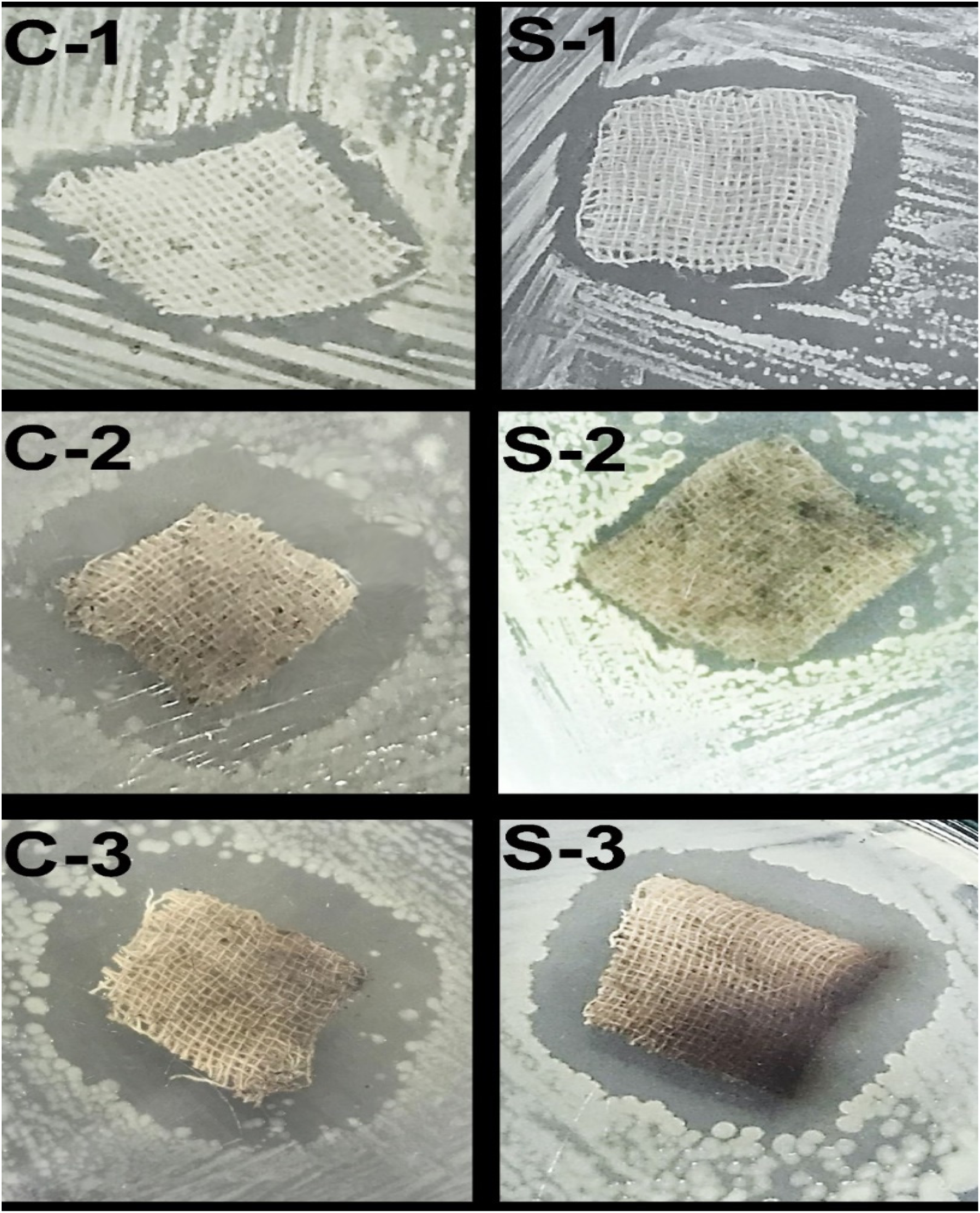
Antimicrobial activity of treated cotton textiles with 1% from myrrh extract (1), and with nano silver synthesized from Myrrh extract (2), and Cht/MR/AgNPs (3) against antibiotic resistant strains from Candida albicans (C) and Staphylococcus aureus (S).
Scanning Electron Microscopy (SEM)
SEM imaging (Figure 5) was utilized to observe the morphological changes in S. aureus and C. albicans following exposure to minimum inhibitory concentrations (MICs) of Cht/MR/AgNPs. Initially (Figure 5- 0), bacterial cells exhibited well-preserved and intact cell walls. However, after 3 to 6 h of exposure, signs of wall softening and notable distortions became evident, accompanied by leakage of internal constituents (Figure 5- 1,2). By the ninth hour mark, some treated S. aureus and C. albicans cells were lysed (Figure 5- 3). After 12 h of exposure, nearly all treated cells were lysed, with residual cell walls appearing in conjunction with leaked internal components and nanomaterials (Figure 5 - 4).

SEM of treated Staphylococcus aureus (S) and Candida albicans (C) with cht/mr/agNPs nanocomposite for 0, 3, 6, 9 and 12 h (form 0 to 4 in figure, respectively).
The cytotoxicity assay of Cht/MR/AgNPs nanocomposite toward normal HDF (C-12302) cells using MTT method illustrated minimal biotoxicity of nanocomposites toward human dermal fibroblasts, up to concentration of 50 mg/mL.
Discussion
The biosynthesis of Cht/MR/AgNPs nanocomposite presents a promising strategy for effectively controlling skin pathogenic microbes. The synthesized nanocomposite exhibited significant antimicrobial activity against a range of microbial strains, including S. aureus and C. albicans using a simple, economical, and environmentally outgoing nanobiotechnological approach. The usage of use nanosilver, despite its potential cytotoxicity and carcinogenicity based on several reasons; their potent antibacterial actions, their synthesis with biogenic materials (MR), and their conjugation with biopolymers (Cht), which are believed to greatly minimize their biotoxicity toward mammalian cells,18,20,25 and this was verified in current study. The TEM imaging of MR-synthesized AgNPs appointed some agglomeration, which occurred due to the constituents of the extract aggregating and adhering to the surface of AgNPs. 25 The MR-synthsized AgNPs apperaed with well-despersion and compositesd with MR residues (Figure 3). However the biosynthesis process is suggested to be obtimized via incorporating of further stabilizing materials (eg biopolymers, thickning agents, …), and by managing the zeta potential of synthsizing nanometals through adjusting process pH and other conditions, to diminsh the potential agglumeration of nanomaterials17,20,25
SEM imaging revealed notable morphological changes in bacterial and fungal cells following exposure to the nanocomposite, indicative of its disruptive effects on cell walls and internal constituents. 26 Moreover, the enhanced antimicrobial potential of the composite fabric highlights its potential for applications in textile coatings aimed at preventing microbial infections. Overall, the findings suggest that the Cht/MR/AgNPs nanocomposite holds considerable promise as a therapeutic agent for combating skin pathogenic microbes.
The FTIR analysis demonstrated the effectiveness of bio-organics derived from myrrh extract as capping or reducing agents for AgNPs. These bio-organics included germacrene B, limonene, curzerene, beta selinene, myrcenol, isocericenine, and spathulenol.25,27 The phytosynthesis of metals nanoparticles was effectually gathered due to the reducing powers of phyto-constituents and their capping potentialities that generates homogenous NPs sizes with minimum agglomeration.28,29
Cht/MR/AgNPs nanocomposite could burst, destroy, or break down bacterial and candidal cells. More research is required to fully understand the mechanisms of such types of Cht/MR/AgNPs nanocomposites’ antibacterial and anticandidal capabilities, additionally to its use as an antibacterial and anticandidal agent. 16 The aim of the current study was to extract and apply a variety of bioactive natural agents, such as Cht, MR, and AgNPs, for the possible prevention of skin infections. This was advised because natural products have a wide range of antimicrobial activities, are expected to be biosafe, and are compatible with the human body. 23
The antimicrobial actions of fabricated nanocomposites are suggested to involve many factors. The ratio between Cht and MR/AgNPs imparts high importance for the net zeta potential of nanocomposites; the higher Cht ratio derives nanocomposites to positivity, whereas the higher MR/AgNPs ratio enforces nanocomposites negativity. The composites charges (especially the positive charges) trigger their attachment and interactions with microbial cells that are negatively charged.9,16,18 Additionally, the high ratio of MR-mediated AgNPs is the suggested main responsible for antimicrobial actions, due to synergistic microbicidal actions of MR and AgNPs (including cellular penetration, disruptions and disturbing their biosystems).7,9,20,25
However, the suggested specific mechanisms that are involved in the destruction of microbial cells by the Cht/MR/AgNPs nanocomposite include the attachment of Cht-based capsules onto cells surfaces (due to their opposite charges) to disrupt cellular permeability, the release of MR/AgNPs from the nanocomposite to enter the cells, and the destructive interactions between each component (Cht, MR and AgNPs) with the cellular segments (eg membranes, enzymes, DNA, RNA, proteins, …).9,16,18
AgNPs that have been produced and their antimicrobial action in human skin cells may be key components of novel anti-inflammatory and sore treatment medicines. The antibacterial properties of green synthesized AgNPs made with myrrh extract show more promise against a variety of harmful microorganisms. 30 For eco-friendly AgNP synthesis, myrrh extract can be utilized as a green reducing and covering agent. Even after four months, there were no discernible alterations to the synthesized AgNPs, demonstrating their high stability. Furthermore, when compared to myrrh extract, AgNPs exhibited enhanced antifungal activity and high bacterial and fungal activity against both S. aureus and C. albicans. AgNPs are adaptable and can be employed in a variety of biological and biomedical applications as antimicrobial agents. 25
Regarding the promising attained antimicrobial activity of MR-mediated AgNPs and their nanoconjugates with Cht, it can be suggested to potentially improve the AgNPs antibacterial actions to make them more effective against more resistant microorganisms or emerging strain via biosynthsizing with more effectual natural derivatives (contained higher amunts of bioactive compouns), and compositing with aditional biopolymers to diminsh their potential biotoxicity toward mammalian cells.31,32
MR was selected due to its well-established benefits; it has astringent, antimicrobial, and anti-inflammatory properties. It is a common ingredient in many skin care products due to its unique characteristics and therapeutic advantages. It has moisturizing and soothing properties and can be used to relieve acne, blisters, dryness, and inflammation. 33 Within this context, the Cht/MR/AgNPs matrix has demonstrated numerous benefits in terms of having favorable biological, chemical, and physical properties for the defense of skin tissues. Because of their bioactivities, compatibility, and structural enhancement of the produced composites, Cht/MR/AgNPs were recommended as ideal nanocomposites for skin protection and were therefore encouraged to be applied in tissue engineering and other skin regeneration sectors. 34
The elevated biosafety of Cht/MR/AgNPs nanocomposites toward normal HDF cells, after validating with MTT method, could advocate their practical usage for human skin protection and disinfection using these innovative natural materials.1,23,34-37 The outstanding properties of Cht/MR/AgNPs components can additionally promote their prospective assessment as candidates for skin regeneration, especially after wounds/burns injuries. Because of its amazing features, such as its anti-inflammatory, biocompatible, antibacterial, and wound-healing properties (such as cytokine production, fibroblast activation, and promotion of My synthesis), Cht was also recommended for skin protection. 35 Cotton fiber coated with Cht has the potential to accelerate wound healing by stimulating PMN (polymorphonuclear) cells to infiltrate the injured skin. 36
One important consideration for the bioactivity of treated textiles’ potential uses in the medical field is the longevity of their antibacterial action. 6 Treated textiles containing Cht/MR/AgNPs maintained much higher antimicrobial potentialities than those reported in other pertinent studies involving textiles loaded with AgNPs or other individual antimicrobial agents.36-38 This is likely due to their synergistic composition, which strengthened the cross linkage with cotton fibers and prevented their rapid loss.39-41 The main limitations of this study may include the need for assessing nanomaterials biosafety using animal models, the evaluation of antimicrobial durability of fabricated textiles after several washing cycles, the probable inflammation consequence when applying coated textiles with nanomaterials, and the coast effectiveness evaluation of produced materials.
Conclusion
The AgNPs was perfectly biosynthesized using MR with minute size and homogenous dispersal. The Cht/MR/AgNPs nanocomposite was innovatively fabricated and characterized; the antimicrobial actions of this nanocomposite were evidenced toward different bacterial and mycotic skin pathogens. The loaded textiles with Cht/MR/AgNPs nanocomposite exhibited remarkable antimicrobial actions, where the nanocomposite could destruct microbial cells and prevent their development. The fabricated natural nanocomposites and their loaded hygienic textiles are promisingly suggested for controlling pathogenic skin microbes. The prospective role of fabricated nanocomposites in tissue regeneration is recommended to be investigated using in vivo, in situ and molecular studies. It is also suggested to evaluate the stability of the nanocomposite under different environmental conditions, such as prolonged exposure to sunlight or extreme temperatures.
Footnotes
Acknowledgements
The authors declare their appreciation and foremost gratefulness to ALLAH for the merciful and generous guiding throughout this work.
Author Contributions
Data Availability
“The datasets used and analyzed during the current study are all provided in the manuscript.”
Declaration of Conflicting Interests
The authors declared no potential conflicts of interest with respect to the research, authorship, and/or publication of this article.
Ethical Approval
“Ethical Approval is not applicable for this article.”
Funding
The authors received no financial support for the research, authorship, and/or publication of this article.
Statement of Human and Animal Rights
“This article does not contain any studies with human or animal subjects.”
Statement of Informed Consent
“There are no human subjects in this article and informed consent is not applicable.”