
Abstract
Background
The aim of this study was to explore the preventive and therapeutic potential of salvianolic acid B in inhibiting senile cataracts through network pharmacology and experimental validation.
Methods
Disease-related genes were obtained from the DisGeNET and GeneCards databases. Drug targets were identified from the Swiss Target Prediction and PharmMapper databases, shared genes were identified via the Venny website, links between genes were identified via a protein–protein interaction (PPI) network, and GO and KEGG analyses were subsequently performed via R software. The key genes were identified via Cytoscape software, and their binding with Sal-B was demonstrated by molecular docking. Then, the results were verified by cell experiments. A CCK-8 assay was used to assess the activity of human LECs with or without H2O2 or Sal-B treatment, and the cell apoptosis rate of each group was determined by flow cytometry. The gene expression levels of caspase 3, TNF-α and MMP-9 in human LECs treated with or without H2O2 or Sal-B were determined by qPCR.
Results
A total of 705 and 152 cataract-related genes and salvianolic acid B-related genes, respectively, were identified, with 37 shared genes. The PPI results showed that MMP9, IL-2, JUN, TNF-α and caspase 3 were the core genes. GO data analysis revealed that the biological process, cell component and molecular function terms were most enriched in the categories “apoptosis progress”, “cytosol” and “protein binding”. Through KEGG enrichment analysis, we found that the core genes were related to the IL-17 and TNF signalling pathways. Cytoscape results showed that MMP9, TNF-α and caspase 3 were the key genes, and molecular docking showed that the drugs interacted well with the target molecules. The experimental results showed that salvianolic acid B inhibited H2O2-induced decreases in LEC activity and apoptosis and inhibited the gene expression of MMP9, TNF-α and caspase 3.
Conclusion
Network pharmacology and molecular docking results showed that salvianolic acid B has the potential to prevent and treat senile cataracts. The experimental results verified the finding that salvianolic acid B can inhibit the decrease in LEC activity and apoptosis induced by H2O2 and verified the expression of the key molecules MMP9, TNF-α and caspase 3.
Introduction
Senile cataracts, also known as age-related cataracts, mainly manifest as a decrease in lens transparency with age, and turbid lenses prevent the clearly visualization of objects on the retina, which is the main reason for the gradual decline in vision in elderly people. 1 As the world population ages, the number of people with visual dysfunction and blindness caused by cataracts is also increasing. 2 The prevalence of cataracts in elderly people over 80 years old is reportedly as high as 92.6%, 3 which is related to oxidative stress, ageing and ultraviolet rays. 4 Although phacoemulsification intraocular lens implantation is currently an effective method for the treatment of cataracts, cataract surgery also has some potential risks, such as postoperative retinal detachment, macular oedema, corneal endothelial decompensation and dry eye aggravation. 5 Therefore, nonsurgical therapy or drugs to treat or delay the progression of cataracts are still the focus of current research.
Salvianolic acid B is extracted from the traditional Chinese medicine Salvia miltiorhiza Bunge (Lamiaceae). Salvia miltiorrhiza has been gradually accepted as a healthy product worldwide, 6 and Sal-B is the main active ingredient. Salvianolic acid B has been used to treat age-related degenerative diseases such as Alzheimer's disease and Parkinson's disease 7 because of its extensive anti-inflammatory, antioxidant and antiapoptotic effects.8,9 Xiaobin Liu et al reported that Sal-B may prevent and treat dry AMD by protecting RPE cells from H2O2-induced oxidative damage. 10 However, the effect of Sal-B on senile cataracts is unknown.
Network pharmacology is a new and effective method for studying the effects of drugs on specific diseases, especially traditional Chinese medicines.11,12 In this study, first, the therapeutic potential and targets of Sal-B in the treatment of senile cataracts were explored through network pharmacology and molecular docking. Then, at the experimental level, an oxidative damage model of human lens epithelial cells (HLECs) was constructed by hydrogen peroxide (H2O2) treatment, and Sal-B pretreatment of LECs was used to observe whether Sal-B inhibits H2O2-induced oxidative damage in HLECs and further explore the underlying mechanism.
Materials and Methods
Network Pharmacology
Identification of Drug Targets and Disease-Related Genes
Disease-related genes were obtained with the keywords senile cataract from the DisGeNET database (https://www.disgenet.org/web/DisGeNET/menu/home) and the GeneCards database (https://www.genecards.org). The molecular formula of salvianolic acid was obtained from the PubChem database, and drug targets were obtained from the PharmMapper database (http://www.lilab-ecust.cn/pharmmapper/) and the SwissTargetPrediction database (http://www.swisstargetprediction.ch/). The Venny website was used to identify common targets.
Construction of the PPI Network
PPI networks showing the links between 37 proteins were generated through the online STRING website (https://string-db.org/).
Analysis via Cytoscape Software
Through the plug-in cyto NCA in Cytoscape 2.3.1, the key genes were screened stepwise by topological analyses according to the average betweenness, closeness and degree of genes.
Gene Pathway Enrichment Analysis
The enriched Kyoto Encyclopedia of Genes and Genomes (KEGG) signalling pathways and Gene Ontology (GO) terms, including biological process (BP), cellular component (CC), and molecular function (MF), of potential targets were clustered using the ClusterProfiler package in R software (version: 3.18.0). 13 Bar plots and dot plots were generated to display the top 20 enriched BPs, CCs, MFs and KEGG pathways. The colour and size of the bubbles represent the p value of the term and the enriched gene counts, respectively.
Molecular Docking
Molecular docking was carried out with AutoDock Vina v.1.1.2 software. The structure of the ligand was downloaded from the ZINC database (http://zinc.docking.org/) and saved in mol2 format. The 3D structures of tumour necrosis factor (TNF), MMP 9 and caspase 3 were downloaded from the RCSB PDB database (https://www.rcsb.org/). Here, we used PyMOL v.0.99 software to visualize the 3D structures of the docking results.
Experimental Verification
Cell Culture
HLEC-SRA01/04 cells were purchased from Saiku Biotechnology Co. Ltd (#CC4022, Guangzhou, China), incubated in a cell incubator at 37 °C with 5% carbon dioxide and cultured in high-glucose DMEM (Gibco, Carlsbad, CA, USA) supplemented with 20% foetal bovine serum (Zeta Life, CA, USA). The effects of different drug treatments on SRA cells were evaluated in detail in the following experiments.
CCK-8 Assay
The CCK-8 assay was used to assess cell viability of SRA after being treated with H2O2 with or without Sal-B. The cells were inoculated in a 96-well plate (5000 cells per well). The cells were pretreated with Sal-B (Selleck, Shanghai, China) for 24 h with or without H2O2 (Lircon, Shandong, China), incubated for another 24 h, and 10 μl of CCK-8 solution (Zeta Life, CA, USA) was added and the sample was incubated for 2 h. The OD values of each well were measured by a spectrophotometer at 450 nm using a microplate reader (BioTek, Vermont, USA).
Cytoflow Assay
A Cytoflow Assay Kit (BD, Franklin Lakes, NJ, USA) was used to determine the apoptosis rate of SRA cells pretreated with Sal-B for 24 h with or without H2O2 and incubated for another 24 h. The cells were digested with trypsin without EDTA, centrifuged, and then resuscitated with 4 °C PBS and centrifuged again. Then, 500 µl of 1× binding solution was added to each tube to resuspend the cells. Then, 100 μl of cell suspension (number of cells > 1 × 10⁵), 5 μl of Annexin-FITC and 5 μl of PI were added to a 1.5 ml sterilized EP tube and placed in a dark room for 15 min. Then, 400 μl of 1× binding solution was added. Finally, the bacteria were filtered into a flow tube and analysed on a computer. The results were calculated by FlowJo software (the apoptosis rate was calculated by adding the Q2 and Q3 quadrants).
qPCR Assay
After 24 h of treatment with or without salvianolic acid B or H2O2, the cells were cleaved, and RNA was extracted with a Tissue RNA Purification Kit (EZBioscience, Roseville, USA). Then, reverse transcription and cDNA amplification were carried out using a reverse transcription kit (TransGen Biotech, Beijing, China). The results were analysed with LightCycler 96 software and compared with those obtained using GAPDH as the internal control. The sequences of the primers used in the experiment are shown in Supplementary Table 1.
Statistical Analysis
All experiments were performed in triplicate, and the data were analysed and plotted with GraphPad Prism 9 software. ANOVA data analysis method was used for statistical analysis, and then t tests were used to compare two groups, Tukey test was used to determine the difference between more groups. P < 0.05 was considered to indicate statistical significance.
Results
Identification of Sal-B Targets and senile Cataract-Related Genes
Disease-related genes were obtained, and the search keyword used was “senile cataract”. Ninety-two genes were obtained from the Disgenet database, 662 genes were obtained from the GeneCards database according to a relevant score greater than 2.1 and protein coding; then, the 705 genes were subsequently merged. The molecular formula of Sal-B was obtained from the PubChem database (Figure 1A). Then, 100 drug targets were identified through the SWISS Target Prediction database, 59 targets were identified via the Phammapper database according to the molecular formula, and 152 genes were identified from the two databases. Thirty-seven drug- and disease-related target genes were obtained from the Venny website (Figure 1B).

Acquisition of 37 common Sal-B and senile cataract targets.
Construction of the PPI Network
The PPI network was used to show the connections between proteins (Figure 2A). The greater the number of connections, the more closely connected that target is to other proteins; MMP-9, TNF, and IL-2 had the highest degree of connection, indicating their core role (Figure 2B).

PPI network construction: A: PPI network of 37 shared genes. The filled nodes and connecting lines represent the protein structure and the connections between proteins, respectively. B: Thirty-seven protein node connections are shown in the PPI network.
GO and KEGG Enrichment Analyses
According to the GO enrichment results for biological process, molecular function, and cell component; in BP, the number of enriched genes related to apoptosis was greatest. Similarly, in CC and MF, the number of enriched genes related to both cytosol and protein binding was greatest (Figure 3A-F). KEGG enrichment analysis revealed that this enrichment was related to the IL-17 and TNF signalling pathways (Figure 4A, B). We further found that genes related to the MAPK signalling pathway were obviously enriched, according to the map of the IL-17 and TNF pathways (Figure 4C).

GO analysis results of the top 20 BC, CC and MF terms are displayed in a bar plot and dot plot. (A) Bar plot: BP, (B) dot plot: BP, (C) bar plot: CC, (D) dot plot: CC, (E) bar plot: MF, (F) dot plot: MF. The width of the bar represents the enriched gene count, and the bubble colour and size of the dot plot represent the P value of the term.

Enrichment of the top 20 KEGG pathway terms displayed by bar plots and dot plots. A: Bar plot. B: Dot plot. The width of the bar, the bubble colour and the size of the dot represent different trends. C: Il-17 and TNF pathway map. The genes marked in red represent the shared genes enriched in pathways.
Topological Analyses
Topological analyses were performed through cyto NCA in the Cytoscape plug-in according to the average betweenness, closeness, and degree values (31.5135, 0.5554 and 11.1892, respectively). Eleven genes satisfying these three criteria were selected to create subnetworks. Then, according to the above method (the average values of betweenness, closeness, and degree genes were 1.0909, 0.9102, and 8.9091, respectively), 5 genes were identified, namely, caspase 3, TNF, MMP 9, JUN, and kinase insert domain protein receptor (KDR). The top 3 genes utilized for further research were caspase 3, TNF and MMP 9 (Figure 5).
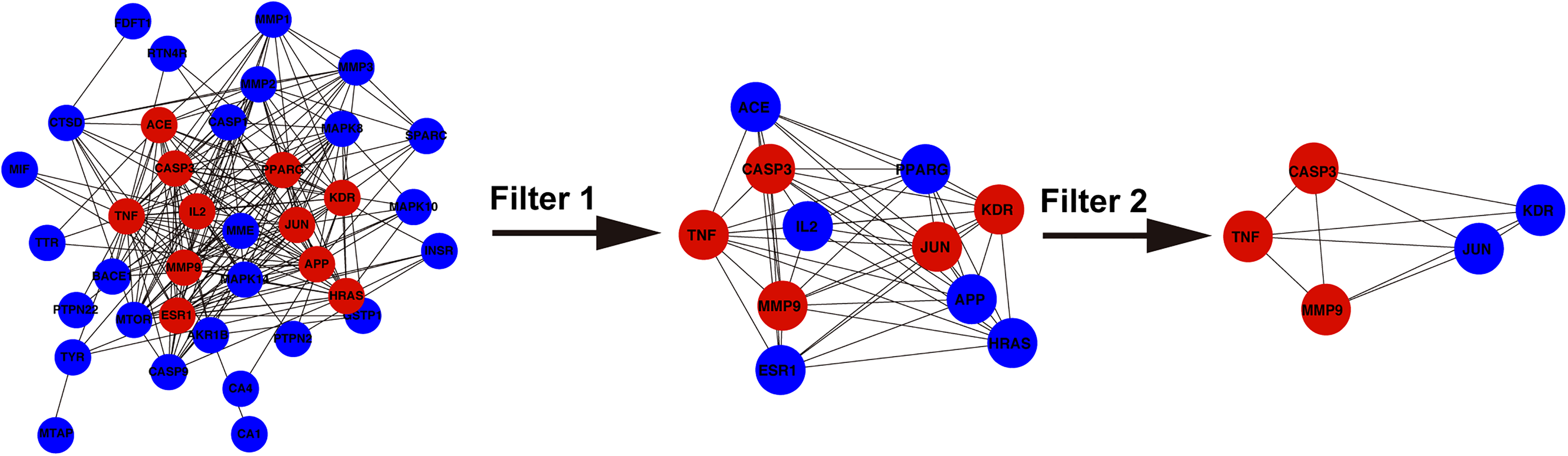
Topological analyses of the 37 shared genes. Red and blue represent genes that met or did not meet the screening conditions, respectively.
Molecular Docking Verification
The binding forces of the key molecules TNF, MMP9 and caspase 3 to Sal-B were determined by molecular docking software. The results showed that Sal-B can easily interact with TNF, MMP-9 and caspase-3 and showed the 3D structures and binding abilities (Figure 6).

Molecular docking of key genes with sal-B and a 3D model diagram. A: TNF. B: MMP-9. C: Caspase 3.
Salvianolic Acid B Inhibits the Decrease in HLEC Activity Induced by H2O2
Through a CCK-8 assay, we found that 2.5–20 μM Sal-B had no cytotoxic effect on HLECs (Figure 7A), while 300 μM H2O2 inhibited the activity of human lens epithelial cells (Figure 7B). We utilized 2.5–10 μM Sal-B to evaluate whether it could reverse the decrease in HLEC activity induced by H2O2. The results showed that 2.5–7.5 μM Sal-B reversed the decrease in cell activity induced by H2O2 in a dose-dependent manner (Figure 7C), and this concentration was used for the following experiments.

Sal-B reversed the decrease in human LEC activity induced by H2O2. A-C: CCK-8 assays were used to measure HLEC activity after treatment with different concentrations of Sal-B or H2O2. NS: No significant significance, ****P < 0.0001VS H2O2 group.
Salvianolic Acid B Inhibits H2O2-Induced HLEC Apoptosis
Flow cytometry showed that the percentage of apoptotic cells in the Sal-B-pretreated group was lower than that in the H2O2-treated group (Figure 8A and B). Moreover, the qPCR results showed that the expression of caspase 3 in the Sal-B-pretreated group was lower than that in the H2O2-treated group (Figure 9 A). In conclusion, Sal-B reversed H2O2-induced apoptosis in a dose-dependent manner.

Sal-B reversed H2O2-induced HLEC apoptosis. A: Flow cytometry was used to assess the apoptosis rate of HLECs treated with or without H2O2 or Sal-B. ****P < 0.0001.

Expression of the caspase 3, MMP-9 and TNF-α genes in HLECs treated with or without H2O2 or Sal-B. A-C: qPCR was used to determine the gene expression of caspase 3, MMP-9 and TNF-α in HLECs treated as described above. **P < 0.01, ****P < 0.0001VS H2O2 group.
Salvianolic Acid B Inhibits Oxidative Damage by Inhibiting H2O2-Induced TNF-α and MMP-9 Gene Expression in HLECs
The network pharmacology results showed that TNF-α and MMP-9 play potentially important roles in the inhibition of senile cataracts by salvianolic acid B; the qPCR results showed that the gene expression levels of TNF-α and MMP-9 increased in HLECs after treatment with H2O2, while Sal-B pretreatment inhibited this increase (Figure 9B, C).
Discussion
Senile cataracts, also known as age-related cataracts, are the main cause of vision decline in elderly individuals. The lens of normal people is transparent and can clearly visualize external objects on the retina as part of the intraocular refractive system. However, with increasing age, the lens will experience varying degrees of opacity, and a decrease in transparency will lead to a decrease in the vision of cataract patients. 14 The gradual worsening of cataracts can even lead to blindness. Phacoemulsification with intraocular lens implantation is an effective surgical treatment, 15 but for patients who are unable to undergo surgery or have a high risk of complications, researchers have paid increasing attention to how to use drugs to prevent cataracts or delay progression. Salvianolic acid B has been reported for the treatment of other ocular diseases, and Xiaobin Liu et al reported that Sal-B may have the potential to prevent and treat dry age-related macular degeneration through the inhibition of oxidative damage to RPE cells. 10 Age-related cataracts are related to oxidative damage to lens epithelial cells. Network pharmacology can be utilized for bioinformatics analysis by identifying common genes related to diseases and drugs and is the most recently developed method to explore the preventive and therapeutic potential of drugs in the treatment of diseases. Therefore, we explored whether salvianolic acid B has the potential to inhibit age-related cataracts through network pharmacology and then investigated the inhibitory effect of salvianolic acid B on oxidative damage in human lens epithelial cells and its potential mechanism.
In this study, 152 target genes of salvianolic acid B and 705 genes related to age-related cataract diseases were identified via network pharmacology, 37 shared genes were obtained via a Venn diagram, and the relationships between 37 genes were visualized via a PPI network. Among them, we found that TNF, IL-2, MMP 9, caspase 3 and JUN occupied core positions, indicating that they mediate the important relationship between salvianolic acid B and senile cataracts. three key genes, TNF, MMP 9 and caspase 3, were identified via Cytoscape software, and molecular docking showed that salvianolic acid B had superior binding energy. The visualization results also showed that salvianolic acid B could interact well with these genes.
Previous study reported that MAPK signalling pathway were involved in corneal wound healing and corneal vascularization after alkali burn injury,16,17 Guanghai Guo et al reported that inhibiting the mitogen-activated protein kinase (MAPK) signalling pathway can reduce oxidative damage and apoptosis in LECs. 18 In our study, GO analysis revealed that salvianolic acid B inhibited age-related cataracts in terms of the biological process apoptosis, the molecular function protein binding, and the extracellular region cellular component. Similarly, KEGG pathway analysis revealed that the TNF and IL-17 signalling pathways are also involved in this inhibitory effect, and we identified the specific molecules involved in these pathways (Figure 6) via the KEGG database. Our results show that this effect is strongly related to the MAPK signalling pathway,This result may suggest that the MAPK signalling pathway mediates the inhibition of senile cataracts by Sal-B. in the next cellular experiments, Salvianolic acid B inhibited the decrease in human LEC activity induced by H2O2 and reduced HLEC apoptosis. Which further verified the GO data analysis.
In age-related cataract formation, caspase-3 is involved in the oxidative stress-induced apoptosis of lens epithelial cells.19–21 Hangping Yao et al reported that inhibiting the activation of caspase 3 can reduce oxidative damage in HLECs. 21 MMP9 belongs to the class of matrix metalloproteinases whose substrates can be extracellular matrix molecules, secreted cytokines and cell surface molecules 22 ; at the same time, previous studies have shown that the expression level of MMP9 is the highest in cortical cataract patients and increases with age. 23 Previous studies have also shown that MMP9 is involved in the decomposition of extracellular matrix of LECs and plays a role in diabetic cataracts. 24 Furthermore, previous studies have also shown that stimulation of pig lenses with H2O2 can induce the production of MMP9. 25 In this study, we found that Sal-B inhibited the increase in MMP9 induced by H2O2. TNF is a prototypic member of the TNF ligand family that plays a role in regulating inflammation through the interaction between its receptors TNFR1 and TNFR2. 26 In this study, we also demonstrated the role of the TNF pathway (Figure 4). In previous studies, TNF-α has often been targeted in the treatment of other ocular diseases, such as glaucoma, uveitis and diabetic retinopathy.27–29 Dong Xu et al reported that H2O2 can increase the expression of TNF-α in LECs during oxidative damage. 30 In this study, we found that Sal-B pretreatment inhibited TNF-α expression induced by H2O2, which may be related to the inhibition of apoptosis, but some studies have also shown that the increase in TNF-α is related to lens capsule opacification. 31 Above all, caspase 3, MMP-9 and TNF were key molecules during the development of cataracts.
There are still some limitations in this experiment. Firstly, the potential of salvianolic acid to inhibit senile cataract was initially explored through network pharmacology, and finally Sal-B was used to inhibit oxidative stress in HLECs and detect related gene expression. However, HLECs used in our experiments are immortalized, which is related to the difficulty in obtaining primary cells, further verification on primary lens epithelial cells may make the results more convincing. And then, the future dosage form of Sal-B for the treatment of senile cataract, whether it is eye drops, subconjunctival injection or oral administration, still needs to be further explored.
Conclusion
Network pharmacology and molecular docking results showed that Sal-B has therapeutic potential for age-related cataracts; the therapeutic effect is related to the inhibition of apoptosis and inflammation. Furthermore, the results also showed that MMP-9, Caspase-3 and TNF may play key roles in this process. At the cellular level, the results confirmed that Sal-B could inhibit the H2O2-induced decrease in HLEC activity and apoptosis, and the expression of key molecular genes was verified. We hope to apply these findings to the clinical treatment of cataract patients in the future.
Supplemental Material
sj-docx-1-npx-10.1177_1934578X241272458 - Supplemental material for Exploration of the Therapeutic Potential of Salvianolic Acid B Against senile Cataracts Based on Network Pharmacology and Experimental Validation
Supplemental material, sj-docx-1-npx-10.1177_1934578X241272458 for Exploration of the Therapeutic Potential of Salvianolic Acid B Against senile Cataracts Based on Network Pharmacology and Experimental Validation by Yongxiao Dong, Jin Zhao, Xueli Zheng, Tao Xue, Wenting Ma, Panpan Cao, Ling Wang and Xiaoyong Yuan in Natural Product Communications
Footnotes
Acknowledgements
The study was supported by the Tianjin Key Laboratory of Ophthalmology.
Author Contributions
Yongxiao Dong: Conceptualization, Methodology, Software, Writing- Original draft preparation, Jin Zhao: Data curation, Writing- Original draft preparation, Xueli Zheng: Resources, Supervision, Tao Xue: Supervision Software, Wenting Ma: Software, Panpan Cao: Methodology, Ling Wang and Xiaoyong Yuan: Concepyuation, Funding Acquisition, Resources, Supervision, Writing- Reviewing and Editing.
Data Availability Statement
This article contains all the data generated or analysed during this study. Further enquiries can be directed to the appropriate author.
commercial relationship disclosures: None.
Declaration of Conflicting Interests
The authors declared no potential conflicts of interest with respect to the research, authorship, and/or publication of this article.
Ethics Statement
All studies were analysed by previous articles and bioinformatics software, excluding the original overview of clinical trials, so there was no need for ethical approval.
Funding
The authors disclosed receipt of the following financial support for the research, authorship, and/or publication of this article: This work was supported by the National Natural Science Foundation of China (No. 81970772, 82371033), The Tianjin Natural Science Foundation (21JCZDJC01250), the Tianjin Key Medical Discipline (Specialty) Construction Project (TJYXZDXK-016A) and the Xianyang Science and Technology Plan Project (2021ZDYF-SF-0056).
Supplemental Material
Supplemental material for this article is available online.
Abbreviations
References
Supplementary Material
Please find the following supplemental material available below.
For Open Access articles published under a Creative Commons License, all supplemental material carries the same license as the article it is associated with.
For non-Open Access articles published, all supplemental material carries a non-exclusive license, and permission requests for re-use of supplemental material or any part of supplemental material shall be sent directly to the copyright owner as specified in the copyright notice associated with the article.